Validation of transit-time flowmetry
for chronic measurements of regional
blood flow in resting and exercising rats
Departamento de Fisiologia e Biofísica, Instituto de Ciências Biomédicas, Universidade de São Paulo, 05508-900 São Paulo, SP, Brasil
S.L. Amaral and L.C. Michelini
Abstract
The objective of the present study was to validate the transit-time technique for long-term measurements of iliac and renal blood flow in rats. Flow measured with ultrasonic probes was confirmed ex vivo using excised arteries perfused at varying flow rates. An implanted 1-mm probe reproduced with accuracy different patterns of flow relative to pressure in freely moving rats and accurately quantitated the resting iliac flow value (on average 10.43 ± 0.99 ml/min or 2.78 ± 0.3 ml min-1 100 g body weight-1). The measurements were stable over an
experimental period of one week but were affected by probe size (resting flows were underestimated by 57% with a 2-mm probe when compared with a 1-mm probe) and by anesthesia (in the same rats, iliac flow was reduced by 50-60% when compared to the conscious state). Instantaneous changes of iliac and renal flow during exercise and recovery were accurately measured by the transit-time technique. Iliac flow increased instantaneously at the beginning of mild exercise (from 12.03 ± 1.06 to 25.55 ± 3.89 ml/min at 15 s) and showed a smaller increase when exercise intensity increased further, reaching a plateau of 38.43 ± 1.92 ml/min at the 4th min of moderate exercise intensity. In contrast, exercise-induced reduction of renal flow was smaller and slower, with 18% and 25% decreases at mild and moderate exercise intensities. Our data indicate that transit-time flowmetry is a reliable method for long-term and continuous measurements of regional blood flow at rest and can be used to quantitate the dynamic flow changes that characterize exercise and recovery.
Correspondence L.C. Michelini
Departamento de Fisiologia e Biofísica, ICB-USP Av Prof. Lineu Prestes, 1524 05508-900 São Paulo, SP Brasil
Fax: 55 (011) 818-7285 and 813-0845
E-mail: [email protected] Research supported by CNPq and FAPESP. S.L. Amaral was the recipient of a CNPq fellowship.
Received October 9, 1996 Accepted April 29, 1997
Key words
•Ultrasonic transit-time •Iliac blood flow •Renal blood flow •Exercise •Anesthesia •Conscious rats
Introduction
There is increasing interest in the con-tinuous measurement of total and regional blood flow in conscious animals. Electro-magnetic (1-7) and pulsed Doppler ultra-sonic flow probes (4,5,8-10) have been widely used for these measurements but they pres-ent some disadvantages. Electromagnetic
volumet-ric blood flow. Perivascular transit-time probes measure blood flow by emitting a plane of ultrasonic sound that transits the cross section of the vessel in both the up-stream and downup-stream directions. The dif-ference between the integrated transit-times of accelerated (directed downstream) and decelerated (directed upstream) ultrasonic signals, that interact with the volume flow occurring within the vessel, is a measure of volume per minute (ml/min or l/min). Since the transit-time technique gives absolute flow, independently of vessel size, and is easily implantable due to the reduced dimensions of the apparatus, it is adequate for chronic studies. In the rat, transit-time flowmetry has been widely used to quantitate blood flow in gastric (11,12), renal (13,14), mesenteric (13,15), carotid (13), and femoral (13) arter-ies and in the aorta (13,15) but only under acute conditions.
The validation of transit-time probes has been done in large animals under a variety of experimental conditions (3,7,16,17). In the rat, renal (14), mesenteric and aortic flows (15) were measured with Transonic probes and compared with known blood/saline flows obtained from pump-perfused circuits (15) or by clearance of p-aminohippuric acid (14). These data, obtained in acute experiments, have demonstrated the accuracy of the tran-sit-time method for blood flow measure-ments. No information on the stability of chronic measurements by the transit-time probe is available for conscious freely moving rats. All previous studies concerning regional blood flow changes induced by exercise in rats (18-21) were conducted using the mi-crosphere technique, which only provides information about static but not dynamic flow behavior. There is no information about continuous measurements and/or time-course changes of regional blood flow during main-tained exercise and recovery. Studies of con-tinuous blood flow changes simultaneous to pressure/heart rate changes during exercise are also lacking. In order to standardize the
transit-time flowmetry for recording regional blood flow in rats, the specific objectives of the present study were 1) to validate true flow values obtained in vitro with 1-mm and 2-mm probes, 2) to determine the sensitivity of this technique in reproducing the instanta-neous changes of flow simultainstanta-neously with pressure in freely moving rats, 3) to evaluate the accuracy of the 1-mm probe in recording sequentially the basal iliac flow (IF) after chronic implantation and 4) to describe the dynamic behavior of hindlimb and renal flow at rest, during exercise and recovery.
Material and Methods
In vitro perivascular flow probe testing
The flow probes were submitted to bench tests before each implantation to confirm their performance. The values of the test signal were high (0.9 ± 0.06 to 1.02 ± 0.05 V) as recommended by the manufacturer and did not change throughout the experiment.
In vivo studies
All surgical procedures and protocols used were in accordance with the Institutional Guide-lines for Animal Experimentation.
Male Wistar Kyoto rats (250-300 g, 2-3 months old) were anesthetized with sodium pentobarbital (50 mg/kg, ip). Two 1-cm dor-sal incisions were made in the skin overlying the midscapular and iliac regions, and a blunt hemostat was used to create a subcutaneous tunnel between them. A midline incision was performed in the abdominal wall and the left iliac artery just below the aortic bifurca-tion (hindlimb studies) or the left renal artery (renal blood flow studies) was exposed and isolated for probe placement. All fatty tissue was carefully removed from the isolated seg-ment of the vessel to avoid obstruction of the ultrasonic signal. The 1-mm (or 2-mm in one of the protocols) transit-time ultrasonic flow probe (model 1RB) was placed on the cleared section of the vessel. The probe was sutured to the underlying musculature and the seg-ment of the artery containing the probe was covered with Lyostypt (Braun, Melsungen, Germany) to maintain vessel/probe align-ment and to inhibit fat infiltration. The probe cable was passed through a small incision made in the retroabdominal wall and tun-neled subcutaneously to the dorsal midscapu-lar region. The probe was tested for the presence of a good acoustic signal, and blood flow during anesthesia was recorded for a period of 10-20 min (Gould recorder, model 4000, Cleveland, OH). The abdominal inci-sion was then sutured and the connector end was fixed to the skin. The rats were treated with 60,000 units (0.1 ml, im) of penicillin (Pentabiótico Veterinário Fontoura, São Paulo, SP, Brazil). To record blood pressure, a clear vinyl catheter (3 cm of #10 tubing connected to 13 cm of #90 tubing) was in-serted into the right iliac artery through the femoral artery of ether-anesthetized rats. The external part of the cannula was tunneled subcutaneously through the midscapular
re-gion. The rats were allowed to recover for a period of 24 h.
Experimental protocols
Other groups of rats chronically implanted with a 1-mm probe in the iliac artery (group II, N = 7) or in the renal artery (group III, N = 13) were tested for regional blood flow changes during exercise. Before probe im-plantation the rats were adapted to 5-min treadmill running episodes of 0.3 to 0.8 km/ h, 0% degree for one week. The regional flow during rest, running and recovery was recorded on 3 successive days: on the first and second recording sessions, held 24 and 48 h after probe implantation, blood flow and heart rate were measured continuously. After stabilization of the variables, the rat ran on a treadmill (Inbramed, Porto Alegre, RS, Brazil) adapted for rats. The exercise test consisted of 4 min of graded exercise: 2 min at 0.4 km/h and 2 min at 0.8 km/h, 0% degree. On the second day the rats were implanted with the arterial cannula into the contralateral femoral artery and the running protocol was repeated 24 h later. To assess the reproducibility of IF, arterial pressure (AP) and HR changes during exercise, 2 sessions of running episodes were held for each rat, 40-60 min apart. Before the running protocol, IF, HR and AP were recorded for
15 to 30 min to permit stabilization of the variables and to obtain true resting values.
Group IV consisted of 8 rats with a 2-mm probe (model 2SB) chronically implanted around the iliac artery. IF records were re-peated on 3 consecutive days (40-60 min experimental sessions) in freely moving rats.
Statistical analysis
Data are reported as mean ± SEM. The differences between groups were evaluated by the paired t-test or by analysis of variance for repeated measurements as appropriate. The level of significance was set at P<0.05.
Results
Standardization of 1-mm and 2-mm transit-time probes: in vitro validation and in vivo
stability
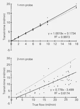
Figure 1 provides the pooled data for relationships between true flow rate and measured flow with both probe sizes in the excised carotid artery. Under these experi-mental conditions the slopes for the 1-mm and 2-mm probes were quite different: 1.002 with a correlation coefficient of 0.997, and 0.778 with a correlation coefficient of 0.617 for probes 1R and 2S, respectively. Although data of carotid flow obtained with the 1-mm flow probe were superimposed to the line of identity, those obtained with the 2-mm probe were not. These results indicate that flow in the common carotid and iliac arteries is bet-ter quantitated by the 1-mm flow probe.
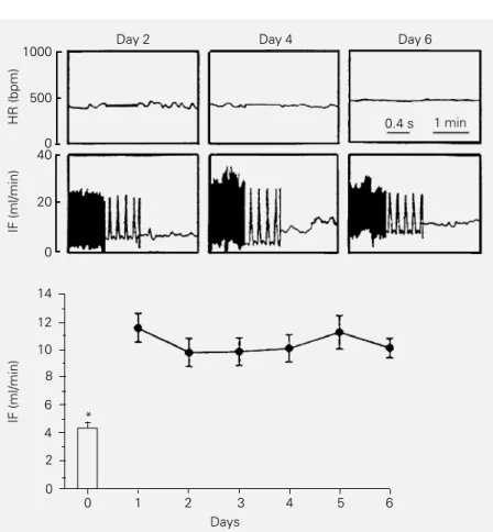
Iliac flow measurements after chronic implantation presented excellent stability. As observed in Figure 2 (upper panel for one conscious rat), both baseline HR and resting IF (pulsatile and mean) were stable and closely similar from day 2 up to day 6 after implantation. For this group, mean IF was on average 10.43 ± 0.99 ml/min, a value that was almost constant throughout the experi-mental period (Figure 2, lower panel). At
Transit-time (ml/min)
20
15
10
5
0
-5
Transit-time (ml/min)
15
10
5
-5 0 20
18
5 10 15 20 25 30
1-mm probe
y = 1.0019x + 0.1734
R2 = 0.9973
y = 0.778x - 3.499
R2 = 0.6174
True flow (ml/min)
Figure 1 - In vitro calibration of 1-mm (upper panel) and 2-mm (lower panel) probes implanted around the rat common carotid artery. Symbols represent ex-perimental data obtained with 1-mm (N = 4) and 2-1-mm (N = 2) probes. The solid line represents the slope of the data and the dotted line, the line of identity. Parameters of linear regression analysis are shown.
0 2 4 6 8 10 12 14 16
0 30
25
rest, HR oscillated between 367 ± 15 and 398 ± 24 bpm during the 6 days of the experimental period. On the 6th day basal mean arterial pressure (MAP) was 110 ± 3 mmHg. It should be noted that at that time (after catheterization of the contralateral femoral artery) mean resting IF was 10.10 ± 0.7 ml/min. In contrast, the IF of conscious rats at rest differed markedly from the IF measured during surgery after acute implan-tation of the probe into pentobarbital-anes-thetized rats. As illustrated in Figure 2, the IF of the same rats (4.46 ± 0.56 ml/min) was significantly reduced by 50-60% during an-esthesia when compared to the conscious state.
Accuracy of probe measurements
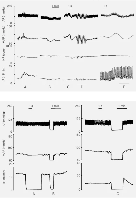
The sensitivity of the transit-time probe in reproducing physiological changes of IF was analyzed in chronically implanted rats also instrumented with an arterial catheter. As shown in Figure 3, IF records reproduced not only tonic but also phasic instantaneous absolute changes of pulsatile and mean flow, i.e., all the physiological patterns of flow changes such as increases/decreases simul-taneous to elevations/falls in pressure (pan-els A and B), no changes regardless of pres-sure changes (panel C) and phasic flow os-cillations opposite to pressure osos-cillations (panel D) were observed. Maintenance of elevated flow (tonic effect) simultaneously to pressure oscillation was also observed (panel E). Most of these flow patterns were presented by the 10 freely moving rats of group I. The flow changes related or not to pressure modification seemed to depend on the behavioral activities and demonstrated that the probe was able to detect with accu-racy all patterns of IF changes.
Zero flow baseline measurement
The accuracy of flow measurements was tested in situ after median laparotomy and
exposition of the distal abdominal aorta and iliac artery implanted with the probe (ether anesthesia). Figure 4 shows a typical record-ing with the 1-mm probe taken from 2 rats. Brief occlusions (20 to 60 s, panels B and C, respectively) actually caused zero pressure and true zero flow in the iliac artery which were properly detected by the probe. Mean flow measured after abdominal aortic occlu-sion in the group of rats as a whole was 0.33 ± 1.3 ml/min, which was not different from zero. Also indicative of probe accuracy in detecting flow changes was the observation of the transient increase that characterizes the reactive hyperemic response after 1-min occlusion of the abdominal aorta (Figure 4,
Figure 2 - Upper panel, Sequential recordings of heart rate (HR) and pulsatile and mean iliac flow (IF) at rest in a conscious rat with a perivascular 1-mm probe chronically implanted around the iliac artery. Lower panel, Mean values of resting iliac flow obtained on 6 consecutive days in the group of 10 rats. The bar represents the flow of the same rats immediately after probe implantation and during pentobarbital anesthesia. *P<0.05 com-pared to the conscious state (paired t-test).
0.4 s 1 min
Day 2 Day 4 Day 6
HR (bpm)
1000
IF (ml/min)
500
0 40
20
0
IF (ml/min)
14
12
10
8
6
4
2
0 *
0 1 2 3 4 5 6
Figure 3 - Records of pulsatile (AP) and mean arterial pressure (MAP), heart rate (HR) and mean or pulsatile iliac flow (IF) in freely moving rats with a 1-mm probe chronically implanted around the iliac artery. See text for explana-tion.
250
0
AP (mmHg)
125
50
MAP (mmHg)
150
100
20
0 10
IF (ml/min)
1 s 1 min 1 s 1 min
A B C
250
0 125
50 150
100
40
0 20
Figure 4 - Simultaneous record-ings of arterial pressure (AP, MAP) and mean iliac flow (IF) in ether-anesthetized rats during flowmeter zero flow measure-ments without interruption of blood flow (A) and the true zero flow measurements (B and C).
panel C). Moreover, the flowmeter zero flow function (Figure 4, panel A) produced zero flow recordings similar to true zero flow measurements (panels B and C). Since no
differences between the two methods for measuring zero blood flow were observed, the zero baseline function was used for cali-bration during the chronic experiments. We
AP (mmHg)
250
0
MAP (mmHg)
150
50 1000
0 40
0
HR (bpm)
IF (ml/min)
1 min 1 s 1 s
1000
also confirmed true zero flow in some ani-mals by measuring IF in situ after sacrifice at the end of the experiments.
Regional flow changes during exercise
The behavior of IF in response to dynam-ic exercise was tested simultaneously with AP records in another group of chronically instrumented rats during short 4-min runs on a treadmill. As illustrated in Figure 5, AP showed a small but similar increase (+10 to +15 mmHg) during the running period in rats implanted with iliac or renal probes. HR and IF increased quickly during the first 15-30 s of mild exercise (0.4 km/h), with an addi-tional increase being observed at the begin-ning of moderate exercise intensity (0.8 km/ h), and remained at this level until the end of the exercise. In contrast, renal flow (RF) showed opposite and smaller changes, i.e., it was almost unchanged at the beginning of exercise and showed a slow and continuous decrease until the end of the exercise proto-col.
Table 1 shows the baseline values of MAP, HR and regional blood flow obtained for the iliac and renal groups (protocols II and III). Baseline MAP, HR and absolute iliac and renal flow were similar for both groups. The renal group also had a smaller body weight (366 ± 17 vs 458 ± 18 g in the iliac group) with a similar relative flow (3.04 ± 0.5 vs 2.78 ± 0.3 ml min-1 100 g-1). On the
other hand, calculations of flow index by correcting for kidney and hindlimb muscle
Figure 5 - Recordings of pulsa-tile (AP) and mean arterial pres-sure (MAP), heart rate (HR) and mean regional flow (BF) for 2 rats with a chronically implanted 1-mm probe (left panel, iliac flow and right panel, renal flow) during rest, exercise of mild (0.4 km/h) to moderate (0.8 km/h) intensity (black bars) and recov-ery.
Table 1 - Resting mean arterial pressure (MAP), heart rate (HR), and iliac and renal flow measured with a 1-mm probe in 2 groups of rats.
Iliac flow Renal flow (N = 7) (N = 10)
MAP (mmHg) 109 ± 3 110 ± 4
HR (bpm) 370 ± 6 388 ± 21
Absolute flow (ml/min) 12.03 ± 1.1 10.86 ± 1.7
Relative flow (ml min-1 2.78 ± 0.3 3.04 ± 0.5
100 g body weight-1)
Flow index 1.7 7.9
(ml min-1 g tissue-1)
0 40
0
HR (bpm)
BF (ml/min)
250
0 150
50
0 20
0 0.4 km/h 0.8 km/h
0 2 4
Rest Exercise Recovery
0.4 km/h 0.8 km/h
0 2 4
Rest Exercise Recovery
Iliac probe Renal probe
1 min
AP (mmHg)
250
0
MAP (mmHg)
150
weight (on average 1.37 g and 7.49 g, respec-tively) showed a higher value for renal tissue (7.93 vs 1.7, renal and hindlimb index, re-spectively).
The flow changes during exercise for both groups are illustrated in Figure 6. Dur-ing mild exercise the first workload mean IF was increased by +15.1 ± 3.1 ml/min, a value that is more than double (114% increase) the resting IF, reaching +20.43 ± 2.45 ml/min (+158%) at the second workload. At the same exercise intensity, RF showed minor
changes compared to baseline, i.e., RF de-creased on average 2 ml/min (-18%) during the second minute of 0.4 km/h and 3-4 ml/ min at 0.8 km/h (-25% below the resting value). As observed in Figure 6, the recovery of both IF and RF was progressive, reaching basal values 3-4 min after the end of exer-cise.
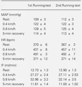
IF changes during acute exercise were reproducible. In two successive runs on a treadmill 40-60 min apart, similar AP, HR and IF responses were observed (Table 2): for the same AP changes, IF was increased 2.1 and 2.6 times for 0.4 and 0.8 km/h in the first test and 2.1 and 2.5 times in the second test and a complete recovery of baseline values occurred 5 min after the end of exer-cise.
Absolute flow measurements: 1-mm vs 2-mm
probes
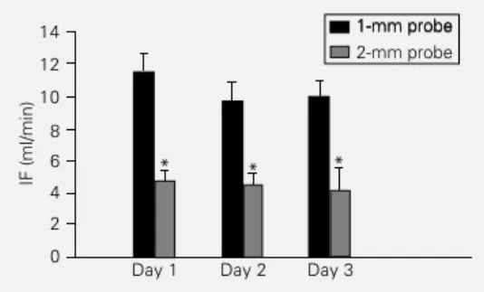
An important and unexpected observa-tion concerning absolute blood flow values is illustrated in Figure 7. Comparing IF val-ues obtained with the 1-mm probe (group I) with the values presented by another group of rats chronically implanted with the 2-mm probe (group IV, N = 8), we observed that probes of different sizes showed different absolute IF values for the same condition, i.e., conscious state at rest. The values ob-tained with the large probe were significant-ly lower (57% on average) than those ob-served with the 1-mm probe. It should be noted that the average value for the 3-day period (4.45 ± 0.19 ml/min) was similar to that observed in vitro for a true flow of 10 ml/min (Figure 1).
Postmortem findings
No macroscopic evidence of iliac or re-nal artery damage was found in any group of rats instrumented with the transit-time flow probes. There was also no evidence of dis-turbance of hindlimb or renal blood flow
BF (ml/min)
40
30
20
10
0
Iliac flow Renal flow
0.4 0.8 km/h
-4 -2 0 2 4 6 8 10 12 14 16 min
Rest Exercise Recovery
Figure 6 - Time-course changes of absolute regional flow (BF) (iliac, N = 7 and renal, N = 10) during 4 min of exercise (black bar) and during recovery.
Table 2 - Comparison of mean arterial pressure (MAP), heart rate (HR) and iliac flow (IF) values in 2 running tests involving 7 normotensive rats.
Resting values, maximal changes at 2 different intensi-ties and recovery values are presented.
1st Runningtest 2nd Runningtest
MAP (mmHg)
Rest 109 ± 3 112 ± 3
0.4 km/h 122 ± 4 122 ± 3
0.8 km/h 128 ± 5 125 ± 4
5-min recovery 114 ± 4 113 ± 4
HR (bpm)
Rest 370 ± 6 367 ± 3
0.4 km/h 431 ± 8 457 ± 11
0.8 km/h 491 ± 3 492 ± 7
5-min recovery 371 ± 12 371 ± 14
IF (ml/min)
Rest 12.73 ± 1.3 12.90 ± 1.3
0.4 km/h 27.27 ± 2.4 27.11 ± 2.53
0.8 km/h 32.96 ± 3.2 32.14 ± 3.5
since the performance of rats on the tread-mill after probe implantation was similar to that observed 1-2 days before surgery.
Discussion
In the present study we demonstrate for the first time that a transit-time probe per-mits sequential and accurate measurement of regional blood flow responses during rest, exercise and recovery. In addition, we showed that after chronic implantation the technique provided stable and reliable measurements, thus permitting continuous (and simulta-neously to pressure) recording of tonic flow as well as its phasic changes. In contrast to previous reports that blood flow is inde-pendent of the size of the transit-time probe (14,15), we showed that 1-mm and 2-mm probes gave different absolute flow values, both in vitro and when chronically implanted, indicating that volumetric flow measurements are limited by the window of accuracy, which varies with probe size.
The transit-time technology for measur-ing dynamic changes of regional blood flow has several advantages over other methods such as electromagnetic, microsphere and Doppler system flowmetry: 1) small probe size permitting easy implantation; 2) con-tinuous measurement of absolute blood flow representing an enormous advantage over phasic (microspheres, dye dilution) (3,22,23) or relative measurements (3,4,8-10); 3) probes are nonconstrictive and therefore do not interfere with the pulse pressure or per-centage of diastolic flow as electromagnetic devices do (7,14); 4) the probes are suitable for long-term studies (17) since the quality of the received signal improves with reactive infiltrating tissue growth around the probe/ vessel, providing the necessary acoustic con-duction (3,15), and 5) the flowmeter zero offset function permits frequent calibration in conscious freely moving rats. The perivas-cular transit-time probe has been success-fully used for continuous assessment of flow
in coronary (17), mesenteric (13,15-17), re-nal (13,14,17,24), carotid (13), gastric (11,12), hindlimb (13,17,25,26) and pulmo-nary arteries (7) and also for measurement of cardiac output (3,8,13,15,24). Most of these studies, however, were carried out after acute implantation and few were conducted after chronic implantation. Chronic implantation, however, was employed to study drug ef-fects on cardiac output and regional flows (8) and did not attempt to describe the daily flow profile of resting animals (17,25). The results of the present experiments showed that implantable transit-time probes provid-ed stable resting iliac flow values throughout the experimental period of 1 to 6 days. An-other important result was the fact that the mean iliac flow of awake rats was not af-fected by arterial catheter placement in the contralateral femoral artery. These observa-tions indicate that perivascular transit-time probes are suitable for chronic flow/pressure studies from the first day after implantation. In addition, the flow records obtained simul-taneously to blood pressure records in awake rats demonstrated that transit-time probes reliably reproduce different flow patterns (mean and/or pulsatile) conditioned by pres-sure oscillations, opposite to prespres-sure changes or bearing no relationship to them. Brief pulsatile records of femoral flow have al-ready been reported for conscious rats (25), but they were obtained immediately before tail suspension and did not represent the true baseline flow. Bednarik and May (17) also employed this technique to measure mesen-teric, renal, iliac and coronary blood flows
Figure 7 - Comparison of abso-lute iliac flow values (IF) ob-tained in the conscious state at rest (day 1 up to day 3) for rats implanted with a 1-mm (group I, N = 10) or 2-mm probe (group IV, N = 8). *P<0.05 compared to the 1-mm probe (ANOVA).
IF (ml/min)
14 12 10
8 6 4 2 0
1-mm probe 1-mm probe 2-mm probe
Day 1 Day 2 Day 3
simultaneously to pressure records in con-scious sheep 16 months after probe implan-tation, but no time-course plots for resting flow values were presented. These observa-tions, taken together with the present data, indicate that transit-time flowmetry is an effective and accurate technique for the quantitation of absolute baseline flow values over time. It is better than electromagnetic devices (1,2) because it is nonconstrictive and does not show drift of the flow signal (6). The present protocol also included com-parisons of zero blood flow obtained by the zero flow baseline function of the flowmeter and by measurement of zero blood flow after complete occlusion of the vessel. The two procedures provided similar measurements and, since there were no detectable changes in zero baseline function during the experi-mental protocol, we confirmed the advan-tage of the ultrasonic method over electro-magnetic flowmetry in chronic studies. These observations agree with other studies (13, 15,17).
An important and unexpected observa-tion was the difference between the 1-mm and 2-mm probes concerning the resting iliac flow values of conscious rats. This discrep-ancy could be explained by the different windows of accuracy for the 1-mm and 2-mm probes. D’Almeida et al. (15) reported that true flow for the 2-mm probe is identical to measured flow only for values of about 20 ml/min (ranging from 10.4 to 45 ml/min), with marked deviations occurring at smaller or larger flow values. Our data with the 2-mm probe also showed deviations from true flow, but extending for all ranges of flow tested. On the other hand, our data with the 1-mm probe indicated a perfect correlation between measured and true flows, confirm-ing previous observations (15) and strongly suggesting that the flows measured with the 1-mm probe accurately represent the true flow rates. Based on these observations, we suggest that the resting iliac flow of awake normotensive rats is about 2.8 ml min-1 100
g body weight-1 or 1.7 ml min-1 g tissue-1.
We also showed that measurements in conscious and anesthetized rats yielded dif-ferent values even when using the same probe. It is well known that pentobarbital anesthe-sia depresses respiratory function, decreases sympathetic vasoconstrictor tone and in-creases vasodilator tone, thus reducing stroke volume, central venous pressure and arterial pressure (27). Accordingly, during anesthe-sia the iliac flow was proportionally de-creased when compared to the conscious state. In keeping with these observations, care should be taken when comparing flow profiles obtained with different transit-time probe sizes during different states (conscious vs anesthetized) and by different methods. Our absolute values are also in agreement with those obtained by Iriuchijima (1) who found a resting terminal aortic flow of 5.8 ml min-1 100 g-1, a value similar to that obtained
here for iliac flow if we consider that aortic flow is shared by each iliac artery. It should be noted that the flow index calculated from the present data confirmed the well-known concept of flow distribution, i.e., that the renal circulation receives a larger portion of cardiac output than the skeletal muscle cir-culation under resting conditions.
unex-pected due to autoregulatory responses (29). Whether renal flow is maintained or de-creased during exercise remains controver-sial (30). Most evidence suggests that redis-tribution of renal flow to exercising muscles only occurs when the exercise intensity is sufficiently severe and/or when a decrease in oxygen delivery to working muscles is imposed (30). Dynamic regional flow pat-terns during exercise have not been studied previously mainly due to the lack of an ap-propriate technique. Blood flow distribution within and among muscles in rats (18-21) and dogs (30) during exercise was deter-mined by means of radiolabeled and radio-active microsphere techniques which permit few and static flow measurements, but do not provide information about the dynamic moment-to-moment flow changes as obtained here with the perivascular ultrasonic probe.
Based on the present observations, we pro-pose that the transit-time technique is ad-equate to reproduce with accuracy any flow profile independent of absolute change (in-crease/decrease) and time-course (instanta-neous or phasic/maintained or tonic).
In summary, the transit-time flow probe provides important advantages over other systems for the measurement of regional blood flow. It provides direct flow volume measurements, has a negligible zero offset and presents a long in vivo life span, thus permitting accurate measurements of regional flow after chronic implantation over long periods. It is a good and reliable method to measure instantaneously and continuously the dynamic changes of blood flow during rest, exercise and recovery in small animals such as rats.
References
1. Iriuchijima J (1983). Regional blood flow in conscious spontaneously hypertensive rats. Japanese Journal of Physiology, 33: 44-51.
2. Iida N (1995). Different flow regulation mechanisms between celiac and mesen-teric vascular beds in conscious rats. Hy-pertension, 25: 260-265.
3. Hartman JC, Olszanski DA, Hullinger TG & Brunden MN (1994). In vivo validation of a transit time ultrasonic volume flow meter. Journal of Pharmacological Meth-ods, 31: 153-160.
4. Janssen BJ, Oosting J, Slaaf DW, Persson PB & Struijker-Boudier HA (1995). Hemo-dynamic basis of oscillations in systemic arterial pressure in conscious rats. Ameri-can Journal of Physiology, 269: H62-H71. 5. Grohs JG, Huber S & Raberger G (1993). Simultaneous assessment of cardiac out-put with pulsed Doppler and electromag-netic flowmeters during cardiac stimula-tion. Journal of Pharmacological and Toxi-cological Methods, 30: 33-38.
6. Gorewit RC, Aramondo MC & Bristol DC (1989). Measuring bovine mammary gland blood flow using a transit-time ultrasonic flow probe. Journal of Dairy Science, 72: 116-119.
7. Grant BJB, Paradowski LJ & Fitzpatrick JM (1988). Effect of perivascular electro-magnetic flow probes on pulmonary he-modynamics. Journal of Applied Physiolo-gy, 65: 1885-1890.
8. Dundore RL, Habeeb PG, Pratt PF, Becker LT, Clas DM & Buchholz RA (1992). Dif-ferential hemodynamic responses to se-lective inhibitors of cyclic nucleotide phos-phodiesterases in conscious rats. Journal of Cardiovascular Pharmacology, 19: 937-944.
9. Hartley CJ & Cole JS (1974). An ultrasonic pulsed Doppler system for measuring blood flow in small vessels. Journal of Applied Physiology, 37: 626-629. 10. Gelsema AJ, Copeland NE, Drolet G &
Bachelard H (1993). Cardiovascular ef-fects of neural activation of the extended amygdala in rats. Brain Research, 626: 156-166.
11. Holzer P, Lippe ITh, Jocic M, Wachter Ch, Erb R & Heinemann A (1993). Nitric oxide-dependent and -inoxide-dependent hyperaemia due to calcitonin gene-related peptide in the rat stomach. British Journal of Phar-macology, 110: 404-410.
12. Holzer P, Wachter Ch, Jocic M & Heinemann A (1994). Vascular bed-de-pendent roles of the peptide CGRP and nitric oxide in acid-evoked hyperaemia of the rat stomach. Journal of Physiology, 480: 575-585.
13. Hoffman A, Grossman E, Öhman KP, Marks E & Keiser R (1989). Endothelin induces an initial increase in cardiac out-put associated with selective vasodilation in rats. Life Sciences, 45: 249-255. 14. Welch WJ, Deng X, Snellen H & Wilcox C
(1995). Validation of miniature ultrasonic transit-time flow probes for measurement of renal blood flow in rats. American Jour-nal of Physiology, 268 (Renal, Fluid and Electrolyte Physiology, 37): F175- F178. 15. D’Almeida MS, Gaudin C & Lebrec D
(1995). Validation of 1- and 2-mm transit-time ultrasound flow probes on mesen-teric and aorta of rats. American Journal of Physiology, 268 (Heart and Circulatory Physiology, 37): H1368- H1372. 16. Barnes RJ, Comline RS, Dobson A &
17. Bednarik JA & May CN (1995). Evaluation of transit time system for the chronic measurement of blood flow in conscious sheep. Journal of Applied Physiology, 78: 524-530.
18. Laughling MH, Armstrong RB, White J & Rouk K (1982). A method for using micro-spheres to measure muscle blood flow in exercising rats. Journal of Applied Physi-ology, 52: 1629-1635.
19. Armstrong RB & Laughling MH (1984). Exercise blood flow patterns within and among rat muscles after training. Ameri-can Journal of Physiology, 246(Heart and Circulatory Physiology, 15): H59-H68. 20. Peterson DF, Armstrong RB & Laughling
MH (1988). Sympathetic neural influences on muscle blood flow in rats during submaximal exercise. Journal of Applied Physiology, 65: 434-440.
21. Mush TI & Terrel JA (1992). Skeletal muscle blood flow abnormalities in rats with chronic myocardial infarction: rest and exercise. American Journal of Physi-ology, 262 (Heart and Circulatory Physiol-ogy, 31): H411-H419.
22. Kobayashi N, Kobayashi K, Kouno K, Horinaka S & Yagi S (1994). Effects of intra-arterial injection of colored micro-spheres on systemic hemodynamics and regional blood flow in rats. American Jour-nal of Physiology, 266 (Heart and Circula-tory Physiology, 35): H1910-H1917. 23. Kissling G, Ross C & Brandle M (1993).
Validity of thermal dilution technique for measurement of cardiac output in rats. American Journal of Physiology, 265 (Heart and Circulatory Physiology, 34): H1007-H1013.
24. Shiraichi H, Silverman NH & Ruldolph AM (1993). Accuracy of right ventricular out-put estimated by Doppler echocardiogra-phy in the sheep fetus. American Journal of Obstetrics and Gynecology, 168: 947-953.
25. Roer DR & Dillaman M (1994). Decreased femoral arterial flow during simulated microgravity in the rat. Journal of Applied Physiology, 76: 2125-2129.
26. Wilkening RB, Boyle DW & Meschia G (1988). Measurement of blood flow and oxygen consumption in the pelvic limb of fetal sheep. Proceedings of the Society for Experimental Biology and Medicine, 187: 498-505.
27. Fragen RJ (1986). Cardiovascular effects of intravenous anesthetics. In: Altura BM & Halevy S (Editors), Cardiovascular Ac-tions of Anesthetics and Drugs Used in Anesthesia. Vol. 1. Karger, New York, 51-73.
28. Mitchell JH (1990). Neural control of the circulation during exercise. Medicine and Science in Sports and Exercise, 22: 141-154.
29. Cowley Jr AW (1992). Long term control of arterial blood pressure. Physiological Reviews, 72: 231-300.