INTRODUCTION
Strains of Aspergillus nidulans with duplicate chro-mosomal segments are unstable at mitosis (Bainbridge and Roper, 1966). Two strains (A and B) which carry the same chromosomal duplication, Dp(I-II), have been widely used to study the main features of mitotic instability (Nga and Roper, 1968; Azevedo and Roper, 1970; Azevedo, 1975; Menezes and Azevedo, 1978; Marjefeld and Roper, 1978). Colonies from strains carrying this duplication have re-duced growth rates and mainly produce two types of tors, designated improved and deteriorated. Improved sec-tors occur frequently and arise from nuclei which have lost a variable part or all of the duplicated segment (Nga and Roper, 1968). Deteriorated sectors are regularly but less frequently found. These sectors were postulated to result from tandem duplications on duplicated segments or from transposition to non-duplicated regions, with the latter showing a reduced degree of instability (Azevedo and Roper, 1970).
In the last decade, A. nidulans has been manipulated by recombinant DNA techniques. This fungus has been used for gene cloning, expression and regulation studies (Bowyer et al., 1994; Lee and Adams, 1994). The karyo-type of A. nidulans has also been resolved by electro-phoresis (Brody and Carbon, 1989). The size of the indi-vidual chromosomes was estimated to be 2.9-5.0 Mb, with a total size of 31 Mb. The development of pulsed-field gel electrophoresis techniques provides new possibilities for studying how chromosomal duplications affect mitotic instability. We used pulsed-field gel electrophoresis to
determine the electrophoretic karyotype of A. nidulans strains carrying Dp(I-II) and variants derived from them.
MATERIAL AND METHODS
The A. nidulans strains were derived from Glasgow stocks. Strains A and B were derived from a spontaneous duplication strain (Pritchard, 1956; Nga and Roper, 1968). Deteriorated strains (V5, V9 and V17) were isolated as sectors from strains A and B (Azevedo and Roper, 1970). Two non-duplication strains, pabaA1 and MSE (McCully and Forbes, 1965), were used as controls. One improved strain (AM) obtained as a spontaneous yellow sector from strain A was also used. The culture media were solid and liquid minimal medium (MM) as described by Pontecorvo et al. (1953) supplemented with proline, p-aminobenzoic acid, adenine, biotin, riboflavin, nicotinamide, methionine and pyridoxine.
Chromosomal DNA of A. nidulans was prepared ac-cording to Brody and Carbon (1989), with some modifi-cations. About 109 conidia from each strain were
inocu-lated in 800 ml of supplemented liquid MM in two-liter flasks. These were incubated for 12-15 h at 25°C, with vigorous shaking. Mycelia were harvested by filtration, washed with OM buffer (1.2 M MgSO4, 10 mM sodium
phosphate, pH 5.8) and 1-3 g was then suspended in 10-20 ml of OM buffer. Then 20-40 mg of lysing enzyme (Sigma) and 10-20 mg of bovine serum albumin were added. To produce protoplasts, a mycelium from each strain was in-cubated for 2 h at 30°C, with gentle swirling. The suspen-sion was filtered through miracloth and 10 ml of filtrate
Electrophoretic characterization of
Aspergillus nidulans
strains with chromosomal duplications
Marisa V. de Queiroz1, Aline Aparecida Pizzirani-Kleiner2 and João Lúcio Azevedo2
Abstract
Pulsed-field gel electrophoresis was used to characterize strains of Aspergillus nidulans with a chromosomal duplication Dp(I-II). Morphologically deteriorated and improved variants of these strains were also analyzed. The electrophoretic karyotype demonstrated that in two duplicated strains (A and B) the 4.2 Mb band, which corresponds to chromosome II, was absent and a new band was observed. Hybridization studies using the uapA (chromosome I) and wA (chromosome II) genes demonstrated that the new band corresponded to chromosome II plus the duplicated segment of chromosome I. The size of the chromosomal duplication was approxi-mately 1.0 Mb. Analysis of the chromosomal bands of a morphologically improved strain showed that the duplicated segment of chromo-some I was completely lost. The morphologically deteriorated variants V9 and V17 had the same karyotype as the duplicated strains. However, the deteriorated variant V5 lost part of chromosome I and had a rearrangement involving chromosome V. This rearrangement may have resulted from the mutagenic treatment used to obtain the genetic markers. Pulsed-field gel electrophoresis was found to be an excellent tool for locating chromosomal rearrangements.
containing the protoplasts was transferred to centrifuge tubes and carefully overlaid with 10 ml of ST buffer (0.6 M sorbitol, 10 mM Tris-HCl, pH 7.0). The tubes were cen-trifuged at 4,000 g for 15 min. The banded protoplasts were removed using a bent Pasteur pipette and mixed with an equal volume of 1 M sorbitol, 10 mM Tris-HCl, pH 7.5 buffer. The protoplasts were pelleted at 4,300 g for 10 min and washed twice with 5 ml of 1 M sorbitol, 10 mM Tris-HCl (pH 7.5) buffer containing 10 mM CaCl2. The
pellet was resuspended in GMB buffer (0.125 M EDTA, pH 7.5, 0.9 M sorbitol) to give a concentration of 108
pro-toplasts/ml. An equal volume of molten 1.4% InCert aga-rose (FMC) in GMB buffer precooled to 42°C was added, and the agarose/protoplast mixture was poured into a plug mold and solidified on ice for 10 min. The agarose plugs were immersed in NDS buffer (0.5 M EDTA, 10 mM Tris-HCl, pH 9.5, 1% sodium N-lauroylsarcosinate) contain-ing proteinase K (1 mg/ml) at 50°C for 24 h. The plugs were washed three times in 50 mM EDTA, pH 8.0, at 50°C and stored at 4°C.
Gel electrophoresis was performed using a Bio-Rad contour-clamped homogeneous electric field (CHEF) sys-tem at 45 V. The running buffer was 0.5X TBE or 0.5X TAE buffer at 14oC (Sambrook et al., 1989). The
separa-tion was done on agarose gels, as described by Geiser et al. (1994), using three pulse intervals of 50 min, 45 min and 37 min with durations of 73 h, 18 h and 73 h, respec-tively. The gels were stained with ethidium bromide (1 mg/ ml) for 30 min and destained in distilled water for at least 1 h. After destaining, the gels were soaked successively in 0.25 M HCl (two 15-min periods) and 0.5 M NaOH, 1.0 M NaCl (two 30-min periods) at room temperature, fol-lowed by neutralization in 1 M ammonium acetate, 20 mM NaOH (two 30-min periods). Southern transfer was per-formed as described by Sambrook at al. (1989). The DNA was transferred to a nylon membrane (Hybond-N, Amersham) for 15 h in 20X SSC. The fragments contain-ing the genes uapA (Diallinas and Scazzocchio, 1989), wA (Tilburn et al., 1983), pyrG (Oakley et al., 1987) and hxA (Glatigny and Scazzocchio, 1995) used as probes in hy-bridization experiments were p-dCTP labeled according to the manufacturer’s instructions (Multiprime DNA la-beling system, Amersham).
RESULTS AND DISCUSSION
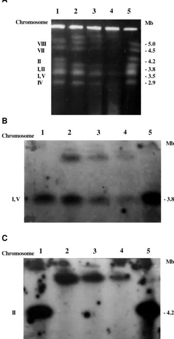
Separation of the chromosomes of A. nidulans strains A and B by a pulsed-field electrophoretic technique (CHEF) produced a different electrophoretic karyotype than that described by Brody and Carbon (1989). In both strains, the band corresponding to chromosome II was not present while a new band of approximately 5.2 Mb was observed (Figure 1A, lanes 2 and 3). Classic genetic tech-niques have shown that strains A and B carry part of chro-mosome I duplicated and translocated to chrochro-mosome II (Nga and Roper, 1968). To locate chromosome II in these
Figure 1 - Separation of Aspergillus nidulans intact chromosomal DNA on a CHEF gel (1A). The DNA was isolated from AM (lane 1), strain A (lane 2), strain B (lane 3), strain V17 (lane 4) and strain pabaA1 (lane 5). B and C, the same DNA samples probed with the labeled genes uapA and wA, respectively.
strains, the chromosomes were hybridized with gene wA of A. nidulans. This gene hybridized with the new band of 5.2 Mb in the duplicated strains (Figure 1C, lanes 2, 3 and 4). The chromosomes of the duplicated strains were also hybridized with the uapA gene of A. nidulans located in the duplicated portion of chromosome I (Figure 1B). Hy-bridization of the uapA gene was observed in the band cor-responding to chromosome I, as found by Brody and Car-bon (1989), as well as in the new 5.2 Mb band. These
re-A
Chromosome Mb
1 2 3 4 5
- 5.0 - 4.5
- 4.2 - 3.8 - 3.5 - 2.9 VIII
VII
II I, II I, V IV
Chromosome 1 2 3 4 5
Mb
- 3.8 I, V
B
Chromosome 1 2 3 4 5
Mb
- 4.2 II
sults agree with classic genetic data showing that the 5.2 Mb band which appears in the duplicated strains represents chromosome II containing part of chromosome I. The size of the duplicated region was approximately 1 Mb.
Gene yA is found in the duplicated region of strains A and B. The mutant gene yA1 occurs in chromosome I whereas the wild type gene occurs in the duplicated seg-ment of chromosome II. The reverse occurs in strain B. Thus, the duplicated strain A has green conidia and the loss of the duplicated segment linked to chromosome II re-sults in yellow conidia since the wild type gene yA is lost. One yellow sector (improved), obtained spontaneously from strain A, had a electrophoretic karyotype similar to that of the control strain pabaA 1 (Figure 1A, B and C, lane 1), indicating that in the improved strain the dupli-cated segment of chromosome I, which was translodupli-cated to chromosome II, was lost so that chromosome II returned to its original size.
Brody and Carbon (1989) examined the electropho-retic karyotype of A. nidulans strains with known chro-mosomal translocations. They performed the hybridiza-tion with genetically mapped genes to assign electro-phoretic gel bands to specific linkage groups. All translo-cated strains tested showed alterations in their electro-phoretic pattern when compared to non-translocated strains. Geiser et al. (1996) determined the electropho-retic karyotype of A. nidulans strains isolated in various parts of the world. One of the isolates (M-85) has a B-chromosome of about 1 Mb and another (Cambridge) a non-reciprocal translocation of about 1.6 Mb from chro-mosome VI to VIII. Crossing a translocated strain (Cam-bridge) with a wild type strain produced a variety of phe-notypes, including duplicated strains. These authors sug-gested that the duplicated segment in these strains was more stable than that observed by Bainbridge and Roper (1966) in duplicated strains of A. nidulans, since only one reversible sector was observed in 20 plates analyzed.
Sectors considered deteriorated also appear sponta-neously in strains A and B of A. nidulans. The deterio-rated variants have little sporulation and reduced growth compared to the duplicated strains. Azevedo and Roper (1970) suggested that the deteriorated variants could re-sult from tandem duplications in one of the duplicated segments, leading to an increased instability, followed by transposition of this duplication to other genome regions. This suggestion was based on the observation that the fac-tor responsible for the deteriorated phenotype often mapped onto chromosomes which were not involved in chromosomal duplication.
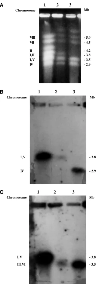
Three deteriorated strains isolated as spontaneous sectors from strains A and B of A. nidulans by Azevedo and Roper (1970) were analyzed. The mutation respon-sible for the deteriorated phenotype in both strains was mapped to different chromosomes. The deteriorated vari-ants V5, V9 and V17 had their deteriorated determinvari-ants mapped to chromosomes VIII, III and VII, respectively
(Azevedo and Roper, 1970). Variants V9 and V5 were iso-lated as sectors of strain A and the variant V17 as a sector of strain B. Variants V9 (data not shown) and V17 (Figure 1A, lane 4) had the same electrophoretic karyotype as strains A and B. However, variant V5 had a distinct elec-trophoretic karyotype. Only five chromosomal bands were found in variant V5 (Figure 2A, lane 3). The missing band was characterized by Brody and Carbon (1989) as being formed by chromosomes I and V. The reduction in size of chromosome I in variant V5 may reflect a deletion in the duplicated region present in this chromosome, for this would lead to the loss of approximately 1 Mb, causing chromosome I to migrate together with chromosome IV. This, however, does not explain the disappearance of the band corresponding to chromosome V. To locate these two chromosomes (I and V) in variant V5, hybridization was performed with the gene hxA, present in chromosome V, and with the gene pyrG, present in the non-duplicated re-gion of chromosome I. Chromosome I of strain V5 was confirmed to have a deletion of around 1 Mb and thus mi-grated together with chromosome IV (Figure 2B, lane 3). Both strains A and V5 have a deletion or translocation of a fragment of chromosome V, causing this chromosome to migrate with chromosomes III and VI (Figure 2C, lane 3). This result was surprising since this rearrangement had not been detected by classic genetic experiments. These find-ings also confirmed that deletion of the duplicated region in chromosome I was possible, as previously described (Nga and Roper, 1968).
Montenegro et al. (1992) showed that A. nidulans strain ATC 2890 had a rearrangement probably involving duplication of some fragment or translocation. This strain had a larger chromosome III than that observed in other A. nidulans strains, which allowed the separation of chromo-somes III and VI during electrophoretic karyotyping. These authors have attributed this rearrangement to the ultraviolet irradiation to which this strain was exposed to increase peni-cillin production. The rearrangement in chromosome V of strains A and V5 noted in our experiments may also have resulted from the mutagenic treatment used to obtain the genetic markers.
ACKNOWLEDGMENTS
The authors thank Annie Charbonnier-Glatigny for techni-cal assistance and Professor Claudio Scazzocchio for having re-ceived Marisa V. de Queiroz in his laboratory. Marisa V. de Queiroz was supported by a studentship from Conselho Nacional de Desenvolvimento Científico e Tecnológico (CNPq)/RHAE. Publication supported by FAPESP.
RESUMO
Linhagens de Aspergillus nidulans que apresentam duplicação cromossômica Dp(I-II) foram caracterizadas por eletroforese em campo pulsado. Foram analisados variantes morfologicamente deteriorados e melhorados. O cariótipo ele-troforético demonstrou que em ambas as linhagens duplicadas (A e B) a banda de 4,2 Mb, que corresponde ao cromossomo II, não estava presente e foi encontrada uma nova banda. Foram feitas hibridizações usando os genes uapA (cromossomo I) e wA
(cromossomo II), que demonstraram que a nova banda corres-ponde ao cromossomo II mais o segmento duplicado do cro-mossomo I. O tamanho da duplicação foi determinado como aproximadamente 1,0 Mb. A análise das bandas cromossômicas da linhagem morfologicamente melhorada mostrou que o segmento duplicado do cromossomo I foi completamente perdido. Os va-riantes morfologicamente deteriorados V9 e V17 mostraram o mesmo cariótipo eletroforético apresentado pelas linhagens du-plicadas. Contudo, o variante deteriorado V5 perdeu parte do cromossomo I e apresentou um rearranjo envolvendo o cromos-somo V. Esse rearranjo pode ter resultado do tratamento muta-gênico usado para a obtenção dos marcadores genéticos. Os re-sultados obtidos nesse trabalho demonstram que a técnica de eletroforese em campo pulsado é uma ferramenta excelente para a localização de rearranjos cromossômicos.
REFERENCES
Azevedo, J.L. (1975). Altered instability due to genetic changes in a duplica-tion strain of Aspergillus nidulans. Genet. Res. 26: 55-61.
Azevedo, J.L. and Roper, J.A. (1970). Mitotic non-conformity in Aspergillus nidulans: successive and transposable genetic changes. Genet. Res. 16:79-93.
Bainbridge, B.W. and Roper, J.A. (1966). Observations on the effects of a chromosome duplication in Aspergillus nidulans. J. Gen. Microbiol. 42: 417-424.
Bowyer, P., Osbourn, A.E. and Daniels, M.J. (1994). An “instant gene bank” method for heterologous gene cloning: complementation of two As-pergillus nidulans mutants with Gaeumannomyces graminis DNA. Mol. Gen. Genet.242: 448-454.
Brody, H. and Carbon, J. (1989). Electrophoretic karyotype of Aspergillus nidulans. Proc. Nat. Acad. Sci. USA 86: 6260-6263.
Diallinas, G. and Scazzocchio, C. (1989). A gene coding for the acid-xan-thine permease of Aspergillus nidulans: inactivational cloning, charac-terization, and sequence of a cis-acting mutation. Genetics122: 341-350.
Geiser, D.M., Arnold, M.L. and Timberlake, W.E. (1994). Sexual origins of British Aspergillus nidulans isolates. Proc. Natl. Acad. Sci. USA 91: 2349-2352.
Geiser, D.M., Arnold, M.L. and Timberlake, W.E. (1996). Wild chromosomal variants in Aspergillus nidulans. Curr. Genet. 29: 293-300.
Glatigny, A. and Scazzocchio, C. (1995). Cloning and molecular character-ization of hxA, the gene coding for the xanthine dehydrogenase (purine hydroxylase) of Aspergillus nidulans. J. Biol. Chem. 270: 3534-3550.
Lee N.B. and Adams, T.H. (1994). The Aspergillus nidulans fluG gene is
1 2 3
Mb
- 5.0 - 4.5
- 4.2 - 3.8 - 3.5 - 2.9 Chromosome
VIII VII
II I, II I, V IV A
1 2 3
C
Chromosome Mb
- 3.8
- 3.5 I, V
III, VI
1 2 3
B
Chromosome
- 3.8
- 2.9 I, V
IV
Mb
required for production of an extracellular developmental signal and is related to prokaryotic glutamine synthetase I. Genes Devel. 8: 641-651.
Marjefeld, I.J. and Roper, J.A. (1978). The effects of coumarin on the fre-quency of deletions in a duplication strain of Aspergillus nidulans. Mol. Gen. Genet. 159: 203-206.
McCully, K.S. and Forbes, E. (1965). The use of p-fluorphenylalanine with “master” strain of Aspergillus nidulans to assign to linkage groups. Genet. Res. 6: 352-359.
Menezes, E.M. and Azevedo, J.L. (1978). Reversion in variant from a duplica-tion strain of Aspergillus nidulans. Mol. Gen. Genet.164: 255-258.
Montenegro, E., Fierro, F., Fernandez, F.J., Gutiérrez, S. and Martín, J.F.
(1992). Resolution of chromosomes III and VI of Aspergillus nidulans by pulsed-field gel electrophoresis shows that the penicillin biosyn-thetic pathway gene pcbAB, pcbC,and penDE are clustered on chro-mosome VI (3.0 megabases). J. Bacteriol.174: 7063-7067.
Nga, B.H and Roper, J.A. (1968). Quantitative intrachromosomal changes arising at mitosis in Aspergillus nidulans. Genetics58: 193-209.
Oakley, B.R., Rinehart, J.E., Mitchell, B.L., Oakley, C.E., Carmona, C.,
Gray, G.L. and May, G.S. (1987). Cloning, mapping and molecular analy-sis of the pyrG (orotidine-5’-phosphate decarboxylase) gene of As-pergillus nidulans. Gene61: 385-399.
Pontecorvo, G., Roper, J.A., Hemons, L.M., MacDonald, K.D. and Bufton, A.W.J. (1953). The genetics of Aspergillus nidulans. Adv. Genet.5: 141-238.
Pritchard, R.H. (1956). A genetic investigation of some adenine-requiring mutants of Aspergillus nidulans. PhD thesis, Glasgow University, UK.
Sambrook, J., Fritsch, E.F. and Maniatis, T. (1989). Molecular Cloning: a Laboratory Manual. Cold Spring Harbor Laboratory Press, New York.
Tilburn, J., Scazzocchio, C., Taylor, G.G., Zabicky-Zissima, J.H., Lockington, R.A. and Davis, R.W. (1983). Transformation by integration in Aspergil-lus nidulans. Gene 26: 205-221.
Zolan, M.E. (1995). Chromosome-length polymorphism in fungi. Microbiol. Rev. 59: 686-698.