www.scielo.br/aabc
Cremophor EL stimulates mitotic recombination in
uvsH//uvsH
diploid strain of
Aspergillus nidulans
CLEVERSON BUSSO and MARIALBA A.A. CASTRO-PRADO
Universidade Estadual de Maringá, Departamento de Biologia Celular e Genética Av. Colombo 5790, 87020-900 Maringá, PR, Brasil
Manuscript received on June 13, 2003; accepted for publication on October 8, 2003; presented byLucia Mendonça Previato
ABSTRACT
Cremophor EL is a solubilizer and emulsifier agent used in the pharmaceutical and foodstuff industries. The solvent is the principal constituent of paclitaxel’s clinical formulation vehicle. Since mitotic recombination plays a crucial role in multistep carcinogenesis, the study of the recombinagenic potential of chemical compounds is of the utmost importance. In our research genotoxicity of cremophor EL has been studied by using anuvsH//uvsHdiploid strain ofAspergillus nidulans. Since it spends a great part of its cell cycle in the G2 period, this fungus is a special screening system for the study of mitotic recombination induced by chemical substances. Homozygotization Indexes (HI) forpabaandbimarkers from heterozygous B211//A837 diploid strain were determined for the evaluation of the recombinagenic effect of cremophor EL. It has been shown that cremophor EL induces increase in mitotic crossing-over events at nontoxic concentrations (0.05 and 0.075% v/v).
Key words:Aspergillus nidulans, mitotic recombination, cremophor EL, Homozygotization Index, antineo-plasm agents.
INTRODUCTION
The organic solvent cremophor EL (CEL) (poly-oxyethyleneglycerol triricinoleate 35) is a viscous liquid produced by the reaction of castor oil (Ricinus communis) with ethylene oxide (Hoffman 1984). CEL is employed as a vehicle for the solubiliza-tion of a wide variety of hydrophobic drugs, in-cluding anesthetics, photosensitizers, sedatives, im-munosuppressive agents and anticancer drugs (Gel-derblom et al. 2001).
Without causing serious toxicity, in vitro cre-mophor EL reverts the Multidrug Resistance (MDR) phenotype in clinically executable concentrations. Multidrug resistance is a mechanism by which
Correspondence to: Castro-Prado M.A.A. E-mail: maacprado@uem.br
cancer cells are able to survive diverse drugs in structurally unrelated groups. Overexpression of the multidrug transporter protein, also known as P-glycoprotein, has often been observed in MDR cells. This transmembrane protein is capable of pumping a wide variety of chemotherapeutic agents out of the cells, protecting them from the agent’s toxic ef-fects (Breier et al. 2000). Cremophor EL afef-fects the metabolism of MDR cells, alters cell membrane properties, and impairs the P-glycoprotein function. Actually the solvent favors the action of antineo-plasm drugs, such as paclitaxel, doxorubicin and vinblastine (Fjällskog et al. 1993, Friche et al. 1993, Woodcock et al. 1990).
as-TABLE I
Genotype and origin of strains.
Strain Genotype Origin
B211 yA2; biA1; AcrA1; wA2; methA17; uvsH77; pyroA4; chaA1 Busso et al. 2001
A837 pabaA1; uvsH77; pyroA4; choA1; chaA1 FGSC
Mutant alleles give the following phetotypes (corresponding genes between parentheses): requirements for 4-aminobenzoic acid (pabaA1), biotin (biA1), methionine (methA17), pyridoxine (pyroA4) and choline (choA1); yellow (y), white (w) and chartreuse (cha) coloring of conidia, respectively; resistance to acriflavine (AcrA1). FGSC – Fungal Genetic Stock Center, University of Kansas Medical Center, Kansas City, USA.
sociated with peroxidation of polyunsaturated fatty acids and with direct disturbing effect in the cell membrane (Nygren et al. 1995, Burton 1991, Bégin et al. 1988).
The transformation of normal human cells into cancer cells is a multistep process, while mitotic recombination is a mechanism involved in bring-ing about such transformation (Nowell 1976, Bar-rett 1993). In heterozygous cells bearing a mutant and normal alleles for a tumor suppressor gene, the somatic recombination may turn up to be a promoter of neoplasms by inducing homozygosis of the mu-tant tumor suppressor allele (Maher et al. 1993, Sengstag 1994).
Mitotic crossing-over has already been re-corded in Drosophila melanogaster, Aspergillus nidulans, Saccharomyces cerevisiae and mam-malian cells in culture, including human cells. It is currently thought to be a common occurrence pro-cess in diploid cells (Ramel et al. 1996, De La Torre et al. 1994, Maher et al. 1993, Kunz et al. 1981, Stern 1936).
The filamentous fungusA. nidulans is an ex-cellent organism for studying mitotic crossing-over. This is chiefly due to two important factors: a)A. nidulans’s mitosis has many common characteris-tics with higher eukaryotes mitosis, and b) the fun-gus spends most of its vegetative cell cycle in G2 phase. At this phase, the presence of two copies of each chromosome favors the occurrence of mitotic crossing-over (Bergen and Morris 1983, Iwanejko et al. 1996).
Since several reports have suggested somatic
recombination in mechanisms leading to carcino-genesis and due to the fact that CEL is used as a solubilizer of hydrophobic drugs, such as antineo-plasm agents, we decided to examine the ability of this solvent to induce mitotic recombination.
MATERIALS AND METHODS
Strains
The genotypes and origin ofA. nidulansstrains are provided in Table I. Diploid strain (B211//A837) (Busso et al. 2001) was prepared according to Roper (1952).
Culture Media
Complete (CM) and minimum medium (MM) were prepared according to Van de Vate and Jansen (1978). Selective medium was prepared with MM and nutritional requirements of each strain. Solid medium was prepared with 1.5% agar; incubation for strain growth was done at 37˚C.
Evaluation of Drug Toxicity
TABLE II
Mitotic segregation ofpabagene among haploid segregants derived frompaba+//paba heterozygous diploid in the presence and in the absence of crossing-over.
Chromosome I
No Crossing-over Crossing-over in the centromere-paba interval
Chromatid
Segregation Diploid genotypes
Chromatid
Segregation Diploid genotypes
1 + 3 paba+// paba 1 + 3 paba+ // paba+
1 + 4 paba+// paba 1 + 4 paba+ // paba
2 + 3 paba+ // paba 2 + 3 paba // paba+
2 + 4 paba+// paba 2 + 4 paba // paba
Segregation after
haploidization 4paba+ : 4 paba 4 paba+ : 2 paba
(*) Homozygous diploid (paba // paba) will not be chosen in MM + pyridoxine plates
Heterozygous diploid
paba
1 +
2
3 4
paba +
1 +
2
3 4
paba
paba + Recombinant
diploid
Cytological Analysis
Colonies of B211 strain were cultivated in dialysis membranes placed aseptically on the surface of petri dishes with CM and CM + 0.05% and 0.075% v/v of CEL. Incubation occurred for 30 h, at 37˚C, and samples taken during 4-28 h period. Membranes were stained with cotton-blue-lactophenol and ana-lyzed under an optic microscope.
Evaluation of the Recombinagenic Potential (Pires and Zucchi1994)
Conidia of the diploid strain were inoculated in MM + pyridoxine + cremophor EL (0.05% and 0.075% v/v) and incubated for 5 days at 37˚C. Treat-ment produced visible diploid sectors, D1-D6, iden-tified by their different morphology from the original diploid. Diploid sectors were submitted to spon-taneous haploidization in CM after purification in MM + pyridoxine. Only haploid segregants were selected for recombinagenesis test (Franzoni et al. 1997). Conidia of each haploid sector were
trans-ferred to 25 defined positions in CM plates (mas-ter plates). Af(mas-ter 48 hours of incubation at 37˚C, colonies were transferred to selective media and the phenotypical analysis of the haploid segregants was carried out.
The treatment with CEL in MM + pyridoxine produces only heterozygous (+//– or –//+) or ho-mozygous (+//+) segregants since the recessive ones (–//–) fail to grow in MM + pyridoxine (Table II). After haploidization of diploids D1-D6 the nutri-tional markers will segregate among the haploids in the proportion of 4+: 4–, if solvent fails to induce recombinagenesis; or 4+: 2–, if solvent induces crossing-over. Values of Homozigotization Indexes (HI) (the ratio between number of prototrophic and auxotrophic segregants) equal or above 2.0 evidence the recombinagenic effects of the substance under analysis (Pires and Zucchi 1994, Chiuchetta and Castro-Prado 2002a,b). Results were compared by Yates corrected Chi-square test.
TABLE III
Time of conidiophore vesicle, metulae and phialides appearance and conidia production of B211 strain in the presence of cremophor EL 0.05 and 0.075% v/v.
Conidiophore Time (hs)
structures and Control* Cremophor EL
conidia production 0.05% v/v 0.075% v/v
Vesicles 22 24 25
Metulae and Phialides 23 25 25
Conidia 25 26 26
*Development without cremophor EL.
evaluate diploid B211//A837 after treatment with cremophor EL.
RESULTS AND DISCUSSION
Aspergillus nidulansreproduces itself asexually by forming multicellular conidiophores and uninucle-ate spores called conidia. We have first studied the effects of CEL on mycelia growth of B211//A837 strain and on conidiophore morphology of B211 strain. Solvent was added to a pyridoxine-supplemented minimal agar medium to obtain final concentrations of 0.05% and 0.075% v/v. Both CEL concentrations had no effect on mycelia growth or on conidiophore morphology (results not shown). Only a slight delay in the timing for conidiophore formation of B211 strain was observed (Table III).
In this study, the B211//A837 diploid strain, ho-mozygous foruvsHmutation, was used as a sensitive system for the evaluation of the genotoxic activity of CEL in nontoxic concentrations.
Various repair mechanisms are mobilized to re-store the original DNA sequence when DNA dam-ages occur. These mechanisms include base exci-sion repair (BER), nucleotide exciexci-sion repair (NER), mutagenic repair and post-replication repair (Gold-man et al. 2002, Hjertvik et al. 1998, Fishel and Kolodner 1995). InA. nidulans the UV-sensitive mutants (uvs) have been classified in differ-ent epistatic groups such as: UvsB, UvsC, UvsF
and UvsI. The study of mitotic intergenic recom-bination in this filamentous fungus may be greatly facilitated by the use of UvsF group mutations: gene mutationuvsH, that operates in the post-replication repair pathway, is responsible for high frequencies of mitotic intergenic recombination in homozygous condition (Osman et al. 1993, Iwanejko et al. 1996). It has been shown that CEL induces a statis-tically significant increase in mitotic crossing-over events inA. nidulans. The treatment of B211//A837 with CEL increased Homozygotization Indexes for pabaandbimarkers (Tables IV and V). This effect, nevertheless, was not dose-dependent.
It is believed that mitotic recombination involv-ing heterozygous cells for a deleterious gene trig-gers carcinogenesis. This process leads towards ho-mozygosis and subsequent expression of the malignant trait (Wijnhoven et al. 2003, Preisler et al. 2000).
Our date demonstrate the recombinagenic ef-fect of CEL inA. nidulanswhen a sensitive strain is used to study mitotic crossing-over. Further anal-yses, using mammalian cells, may be conduced for a better understanding of the carcinogenic potential of this solvent.
ACKNOWLEDGMENTS
TABLE IV
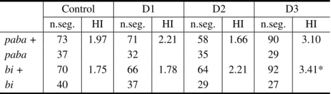
Homozygosity Index (HI) of diploid B211//A837 exposed to 0.05% v/v of cremophor EL.
Control D1 D2 D3
n.seg. HI n.seg. HI n.seg. HI n.seg. HI
paba + 73 1.97 71 2.21 58 1.66 90 3.10
paba 37 32 35 29
bi + 70 1.75 66 1.78 64 2.21 92 3.41*
bi 40 37 29 27
D1, D2 and D3, diploid strains obtained after treatment of the B211//A837strain with 0.05% v/v of cremophor EL; n.seg., number of haploid mitotic segregants; (*) significantly different from control at p<0.05 (Yates corrected Chi-square test, Statistic for Windows Program).
TABLE V
Homozygosity Index (HI) of diploid B211//A837 exposed to 0.075% v/v of cremophor EL.
Control D4 D5 D6
n.seg. HI n.seg. HI n.seg. HI n.seg. HI
paba + 73 1.97 136 3.77* 75 2.20 200 3.03
paba 37 36 34 66
bi + 70 1.75 120 2.30 73 2.02 199 2.97*
bi 40 52 36 67
D4, D5 and D6, diploid strains obtained after treatment of the B211//A837strain with 0.075% v/v of cremophor EL; n.seg., number of haploid mitotic segregants; (*) significantly different from control at p<0.05 (Yates corrected Chi-square test, Statistic for Windows Program).
Sul) and Carmem Boto Querol (Universidade Estad-ual de Maringá) for supplying the cremophor EL; to Mrs. Sonia A. de Carvalho and Mrs. Luzia A. S. Regasse for technical assistance. Cleverson Busso is the holder of a PIBIC/CNPq fellowship.
RESUMO
Cremofor EL (CEL) é um solubilizante e emulsificante amplamente utilizado nas indústrias farmacêuticas e de gêneros alimentícios. É o principal veículo empregado nas formulações clínicas do antineoplásico pacli-taxel. Considerando-se que a recombinação mitótica de-sempenha importante função no processo de carcinogê-nese, o estudo de substâncias químicas com potencial re-combinagênico assume importância crucial, no sentido de se detectar aquelas que eventualmente possam atuar como promotoras de neoplasias. A genotoxicidade do cremofor EL foi estudada no presente trabalho, utilizando-se uma
linhagem diplóideuvsH//uvsHdeAspergillus nidulans. Neste fungo as células vegetativas comumente repousam no período G2 do ciclo celular, facilitando a ocorrência da recombinação mitótica. O efeito recombinagênico do CEL foi avaliado através da determinação dos Índices de Homozigotização para os marcadores nutricionaispabae bido diplóide heterozigoto B211//A837. Os resultados demonstram que CEL é efetivo em induzir crossing-over mitótico em concentrações não tóxicas ao fungo (0.05 e 0.075% v/v).
Palavras-chave: Aspergillus nidulans, recombinação mitótica, cremofor EL, índices de homozigotização, agentes antineoplásicos.
REFERENCES
Bégin ML, Ells G and Horrobin DF.1988. Polyunsat-urated fatty acid-induced cytotoxicity against tumor cells and its relationship to lipid peroxidation. J Natl Cancer Inst 80: 188-194.
Bergen GG and Morris NR.1983. Kinetics of the nu-clear division cycle ofAspergillus nidulans. J Bac-teriol 156: 155-160.
Breier A, Drobná Z, Docolomanský P and Barancík M. 2000. Cytotoxic activity of several unrelated drugs on L1210 mouse leukemic cell sublines with P-glycoprotein (PGP) mediated multidrug resistance (MDR) phenotype. A QSAR study. Neoplasma 47: 100-106.
Burton AF.1991. Oncolytic effects of fatty acids in mice and rats. Am J Clin Nutr 53: 1082-1086.
Busso C, Chiuchetta SJR, Baptista F and Castro-Prado MAA.2001.uvsH//uvsHdiploid strain favors an efficient method to evaluate the recombinagenic effect of chemical and physical agents inAspergillus nidulans(Ascomycetes). Acta Scientiarum 23: 603-607.
Chiuchetta SJR and Castro-Prado MAA. 2002a. Vincristine induces somatic segregation, via mitotic crossing-over, in diploid cells of Aspergillus nidu-lans. Biol Res 35: 31-38.
Chiuchetta SJR and Castro-Prado MAA. 2002b. Recombinagenic effect of Cryptolepine inuvsH+// uvsH+anduvsH//uvsHdiploid strains ofAspergillus nidulans. Folia Microbiol 47: 516-520.
De La Torre RA, Espinosa-Aguirre JJ, Cortinas de Navas C, Izquierdo T and Moron F.1994. Geno-toxic activity of mebendazole in Aspergillus nidu-lans. Mutat Res 305: 139-144.
Fishel R and Kolodner RD.1995. Identification of mismatch repair genes and their role in the develop-ment of cancer. Curr Opin Genet and Dev 5: 382-395.
Fjällskog M-L, Frii L and Bergh J.1993. Is Cre-mophor EL, the solvent for paclitaxel, cytotoxic? Lancet 342: 873.
Franzoni MGM, Castro-Prado MAA and Gebara JS.
1997. On the recombinagenic activity of Norfloxacin in a diploid strain ofAspergillus nidulans. Cytologia 62: 39-45.
Friche E, Demant EJF, Schestad M and Nissen NI.
1993. Effect of anthracycline analogs of
photola-belling of p-glycoprotein by125I-iodomycin and3 H-azidopine: relation to lipophilicity and inhibition of daunorubicin transport in multidrug resistant cells. Br J Cancer 67: 226-231.
Gelderblom H, Verweij J, Nooter K and Sparre-boom A.2001. Cremophor EL: the drawbacks and advantages of vehicle selection for drug formulation. Eur J Cancer 37: 1590-1598.
Goldman GH, McGuirre SL and Harris SD.2002. The DNA damage response in filamentous fungi. Fungal Genet Biol 35: 183-195.
Hjertvik M, Erixon K and Ahnström G.1998. Repair of DNA damage in mammalian cells after treatment with UV and dimethyl sulphate: discrimination be-tween nucleotide and base excision repair by their temperature dependence. Mutat Res 407: 87-96.
Hoffman H.1984. Polyoxythylenglycerol triricinoleat 35 DAC 1979. Pharm Zeit 129: 1730-1733.
Iwanejko L, Cotton C, Jones G, Tomsett B and Strike P.1996. nuvA, anAspergillus nidulansgene involved in DNA repair and recombination, is a homologue of Saccharomyces cerevisiae RAD18 andNeurospora crassa uvs-2. Microbiology 142: 505-515.
Kunz BA, Barcaly BJ and Haynes RH.1981. Phe-nomenology and genetic control of mitotic recombi-nation in yeast. Ann Rev of Genet 15: 57-80.
Maher VM, Bhattacharyya NP, Mah MC, Boldt J, Yang JL and McCormick JJ.1993. Mutations induced by 1-nitrosopyrene and related compounds during DNA replication in human cells and induction of homologous recombination by these compounds. Res Rep Health Eff Inst 40: 41-51.
Nowell P.1976. The clonal evaluation of tumour cell populations. Science 194: 23-28.
Nygren P, Csóka K, Jonsson B, Fridborg H, Bergh J, Hagberg H, Glimelius B, Brodin O, Tholander B and Kreuger A.1995. The cytotoxic activity of Taxol in primary cultures of tumor cells from patients is partly mediated by Cremophor EL. Br J Cancer 71: 478-481.
Osman F, Tomsett B and Strike P.1993. The isola-tion of mutagen-sensitivenuvmutants ofAspergillus nidulansand their effects on mitotic recombination. Genetics 134: 445-454.
detect potential genotoxic agents using mitotic cross-ing over in diploid strains ofAspergillus nidulans. Bras J Gen 17: 371-376.
Preisler V, Caspary WJ, Hoppe F, Hagen R and Stop-per H.2000. Aflatoxin B1-induced mitotic recombi-nation in L5178Y mouse lymphoma cells. Mutage-nesis 15: 91-97.
Ramel C, Cederberg H, Magnusson J, Vogel J and Natarajan AT.1996. Somatic recombination, gene amplification and cancer. Mutat Res 353: 85-107.
Roper JA.1952. Production of heterozygous diploids in filamentous fungi. Experientia 8:14-15.
Sengstag C.1994. The role of mitotic recombination in carcinogenesis. Crit Rev Toxicol 24: 323-353.
Stern C.1936. Somatic crossing-over and segregation inDrosophila melanogaster. Genetics 21: 625-730.
Van de Vate C and Jansen GJO.1978. Meiotic re-combination in a duplication strains ofAspergillus nidulans. Genet Res 31: 29-52.
Wijnhoven SW, Sonneveld E, Kool HJ, Van-Teijlingen CM and Vrieling H. 2003. Chemi-cal carcinogens induce varying patterns of LOH in mouse T-lymphocytes. Carcinogenesis 24: 139-144.