Decreased Levels of KLF2 and Inefficiently Populate the
Peripheral Lymphoid Organs
Lawryn H. Kasper*, Tomofusa Fukuyama¤, Paul K. Brindle*
Department of Biochemistry, St. Jude Children’s Research Hospital, Memphis, Tennessee, United States of America
Abstract
MED23, a subunit of the Mediator coactivator complex, is important for the expression of a subset of MAPK/ERK pathway-responsive genes, the constituents of which vary between cell types for reasons that are not completely clear. MAPK/ERK pathway-dependent processes are essential for T-cell development and function, but whether MED23 has a role in this context is unknown. We generated Med23 conditional knockout mice and induced Med23 deletion in early T-cell development using the lineage specificLck-Cretransgene. While the total cell number and distribution of cell populations in the thymuses ofMed23flox/flox;Lck-Cre mice were essentially normal, MED23 null T-cells failed to efficiently populate the peripheral lymphoid organs. MED23 null thymocytes displayed decreased expression of the MAPK/ERK-responsive genes Egr1,Egr2, as well as of the membrane glycoproteinCd52(CAMPATH-1). MED23 null CD4 single-positive thymocytes also showed decreased expression of KLF2 (LKLF), a T-cell master regulatory transcription factor. Indeed, similarities between the phenotypes of mice lacking MED23 or KLF2 in T-cells suggest that KLF2 deficiency in MED23 null T-cells is one of their key defects. Mechanistic experiments using MED23 null MEFs further suggest that MED23 is required for full activity of the MAPK-responsive transcription factor MEF2, which has previously been shown to mediateKlf2expression. In summary, our data indicate that MED23 has critical roles in enabling T-cells to populate the peripheral lymphoid organs, possibly by potentiating MEF2-dependent expression of the T-cell transcription factor KLF2.
Citation:Kasper LH, Fukuyama T, Brindle PK (2014) T-Cells Null for the MED23 Subunit of Mediator Express Decreased Levels of KLF2 and Inefficiently Populate the Peripheral Lymphoid Organs. PLoS ONE 9(7): e102076. doi:10.1371/journal.pone.0102076
Editor:Laszlo Tora, Institute of Genetics and Molecular and Cellular Biology, France ReceivedApril 25, 2014;AcceptedJune 13, 2014;PublishedJuly 23, 2014
Copyright:ß2014 Kasper et al. This is an open-access article distributed under the terms of the Creative Commons Attribution License, which permits unrestricted use, distribution, and reproduction in any medium, provided the original author and source are credited.
Data Availability:The authors confirm that all data underlying the findings are fully available without restriction. Gene expression microarray data were deposited with GEO (accession number GSE 57061).
Funding:This work was supported by the Cancer Center (CORE) support grant P30 CA021765 (http://cancercenters.cancer.gov/grants_funding/), and the American Lebanese Syrian Associated Charities of St. Jude Children’s Research Hospital (www.stjude.org). The funders had no role in study design, data collection and analysis, decision to publish, or preparation of the manuscript.
Competing Interests:The authors have declared that no competing interests exist. * Email: [email protected] (LHK); [email protected] (PKB)
¤ Current address: University of Tokyo, Tokyo, Japan
Introduction
While certain histone N-terminal tail modifications and coactivator recruitment events correlate well with gene expression on a genome-wide scale, mutagenesis studies to test their roles directly have often produced unexpectedly modest or specific effects (reviewed in [1,2]). This apparent paradox indicates that target gene context is a critical, but still poorly understood aspect of transcriptional regulation. Coactivator context specificity has been evident, for example, since early descriptions of yeast mutants that affect amino acid biosynthesis, mating type switching, and sucrose fermentation; phenotypes that were later ascribed to mutations in ‘‘global’’ coactivators (e.g. GCN5, SWI/SNF). In mammals, the problem of coactivator functional specificity has been illuminated by the use of mice and cells with domain-specific or tissue-specific conditional-null mutations in coactivator genes [3–14].
Multi-subunit coactivator complexes, such as Mediator, repre-sent another dimension of the ‘‘context paradox.’’ The large Mediator complex, and its variants, interacts with RNA polymer-ase II and forms part of the general transcriptional machinery
[15], yet mutation or knock-down of individual subunits in mice and cells have revealed curiously distinct phenotypes [16]. Indeed, it has previously been shown that expression of the same target gene can have a different requirement for the MED23 subunit in different cell types, even in response to the same signal [17]. These kinds of context-dependent functionalities of Mediator and its subunits are perplexing, and understanding how and why they occur remains a challenge [18].
MED23 (SUR2, DRIP130, CRSP3) is a,130 kDa subunit of
Mediator that was initially identified in a screen for suppressors of
an activated RAS induced phenotype inC. elegans where it was
determined to act downstream in the RAF/MAPK pathway [19].
Studies of Med23 knockout ES cells and MEFs confirm that
proliferation of non-small cell lung cancers with activating mutations in RAS [23].
The RTK-RAS-RAF-MEK-ERK axis of the MAPK pathway plays important roles in normal T-cell development and function that include signaling through the T-cell receptor (TCR), regulating thymocyte positive selection, and T-cell homeostasis [24–30]. While MAPK-dependent ETS family transcription factors that require MED23 for full transcriptional activity in MEFs or ES cells are necessary for normal T-cell development [31], cell type context clearly influences which target genes display MED23 dependence [17]. This made it unclear how MED23 deletion would impact T-cells. Using a T lineage specific knock out ofMed23, we found that while MED23 null thymocyte numbers and proportions are essentially normal, the number of MED23 null T-cells in the periphery is reduced. We discovered a novel requirement for MED23 in the expression of KLF2, a T-cell master regulatory transcription factor [32]. Mechanistic experi-ments in MEFs suggest that MED23 is required by the transcription factor MEF2 to directly regulateKlf2expression. Materials and Methods
Mice
Med23conditional knock out mice were generated by inserting LoxP sites into the introns flanking the region containing exons 5,
6 and 7 of Med23 (encoding amino acids 96 to 199) using a
transposon based system reported previously [11]. The Lck-Cre
transgenic mice were originally reported in Hennetet al.[33]. For some experiments, mice also contained an eYFP reporter transgene that was expressed after Cre-mediated recombination [34]. All mouse experiments followed protocols approved by the St Jude Animal Care and Use Committee.
Mouse embryonic fibroblasts (MEFs)
Med23flox/floxembryos homozygous for LoxP conditional alleles ofMed23and wild type littermate control embryos were harvested at e14.5 to produce mouse embryonic fibroblasts (MEFs) that were maintained in 3% oxygen to delay the onset of senescence [35]. Primary MEFs were transduced with adenovirus expressing cre recombinase (MOI 100) for 16 h and experiments were performed four days after transduction. Recombination of the flox sites in
Med23 was confirmed by semi-quantitative genomic PCR and western blot.
Genotyping
PCR to check genotype and allele recombination was carried out with a three primer reaction (Primer 1: ATTCATGGCCAA-CACAGCCC, Primer 2: GCCCAAAGCTGTGTTCTTTCCC and Primer 3: CACTGAGTGTGGCAGCTCATG) using Qia-gen reaQia-gents including Q solution (Cat.#201207) at 94uC 5 min followed by 30 cycles of (94uC 10 sec; 60uC 1 min; 68uC 2 min) with a final extension of 72uC 10 min and bands representing the wild type allele (,1.1 kb), the LoxP flanked unrecombined allele
(,1.35 kb) and the recombined LoxP allele (,1.8 kb) were
resolved on a 1.2% agarose gel.
Flow cytometry, FACS and MACS cell sorting
Flow cytometry was performed on BD Biosciences FACS
Calibur and FACS LSR instruments. FACS of CD4+
or CD8+
single positive thymocytes was performed on a BD Biosciences Aria. All antibodies were from Becton Dickenson. For some
experiments CD4+
single positive thymocytes were enriched using MACS biotin beads (Miltenyi) and anti-CD8 antibody (Miltenyi)
to remove the CD8 expressing single positive and CD4+
/CD8+
double positive thymocytes.
Proliferation assay
Single cell suspensions of total thymocytes were seeded at 0.256106cells per well of a 96 well plate. Anti-CD3 (10mg/ml) and anti-CD28 (10mg/ml) antibodies were immobilized in wells as
noted by incubating for 90 min at 37uC, then the wells were
washed three times with PBS prior to use. Cells were allowed to proliferate for 44 hrs at 37uC, then 1mCi3H thymidine was added to each well and cells were allowed to proliferate a further 18 hrs before the cells were harvested onto a filter, washed and counted.
TUNEL assay
Frozen sections of thymus and spleen were fixed for 20 min at room temperature with 4% paraformaldehyde in PBS, then washed and incubated in PBS for an additional 30 min. Sections were permeabilized (0.1% Triton X-100/0.1% sodium citrate) for
2 min at 4uC, washed twice with PBS and incubated for 1 hr at
37uC with the TUNEL labeling mixture from the Roche In Situ
Cell Death Detection Kit, TMR red (Cat. No. 12156792910). Slides were washed with PBS, stained with DAPI and imaged. As a positive control, one frozen section was incubated with 1mg/ml DNaseI for 10 min at room temperature prior to TUNEL labeling.
Gene expression
RNA was isolated using Trizol (Life Technologies). Microarray platforms used were Affymetrix Murine Genome U74A Version 2 Array and were analyzed using Spotfire software (TIBCO). RNA from thymocytes was isolated immediately following FACS or thymocytes were rested for 4 hrs after harvest then plated for 3 hrs
with plate boundaCD3 antibody as indicated in figure legends.
Array data were deposited with GEO (GSE 57061). Reverse transcriptase reactions were performed using Superscript II (Life Technologies). qPCR was performed on an MJ Research Opticon real time machines using Quantitect SYBR Master Mix (Qiagen).
Antibodies
The KLF2 (LKLF) rabbit polyclonal antibody was provided by Jeffrey Leiden and Laurie Glimcher and was originally published in [36]. The MED23 rabbit polyclonal antibody was generated against a synthetic peptide corresponding to mouse MED23 residues 897–916 (based on human MED23 residue numbering) crosslinked to KLH. It is available from Rockland (DRIP130
antibody, Cat.#100-401-239).
Plasmids and transient transfection assays
The mouse 2215 Klf2 promoter construct (pGL3-Klf2-pro)
and the GAL4-KLF2 construct containing amino acids 1–88 of mouse KLF2 fused to the GAL4 DNA binding domain were
provided by Jerry Lingrel [37,38]. The MEF2 site in the Klf2
promoter reported in Kumaret al.[39] was mutated in the mouse
pGL3-Klf2-pro (ctaAATTtag to ctaTCGGtag) to yield
pGL3-Klf2-pro-mut. The GAL4-MEF2C construct was made using the
Results
MED23 null T-cells develop normally, but are defective in populating the periphery
We generated mice bearing a conditional knockout allele of
Med23by inserting LoxP sites in the introns flanking the region
containing exons 5, 6 and 7 ofMed23encoding amino acids 96
to 199 (Figure S1A). Cre-mediated recombination of the LoxP sites in theMed23floxallele would result in a frameshift if exons
4 and 8 spliced fortuitously. Using an Lck-Cre transgene that
deletes during the DN4 stage of T-cell development [33,41], we
produced Med23flox/flox;Lck-Cre mice to assess the role of
MED23 in T-cells. Efficient recombination of the Med23flox
alleles in the thymuses of Med23flox/flox;Lck-Cre mice (Figure S1B) resulted in a near total loss of MED23 protein (Figure S1C).
In Med23flox/flox;Lck-Cre mice with efficient deletion in the thymus (Figure S1B and S1C), the proportions of CD4 CD8 double negative (DN), double positive (DP) and single positive cells
were normal (Figures 1A and S2A). While theLck-Cretransgene
itself caused a modest reduction in thymocyte number,Med23flox/
flox;Lck-Cre
mice had similar thymocyte counts to mice with the
Lck-Cretransgene alone (Figure 1B).
To further assess the MED23 null T-cell populations in the
thymus and peripheral lymphoid organs, we utilized an eYFP
transgene that is turned on by Cre-mediated recombination [34] and marks cells where theLck-Cretransgene is expressed. We then
determined the percentage of YFP+
cells in the thymus, spleen,
lymph nodes and blood of Med23+/+;Lck-Cre;YFP(control) and
Med23flox/flox;Lck-Cre;YFP mice by flow cytometry (Figure 1C
shows average percentage of YFP+
cells normalized to YFP+
DP thymocytes for each mouse; see Table S1 for unnormalized YFP percentages). InMed23+/+;Lck-Cre;YFPcontrol mice the
percent-age of YFP+
T-cells remained constant between the thymus and
peripheral lymphoid organs (Figure 1C, Table S1,N= 2 control
mice). However, forMed23flox/flox;Lck-Cre;YFPmice, the percent-age of YFP+
(Med23null) T-cells compared to DP thymocytes, was significantly reduced in spleen, lymph nodes and blood (Figure 1C,
Table S1,N= 7 mutant mice; P= 0.0035 to,0.0001). For both
Med23+/+;Lck-Cre;YFP and Med23flox/flox;Lck-Cre;YFPmice the
percentage of YFP+
DP thymocytes was comparable to the
percentage of YFP+
CD4 and CD8 single positives (Figure 1C, Table S1). This indicates that loss of MED23 does not result in a block in thymocyte maturation from double positive to single positive cells. The DN thymocyte compartment had a reduced
percentage of YFP+
T-cells for bothMed23+/+;Lck-Cre;YFP
and
Med23flox/flox;Lck-Cre;YFP mice (Figure 1C), reflecting that the
Lck-Cre transgene does not turn on until the DN4 stage of
development [33,41]. Consistent with this data, in Med23flox/
flox;Lck-Cremice with very high deletion in the thymus, a decrease in the ratio of T-cells (CD3+
) to B-cells (B220+
) in spleen, lymph node and blood compared toMed23flox/floxcontrol mice was evident (Figure 2, Figure S2A). InMed23flox/flox;Lck-Cre mice with more modestMed23deletion in the thymus this deficit in peripheral T-cell numbers was not seen, as the number of undeleted T-T-cells in the thymus was sufficient to completely populate the periphery (Figure
S2A–C, compare mouse with poor deletion (#2) to mice with high
deletion (#1,3 and 4)).
MED23 null T-cells do not display increased apoptosis
The deficit of MED23 null T-cells in the peripheral lymphoid organs, led us to investigate whether there was an increased rate of T-cell apoptosis inMed23flox/flox;Lck-Cremice compared to controls.
Increased apoptosis in situ was not apparent as TUNEL assays
showed no difference betweenMed23flox/flox;Lck-CreandMed23+/
+;Lck-Cre
mice in cryosections from either thymus or spleen (Figure 3).
Figure 1.Med23flox/flox;Lck-Cremice have normal thymic popu-lations, but MED23 null T-cells poorly populate the peripheral lymphoid tissues. A, FACS analysis of thymus from Med23flox/flox
(control) and Med23flox/flox;Lck-Cre mice showing CD4 and CD8
subpopulations.B, Total thymocyte counts from 5–8 week old mice.
Note that theLck-Cretransgene alone decreases the total number of
thymocytes.C, Flow cytometry for deleted (YFP+
) T-cells. Since mice have differing overall deletion efficiencies, deletion percentage for each T-cell type and compartment was normalized to the deletion in double positive (DP) thymocytes for each animal.Med23+/+;Lck-Cre;YFP
, N = 2.
Med23flox/flox;Lck-Cre;YFP, N = 7. Mean +/2SEM. Asterisks indicate significance of two-tailed unpaired t test. * P,0.05, ** P,0.01, *** P,
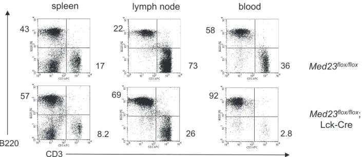
Figure 2. Mice with efficient deletion ofMed23exhibit decreased T-cell numbers in the peripheral lymphoid organs.Flow cytometric analysis of spleen, lymph node and blood fromMed23flox/floxandMed23flox/flox;Lck-Cremice showing proportions of CD3+T-cells and B220+B-cells.
Flow cytometry from mouse shown is representative of phenotype seen in mice with greater than 95% deletion ofMed23in the thymus.
doi:10.1371/journal.pone.0102076.g002
Figure 3.Med23flox/flox;Lck-CreT-cells do not display increased apoptosis.TUNEL assay was performed on cryosections from thymus and
spleen ofMed23flox/flox; Lck-Cre
andLck-Crecontrol mice. A control section was treated with DNaseI prior to TUNEL staining (+TdT) as a positive control, and a cryosection that was not stained (2TdT) serves as a negative control.
MED23 null thymocytes proliferate normally in response to activation, but display abnormal gene expression
The ERK/MAPK pathway is also known to be important in the activation of T-cells including their proliferative response [24,29]. We next harvested total thymocytes fromMed23+/+;Lck-Cre
and
Med23flox/flox;Lck-Cremice and cultured them in the presence of
plate-boundaCD3 antibody, or antibodies against both CD3 and
CD28 (Figure 4A). We found that proliferation in response to aCD3 alone, oraCD3 andaCD28 together, was not significantly different in MED23 null thymocytes compared to controls (Figure 4A).
We next activatedMed23+/+;Lck-CreandMed23flox/flox;Lck-Cre
total thymocytes for 3 hours by exposure to plate-boundaCD3
antibody, followed by isolation of mRNA.Il2mRNA increased in
response to TCR activation as expected and this induction was comparable in MED23 null and control thymocytes (Figure 4B).
The known MED23 target genes,Egr1andEgr2were induced by
T-cell activation and had reduced expression in MED23 null
compared with control thymocytes (Figure 4C,D; P= 0.06 and
0.0002,N= 2). Microarray analysis revealed 26 genes that were
induced at least two fold byaCD3 (Table S2). Seven of these genes
displayed aCD3 inducible expression that was at least twofold
lower inMed23flox/flox;Lck-Crecompared with control thymocytes (Med23+/+;Lck-Cre
and Med23flox/flox), including Egr1, Egr2,
Cd5,Cd6,Nab2,Pcyt1aandIer2.
KLF2 expression is decreased inMed23null single positive thymocytes
Interestingly, microarray analysis of the mock-treated
thymo-cytes revealed thatCd52(CAMPATH-1) mRNA expression was
decreased in MED23 null thymocytes compared to controls (Table
S3). This defect inCd52expression was confirmed by qRT-PCR
(Figure 4E;P= 0.003,N= 2).Cd52has previously been shown to
Figure 4. MED23 null thymocytes proliferate normally in response to stimulation, but have abnormal gene expression.
ATotal thymocytes were cultured in the presence of plate boundaCD3
and aCD28 antibodies (as indicated) for 44 hrs, then allowed to
incorporate3H thymidine for 18 hrs. Counts per minute (CPM) were
normalized to theLck-CreMock counts for each experiment. N = 2–3,
Mean+/2SEM. Statistical analysis shown is Tukey post-test following
one way ANOVA, n.s. not statistically significant.B–E,Il2(B),Egr1(C),
Egr2 (D), and Cd52 (E) mRNA expression analyzed by qRT-PCR in
Med23+/+;Lck-Cre
(control) and Med23flox/flox;Lck-Cre
(Med23 null) total thymocytes rested 4 hours after harvest, then mock treated or treated 3 hours ex vivo with immobilized aCD3.Med23flox/flox;Lck-Cre
thymo-cytes showed greater than 90% deletion. N = 2, Mean+/2SEM.Pvalues
from two-tailed t-tests between aCD3 (B–D) or Mock (E) treated
samples.
doi:10.1371/journal.pone.0102076.g004
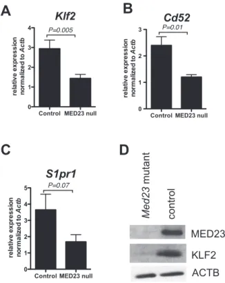
Figure 5. MED23 null single positive thymocytes express decreased KLF2. A–C, Klf2 (A), Cd52 (B) and S1pr1 (C) mRNA
expression in CD4+
single positive thymocytes; YFP+ Med23+/+ ;Lck-Cre;YFPorMed23+/+;Lck-Cre(control) and YFP+Med23flox/flox
;Lck-Cre;YFP
orMed23flox/flox;Lck-Cre
where deletion was greater than 90% (MED23 null) thymocytes. Two-tailed t test; N = 4–8; Mean+/2SEM.D, Western blot of MED23, KLF2 and ACTB (loading control) in whole cell extracts
from YFP+CD4 SP SP thymocytes fromMed23+/+;Lck-Cre;YFP(control)
andMed23flox/flox;Lck-Cre;YFP
be a target of the transcription factor KLF2 [42] and there are distinct similarities between the phenotype ofMed23null T-cells,
and T-cells lackingKlf2 (Lklf) or the KLF2 target gene, S1pr1
(Edg1). All three of these mutant mice retain relatively normal thymic development, but the mutant T-cells have a reduced ability to populate the peripheral lymphocytic organs [36,43–46].
In the thymus, KLF2 is expressed predominantly in single positive (SP) thymocytes [36], so we isolated CD4 SP thymocytes from Med23+/+
;Lck-Cre and Med23flox/flox;Lck-Cre mice and
measured gene expression. We found that expression of Klf2
mRNA was significantly downregulated in MED23 null CD4 SP thymocytes (Figure 5A;N= 8;P= 0.005) as was expression of the
KLF2 target gene,Cd52(Figure 5B;N= 4;P= 0.01). The KLF2
target gene, S1pr1 [42] also tended to be expressed lower in
MED23 null CD4 SP thymocytes (Figure 5C;N= 8; P= 0.07).
Importantly, KLF2 protein levels were also lower in MED23 null CD4 SP thymocytes compared to controls (Figure 5D).
Med23Dflox/Dflox
MEFs also have decreasedKlf2expression
UsingMed23flox/floxMEFs treated with adenovirus expressing
Cre recombinase (Figure S3), we sought to further elucidate the
regulation ofKlf2expression by MED23.Klf2mRNA in MEFs is
induced in response to serum and we found that both the basal (with 0.1% serum) and serum-induced (10% serum) expression of
Klf2 was attenuated by loss of MED23 (Figure 6A; N= 4,
P= 0.02).
We next examined the activity of a luciferase reporter driven by theKlf2promoter (pGL3-Klf2-pro) and found that its activity was significantly decreased in MEFs lacking MED23 (Figure 6B;
N= 6, P= 0.0003). Since there is evidence that expression of
KLF2 target genes,Cd52andS1pr1, is also affected by MED23
loss, we examined the activity of a construct fusing the GAL4 DNA binding domain (DBD) to the first 88 amino acids of KLF2, which includes the activation domain (GAL4-KLF2). We found
that in contrast to the effect of MED23 on Klf2 expression,
MED23 loss does not adversely affect KLF2-dependent transacti-vation (Figure 6C).
It has previously been reported that MEF2 binds to theKlf2
promoter, and that this binding is required for MAPK/ERK5-dependent expression ofKlf2in response to shear stress [39,47]. MEF2C is also reported to be a serum-responsive transactivator [48], further suggesting it contributes to the serum-dependent
expression of Klf2(Figure 6A). We found that mutation of this
MEF2 binding element in ourKlf2promoter luciferase construct
dramatically decreased its activity in both wild type and MED23 null MEFs (Figure 6D). The remaining luciferase activity after mutation of the MEF2 site was MED23-independent as the activity of the mutated construct was the same in wild type and MED23 null MEFs (Figure 6D). Based on this result, we tested the transactivation capacity of a GAL4 DBD full length MEF2C fusion construct (GAL4-MEF2C) and found that it was decreased
by about 40 percent in MEF23 null MEFs (Figure 6E,N= 4–8,
Figure 6. MEFs lacking MED23 show alteredKlf2expression. A,
Klf2mRNA expression by qRT-PCR inMed23+/+(WT) andMed23Dflox/Dflox (MED23 null) MEFs treated with Cre adenovirus, then three days later
starved overnight and treated with serum for 1 hour.N= 4, mean+/2
SEM, two-tailed t test,P= 0.02 for comparison of either mock or serum-treated samples.B, Activity of a luciferase reporter construct driven by
theKlf2promoter (pGL3-Klf2-pro) in WT and MED23 null MEFs (N= 6,
mean+/2SEM, two-tailed t test,P= 0.0003).C, Activity of a GAL4-KLF2 fusion construct containing aa 1–88 of KLF2 in WT and MED23 null MEFs (N= 2, triplicates, mean+/2SEM).D, Mutation of a MEF2 site in theKlf2promoter construct (pGL3-Klf2-pro-mut) dramatically decreases activity of the construct (N= 2, duplicates, mean+/2SEM).E, A GAL4-MEF2C fusion construct is dependent upon MED23 for full activity (N= 4–8, mean+/2SEM, two-tailed t testP= 0.0002). pGL3 Basic and pM1 are empty luciferase and GAL4 DNA binding domain (DBD) vectors respectively. n.s., not statistically significant.
P= 0.0002). This suggests that MED23 could mediate Klf2
expression by providing coactivation function to MEF2 transcrip-tion factors.
Discussion
In this study, MED23 was found to have a rather specific role in T-cell gene expression and development/function, even though it is a subunit of the global coactivator complex Mediator. With regard to cell biology, we found that MED23 is necessary for
T-cells to populate the periphery. We also showed that Med23 is
required for the expression of target genes,Egr1andKlf2, which have both been previously shown to reduce T-cell numbers outside the thymus when mutated (Figures 3 and 4) [36,45,49,50]. MED23 is also required to maintain KLF2 protein levels and KLF2-target gene expression (Cd52), indicating that the reduction inKlf2gene expression has functional consequences for the cells (Figure 5). Finally, the transactivation function of the MEF2 family of transcription factors includes a MED23-dependent component and may contribute toKlf2expression (Figure 6).
The phenotype ofMed23flox/flox;Lck-Cre mice is distinct from the T-cell phenotypes ofElk12/2andElk42/2 mice
Mice null for ELK1, an ETS family transcription factor that is known to utilize Mediator via the MED23 subunit [20], have no obvious phenotype [51]. However, knockout of fellow ETS family member ELK4 (SAP1) results in defective positive selection of
T-cells in the thymus and double-knockout of both Elk1and Elk4
enhances this defect [52]. Despite evidence that both ELK1 and to a lesser degree, ELK4 require the MED23 subunit for coactivation by Mediator [17,20], we did not see a defect in positive selection. Several reasons for this phenotypic disconnect are possible. First,
the timing ofMed23deletion withLck-Cre(DN4) may be too late
to influence positive selection (i.e. MED23 protein may still be present during the critical events). In the future, use of a different Cre transgene that produces earlier deletion in the T-cell compartment could help answer this question. Second, the level of MED23-dependent coactivation provided to ELK4 in T-cells may be insufficient to affect positive selection. In this regard, our own and other studies have shown that loss of MED23 can have the effect of decreasing, but not ablating expression of target genes [17,20]. A third possibility is that the subset of ELK1/ELK4 target genes that are MED23-dependent in T-cells are not those that are required for normal positive selection. Other models support this possibility, as we have found that both the transcription factors CREB and HIF require the coactivators CBP and p300 for expression of some, but not all of their target genes [5,8,12,53]. Likewise, it has been shown that cellular context influences the requirement for MED23 for the expression of target genes in MEFs compared with ES cells, even in response to the same stimulus [17].
MED23 target genesKlf2andEgr1impact the number of peripheral T-cells in knockout mouse models
We have shown that conditional deletion ofMed23in the T-cell lineage of mice produces T-cells with a decreased ability to populate the periphery (Figures 1 and 2). It seems likely that this
defect may be linked to a decrease we observe inKlf2mRNA and
KLF2 protein levels in single positive thymocytes, since Klf2
deletion in T-cells results in a similar, albeit more complete loss of peripheral T-cells [36,45,50].
The reduced expression ofEgr1 in MED23 null T-cells may
also play a role in their decreased ability to populate the peripheral
organs.Egr1has been shown to be necessary for the survival of
recent thymic emigrants in the periphery, although this phenotype was seen only in TCR transgenic mice [49]. Interestingly, other
phenotypes associated with Egr1 deletion, such as increased
thymocyte number [54], were not seen in our study. One possible
explanation is that deletion ofMed23during thymic development
(DN4 stage for the Lck-Cre transgene used in this study) [41]
results in a milder phenotype than would be seen if MED23 were absent from the start of thymic development.
Our data show that MED23 is required for full expression of the
endogenousKlf2gene in both T-cells and MEFs (Figures 5 and 6).
Our in vitro data suggest that MEF2 transcription factors may
play a role inKlf2expression in MEFs, as mutation of a MEF2
binding site in a Klf2 promoter luciferase reporter construct
drastically reduces the activity of the construct (Figure 6D). Likewise, the transactivation function of a GAL4-MEF2C fusion construct is significantly reduced in MED23 null MEFs (Figure 6E), suggesting that MEF2 family members may utilize
MED23-dependent mechanisms of coactivation.Klf2has been previously
reported to be induced in a MAPK/ERK5-dependent manner by shear stress in endothelial cells through the transcription factor MEF2 [47]. However, knockout of ERK5 in mouse T-cells produces no deficit in thymocyte development or peripheral T-cell
numbers, although there was impaired induction ofKlf2mRNA
in response to TCR signaling [55,56]. MEF2C expression has been shown to be critical for commitment to the lymphoid lineage, but does not seem to be necessary for thymocyte development [57]. Thus, whether MEF2 is the key regulator ofKlf2in T-cells remains to be established.
Cell type context in Mediator MED23 subunit utilization
The MED23 subunit was originally implicated as critical for recruitment of the Mediator complex by the transcription factor ELK1, as well as the adenovirus E1A protein [20]. More recently, it has been shown that in at least some gene contexts, the MED23 subunit also appears to be important for the transition from paused to Ser2 phosphorylated elongating RNA Polymerase II via the recruitment of positive elongation factor b (P-TEFb) [58]. In this scenario, the Mediator complex is recruited to target gene promoters equally in the presence or absence of MED23; however, MED23 is required for gene transcription to occur [58]. It is not clear yet what context determines which coactivation mecha-nism(s) MED23 can contribute in specific cell types or at certain promoters. Our study contributes to the findings that the MED23 subunit of Mediator is required for the expression of a narrow, but important subset of genes in a range of cell types [17,20–22,58].
Here we described a newMed23conditional knockout mouse and
its utility in studying this Mediator subunitin vitroandin vivo. Using this model, we established that MED23 has a rather specific biological role in the context of T-cells, and is critical for the expression of the T-cell master regulatory transcription factor, KLF2.
Supporting Information
Figure S1 Conditional allele of Med23 results in a null allele
after cre-mediated recombination. A, Targeting strategy for
Med23 conditional knockout mice. B,C, Southern (B) and
western (C) showing efficient deletion of Med23 floxed alleles
and MED23 protein in thymus from a Med23flox/flox;Lck-Cre
mouse. Western blot performed using an antibody raised against amino acids 906–925.
(PDF)
Figure S2 Mice with efficient Med23 deletion have reduced
peripheral T cell numbers.A, Scatter plots showing CD4 vs CD8
+;Lck-Cre
control mouse and fourMed23flox/flox; Lck-Cremice.B, Southern blot showing thatMed23flox/flox; Lck-Cremice#1, 3 and 4 have efficient recombination of the floxed alleles while#2 has
less than 50% recombination.C, Western blot confirming MED23
protein loss in efficiently deletedMed23flox/flox; Lck-Cremice. (PDF)
Figure S3 Med23Dflox/Dflox MEFs treated with adenovirus expressing Cre recombinase show efficient deletion of MED23. A. Semiquantitative PCR of genomic DNA from Cre adenovirus treated wild type (Med23+/+) andMed23Dflox/DfloxMEFs demon-strating efficient recombination of the conditionalMed23 allele. Bands for the wild type (WT), conditional (flox) and recombined conditional (Dflox) alleles are indicated. B. Western blot of
Cre-adenovirus treated wild type andMed23Dflox/DfloxMEFs showing
loss of MED23 protein. Non-specific band shown as loading control.
(PDF)
Table S1 YFP+
percentages for each cell population in each
mouse that were used to calculate the average normalized YFP+
percentages shown in Figure 1C. (XLSX)
Table S2 Genes induced at least two fold by ex vivo aCD3 treatment of total thymocytes from control (Lck-CreandMed23flox/ flox
) mice based on microarray analysis. (XLSX)
Table S3 Genes expressed at least two fold higher in ex vivo
mock-treated total thymocytes from control (Lck-Creand
Med23-flox/flox
) mice compared withMed23flox/flox; Lck-Cremice based on microarray analysis.
(XLSX)
Acknowledgments
We thank Jerry Lingrel forKlf2constructs and Jeffrey Leiden and Laurie Glimcher for the KLF2 antibody. We also thank Stephanie Lerach, Trushar Jeevan and Miriam Chong for excellent technical assistance. Thanks to the Vector Development and Production Core and Animal Resources Center at SJCRH. The Hartwell Center at SJCRH provided oligonucleotides.
Author Contributions
Conceived and designed the experiments: LHK PKB. Performed the experiments: LHK TF. Analyzed the data: LHK TF PKB. Contributed reagents/materials/analysis tools: LHK TF PKB. Contributed to the writing of the manuscript: LHK PKB.
References
1. Bedford DC, Kasper LH, Fukuyama T, Brindle PK (2010) Target gene context influences the transcriptional requirement for the KAT3 family of CBP and p300 histone acetyltransferases. Epigenetics 5: 9–15.
2. Bedford DC, Brindle PK (2012) Is histone acetylation the most important physiological function for CBP and p300? Aging (Albany NY) 4: 247–255. 3. Kasper LH, Fukuyama T, Lerach S, Chang Y, Xu W, et al. (2013) Genetic
interaction between mutations in c-Myb and the KIX domains of CBP and p300 affects multiple blood cell lineages and influences both gene activation and repression. PLoS One 8: e82684.
4. Bedford DC, Kasper LH, Wang R, Chang Y, Green DR, et al. (2011) Disrupting the CH1 domain structure in the acetyltransferases CBP and p300 results in lean mice with increased metabolic control. Cell Metab 14: 219–230. 5. Kasper LH, Lerach S, Wang J, Wu S, Jeevan T, et al. (2010) CBP/p300 double null cells reveal effect of coactivator level and diversity on CREB transactivation. Embo J 29: 3660–3672.
6. Jin Q, Yu LR, Wang L, Zhang Z, Kasper LH, et al. (2011) Distinct roles of GCN5/PCAF-mediated H3K9ac and CBP/p300-mediated H3K18/27ac in nuclear receptor transactivation. The EMBO journal 30: 249–262.
7. Fukuyama T, Kasper LH, Boussouar F, Jeevan T, van Deursen J, et al. (2009) Histone acetyltransferase CBP is vital to demarcate conventional and innate CD8+T-cell development. Mol Cell Biol 29: 3894–3904.
8. Xu W, Kasper LH, Lerach S, Jeevan T, Brindle PK (2007) Individual CREB-target genes dictate usage of distinct cAMP-responsive coactivation mechanisms. Embo J 26: 2890–2903.
9. Xu W, Fukuyama T, Ney PA, Wang D, Rehg J, et al. (2006) Global transcriptional coactivators CREB-binding protein and p300 are highly essential collectively but not individually in peripheral B cells. Blood 107: 4407–4416. 10. Oliveira AM, Abel T, Brindle PK, Wood MA (2006) Differential Role for CBP
and p300 CREB-Binding Domain in Motor Skill Learning. Behav Neurosci 120: 724–729.
11. Kasper LH, Fukuyama T, Biesen MA, Boussouar F, Tong C, et al. (2006) Conditional knockout mice reveal distinct functions for the global transcriptional coactivators CBP and p300 in T-cell development. Mol Cell Biol 26: 789–809. 12. Kasper LH, Boussouar F, Boyd K, Xu W, Biesen M, et al. (2005) Two transactivation mechanisms cooperate for the bulk of HIF-1-responsive gene expression. Embo J 24: 3846–3858.
13. Kang-Decker N, Tong C, Boussouar F, Baker DJ, Xu W, et al. (2004) Loss of CBP causes T cell lymphomagenesis in synergy with p27(Kip1) insufficiency. Cancer Cell 5: 177–189.
14. Kasper LH, Boussouar F, Ney PA, Jackson CW, Rehg J, et al. (2002) A transcription-factor-binding surface of coactivator p300 is required for haematopoiesis. Nature 419: 738–743.
15. Taatjes DJ (2010) The human Mediator complex: a versatile, genome-wide regulator of transcription. Trends Biochem Sci 35: 315–322.
16. Yin JW, Wang G (2014) The Mediator complex: a master coordinator of transcription and cell lineage development. Development 141: 977–987. 17. Balamotis MA, Pennella MA, Stevens JL, Wasylyk B, Belmont AS, et al. (2009)
Complexity in transcription control at the activation domain-mediator interface. Sci Signal 2: ra20.
18. Carlsten JO, Zhu X, Gustafsson CM (2013) The multitalented Mediator complex. Trends Biochem Sci 38: 531–537.
19. Singh N, Han M (1995) sur-2, a novel gene, functions late in the let-60 ras-mediated signaling pathway during Caenorhabditis elegans vulval induction. Genes Dev 9: 2251–2265.
20. Stevens JL, Cantin GT, Wang G, Shevchenko A, Berk AJ (2002) Transcription control by E1A and MAP kinase pathway via Sur2 mediator subunit. Science 296: 755–758.
21. Wang W, Huang L, Huang Y, Yin JW, Berk AJ, et al. (2009) Mediator MED23 links insulin signaling to the adipogenesis transcription cascade. Developmental cell 16: 764–771.
22. Yin JW, Liang Y, Park JY, Chen D, Yao X, et al. (2012) Mediator MED23 plays opposing roles in directing smooth muscle cell and adipocyte differentiation. Genes & development 26: 2192–2205.
23. Yang X, Zhao M, Xia M, Liu Y, Yan J, et al. (2012) Selective requirement for Mediator MED23 in Ras-active lung cancer. Proceedings of the National Academy of Sciences of the United States of America 109: E2813–2822. 24. Franklin RA, Tordai A, Patel H, Gardner AM, Johnson GL, et al. (1994)
Ligation of the T cell receptor complex results in activation of the Ras/Raf-1/ MEK/MAPK cascade in human T lymphocytes. J Clin Invest 93: 2134–2140. 25. Alberola-Ila J, Forbush KA, Seger R, Krebs EG, Perlmutter RM (1995) Selective requirement for MAP kinase activation in thymocyte differentiation. Nature 373: 620–623.
26. Sharp LL, Schwarz DA, Bott CM, Marshall CJ, Hedrick SM (1997) The influence of the MAPK pathway on T cell lineage commitment. Immunity 7: 609–618.
27. Bain G, Cravatt CB, Loomans C, Alberola-Ila J, Hedrick SM, et al. (2001) Regulation of the helix-loop-helix proteins, E2A and Id3, by the Ras-ERK MAPK cascade. Nat Immunol 2: 165–171.
28. Priatel JJ, Teh SJ, Dower NA, Stone JC, Teh HS (2002) RasGRP1 transduces low-grade TCR signals which are critical for T cell development, homeostasis, and differentiation. Immunity 17: 617–627.
29. D’Souza WN, Chang CF, Fischer AM, Li M, Hedrick SM (2008) The Erk2 MAPK regulates CD8 T cell proliferation and survival. J Immunol 181: 7617– 7629.
30. Kortum RL, Rouquette-Jazdanian AK, Samelson LE (2013) Ras and extracellular signal-regulated kinase signaling in thymocytes and T cells. Trends in immunology 34: 259–268.
31. Costello PS, Nicolas RH, Watanabe Y, Rosewell I, Treisman R (2004) Ternary complex factor SAP-1 is required for Erk-mediated thymocyte positive selection. Nature immunology 5: 289–298.
32. Hogquist KA, Weinreich MA, Jameson SC (2008) T-cell migration: Kruppeled T cells move again. Immunol Cell Biol 86: 297–298.
33. Hennet T, Hagen FK, Tabak LA, Marth JD (1995) T-cell-specific deletion of a polypeptide N-acetylgalactosaminyl- transferase gene by site-directed recombi-nation. Proc Natl Acad Sci U S A 92: 12070–12074.
35. Parrinello S, Samper E, Krtolica A, Goldstein J, Melov S, et al. (2003) Oxygen sensitivity severely limits the replicative lifespan of murine fibroblasts. Nat Cell Biol 5: 741–747.
36. Kuo CT, Veselits ML, Leiden JM (1997) LKLF: A transcriptional regulator of single-positive T cell quiescence and survival. Science 277: 1986–1990. 37. Schrick JJ, Hughes MJ, Anderson KP, Croyle ML, Lingrel JB (1999)
Characterization of the lung Kruppel-like transcription factor gene and upstream regulatory elements. Gene 236: 185–195.
38. Conkright MD, Wani MA, Lingrel JB (2001) Lung Kruppel-like factor contains an autoinhibitory domain that regulates its transcriptional activation by binding WWP1, an E3 ubiquitin ligase. J Biol Chem 276: 29299–29306.
39. Kumar A, Lin Z, SenBanerjee S, Jain MK (2005) Tumor necrosis factor alpha-mediated reduction of KLF2 is due to inhibition of MEF2 by NF-kappaB and histone deacetylases. Molecular and cellular biology 25: 5893–5903. 40. Sadowski I, Bell B, Broad P, Hollis M (1992) GAL4 fusion vectors for expression
in yeast or mammalian cells. Gene 118: 137–141.
41. Bender TP, Kremer CS, Kraus M, Buch T, Rajewsky K (2004) Critical functions for c-Myb at three checkpoints during thymocyte development. Nat Immunol 5: 721–729.
42. Haaland RE, Yu W, Rice AP (2005) Identification of LKLF-regulated genes in quiescent CD4+T lymphocytes. Mol Immunol 42: 627–641.
43. Matloubian M, Lo CG, Cinamon G, Lesneski MJ, Xu Y, et al. (2004) Lymphocyte egress from thymus and peripheral lymphoid organs is dependent on S1P receptor 1. Nature 427: 355–360.
44. Allende ML, Dreier JL, Mandala S, Proia RL (2004) Expression of the sphingosine 1-phosphate receptor, S1P1, on T-cells controls thymic emigration. J Biol Chem 279: 15396–15401.
45. Carlson CM, Endrizzi BT, Wu J, Ding X, Weinreich MA, et al. (2006) Kruppel-like factor 2 regulates thymocyte and T-cell migration. Nature 442: 299–302. 46. Sebzda E, Zou Z, Lee JS, Wang T, Kahn ML (2008) Transcription factor KLF2
regulates the migration of naive T cells by restricting chemokine receptor expression patterns. Nat Immunol 9: 292–300.
47. Parmar KM, Larman HB, Dai G, Zhang Y, Wang ET, et al. (2006) Integration of flow-dependent endothelial phenotypes by Kruppel-like factor 2. J Clin Invest 116: 49–58.
48. Kato Y, Kravchenko VV, Tapping RI, Han J, Ulevitch RJ, et al. (1997) BMK1/ ERK5 regulates serum-induced early gene expression through transcription factor MEF2C. The EMBO journal 16: 7054–7066.
49. Schnell FJ, Kersh GJ (2005) Control of recent thymic emigrant survival by positive selection signals and early growth response gene 1. J Immunol 175: 2270–2277.
50. Weinreich MA, Takada K, Skon C, Reiner SL, Jameson SC, et al. (2009) KLF2 transcription-factor deficiency in T cells results in unrestrained cytokine production and upregulation of bystander chemokine receptors. Immunity 31: 122–130.
51. Cesari F, Brecht S, Vintersten K, Vuong LG, Hofmann M, et al. (2004) Mice deficient for the ets transcription factor elk-1 show normal immune responses and mildly impaired neuronal gene activation. Mol Cell Biol 24: 294–305. 52. Costello P, Nicolas R, Willoughby J, Wasylyk B, Nordheim A, et al. (2010)
Ternary complex factors SAP-1 and Elk-1, but not net, are functionally equivalent in thymocyte development. J Immunol 185: 1082–1092.
53. Kasper LH, Thomas MC, Zambetti GP, Brindle PK (2011) Double null cells reveal that CBP and p300 are dispensable for p53 targets p21 and Mdm2 but variably required for target genes of other signaling pathways. Cell cycle 10: 212–221.
54. Joslin JM, Fernald AA, Tennant TR, Davis EM, Kogan SC, et al. (2007) Haploinsufficiency of EGR1, a candidate gene in the del(5q), leads to the development of myeloid disorders. Blood 110: 719–726.
55. Ananieva O, Macdonald A, Wang X, McCoy CE, McIlrath J, et al. (2008) ERK5 regulation in naive T-cell activation and survival. Eur J Immunol 38: 2534–2547.
56. Weinreich MA, Jameson SC, Hogquist KA (2011) Postselection thymocyte maturation and emigration are independent of IL-7 and ERK5. J Immunol 186: 1343–1347.
57. Stehling-Sun S, Dade J, Nutt SL, DeKoter RP, Camargo FD (2009) Regulation of lymphoid versus myeloid fate ‘choice’ by the transcription factor Mef2c. Nat Immunol 10: 289–296.