Universidade de Aveiro 2017
Departamento de Ciências Médicas
JOÃO ANDRÉ
FERNANDES DE
JESUS
Efeito do Aβ nos complexos da Proteína Fosfatase 1
Aβ effects in Protein Phosphatase 1 complexes
Universidade de Aveiro 2017
Departamento de Ciências Médicas
JOÃO ANDRÉ
FERNANDES DE
JESUS
Aβ effects in Protein Phosphatase 1 complexes
Dissertação apresentada à Universidade de Aveiro para cumprimento dos requisitos necessários à obtenção do grau de Mestre em Biomedicina Molecular, realizada sob a orientação científica da Professora Doutora Odete Abreu Beirão da Cruz e Silva, Professora Auxiliar com Agregação do Departamento de Ciências Médicas da Universidade de Aveiro.
Esta dissertação contou com o apoio financeiro do Instituto de Biomedicina (iBiMED) - UID/BIM/04501/2013 e PTDC/DTP¬PIC/5587/2014 da Fundação para a Ciência e Tecnologia do Ministério da Educação e Ciência, programa COMPETE, do QREN e da União Europeia (Fundo Europeu de Desenvolvimento Regional).
o júri
presidente Professora Doutora Sandra Maria Tavares da Costa Rebelo
ProfessoraAuxiliar Convidada, Universidade de Aveiro
Professora Doutora Maria De Lourdes Gomes Pereira
Professor Associada com Agregação, Universidade de Aveiro
Professora Doutora Odete Abreu Beirão, da Cruz e Silva
agradecimentos Gostaria de agradecer à Professora Doutora Odete da Cruz e Silva pela oportunidade de realizar este trabalho no laboratório de neurociências e por toda a sua orientação, encorajamento e conselhos durante a realização desta dissertação.
À Professora Doutora Sandra Rebelo, e à Filipa por toda a ajuda no aconselhamento e execução de alguns procedimentos laboratoriais.
Ao Márcio por toda a disponibilidade e amizade ao longo deste ano, assim como ao Steven pela companhia constante.
A todos os meus colegas de laboratório que sempre se disponibilizaram a ajudar quando necessário, e por todo o companheirismo e bom ambiente de trabalho criado.
Aos meus amigos por tornarem este longo percurso muito mais fácil.
À Susana por toda a compreensão que teve, e pelo apoio incondicional nos momentos mais difíceis.
E principalmente aos meus pais que tornaram tudo isto possível, e por me apoiarem em tudo ao longo da minha vida.
palavras-chave Aβ, Neurabin-1, Neurabin-2, Phactr3, PP1, Complexos proteicos, Linha celular SH-SY5Y, diferenciação
resumo O cérebro é uma estrutura complexa, que é composta por neurónios capazes de comunicar através de sinapses. Estas normalmente ocorrem entre um terminal axónico e espinha dendritica de neurónios diferentes. As espinhas dendriticas são estruturas dinâmicas que permitem uma rápida adaptação a diferentes estímulos. A PP1 é uma proteína fosfatase que catalisa a maioria das desfosforilações que ocorrem no corpo humano. Tem diversas funções, que variam desde metabolismo de glicogénio a regulação sináptica. As neurorabinas (1 e 2) são duas subunidades reguladores da PP1 que são estruturalmente e funcionalmente idênticas entre si. São muito enriquecidas nas espinhas dendriticas e interagem com diversas proteínas, incluindo a PP1, e guiam as proteínas para receptores, citoesqueleto ou outros compartimentos celulares, regulando assim a morfologia neuronal e transmissão e plasticidade sináptica. A Phactr3 pertence à família de proteínas reguladoras de actina, á qual pertencem quatro membros. A Phactr3 está envolvida na migração celular e regula a dinâmica do citoesqueleto. Interage com a PP1, formando um complexo que é controlado por alterações na concentração de G-actina citoplasmática, regulando assim a dinâmica do citoesqueleto. Doenças neurodegenerativas, são normalmente caracterizadas pela perda de sinapses, morte neuronal, e perda gradual de funções cognitivas e memória. Acredita-se que o principal fator responsável por essas anomalias seja o péptido Aβ. Este resulta de uma clivagem anormal de APP, pela via amiloidogénica, resultando numa sobreprodução do péptido tóxico.
O objectivo desta tese era avaliar os efeitos de Aβ nos níveis de expressão das neurorabinas e Phactr3, assim como os complexos formados com a PP1. Os resultados mostram uma ligeira diminuição nos níveis de expressão das neurorabinas, e num ligeiro aumento na expressão de Phactr3. Os resultados também mostram uma diminuição na interação do complexo Neurorabina-1/PP1, provavelmente devido a um efeito direto de Aβ sobre Neurorabina-1 ou um desequilíbrio nas fosfatases e cinases, e Phactr3/PP1, provavelmente devido a variação nos níveis de G-actina, que compete com a PP1 para interagir com Phactr3. As mesmas alterações não foram verificadas no complexo Neurorabina-2/PP1. Uma explicação possível é que ambas as proteínas são reguladas de forma diferente por diversas cinases. Estes resultados permitem concluir que Aβ interfere na expressão das três proteínas e nos níveis de interação de Neurabina-1/PP1 e Phactr3/PP1. No entanto são necessários mais estudos para obter uma melhor compreensão da relevância fisiológica destes complexos.
keywords Aβ, Neurabin-1, Neurabin-2, Phactr3, PP1, Protein complexes, SH-SY5Y cell line, differentiation
abstract The brain is a complex structure, which is comprised by neurons capable of communication through synapses. These usually occur between an axon terminal and a dendritic spine of different neurons. The dendritic spines are dynamic structures that allow for a rapid adaptation to different stimuli. PP1 is a phosphatase protein that catalyses the majority of dephosphorylation reactions in the human body. It has different functions that vary, from glycogen metabolism to synaptic regulation. Neurabins (1 and 2) are two PP1 regulator subunits that are structurally and functionally similar to each other. They are highly enriched in the dendritic spines, interacting with several proteins, including PP1, and targeting them to receptors, cytoskeleton or other cellular compartments. Thus, regulating neuronal morphology and synaptic transmission and plasticity. Phactr3 belongs to the actin regulatory protein family, which comprises four members. Phactr3 is involved in cell migration and regulates cytoskeleton dynamics. It interacts with PP1 forming a complex that is controlled by changes in cytoplasmic G-actin concentration, thus, regulating actin cytoskeleton dynamics. Neurodegenerative diseases are usually characterised by the loss of synapses, neural death, gradual loss of cognitive functions and memory. It is believed that Aβ is the major culprit in these changes. Aβ results from an abnormal cleavage of APP, via the amyloidogenic pathway, resulting in the overproduction of toxic peptides.
The main aim of this thesis was to evaluate the effects of Aβ on Neurabins and Phactr3 expression, and in PP1 complexes. The results show a slight decrease in neurabins’ expression, and a slight increase in Phactr3 expression. The results also show a decrease in interaction between PP1 and Neurabin-1, possibly due to direct effect of Aβ on Neurabin-1 or the imbalance of phosphatases and kinases, and between PP1 and Phactr3, possibly due to variations of G-actin levels which competes with PP1 to binding with Phactr3. The same changes were not observed in Neurabin-2/PP1 complexes. A possible explanation is that both are differently regulated by several kinases.
The results allow us to conclude that Aβ interferes in the expression of all three proteins and in the interaction of Neurabin-1/PP1. However, additional studies are required in order to better understand the physiological relevance of these complexes.
Index
I. List of figures ... III II. List of tables ... V III. Abbreviations ... VII
1. Introduction ... 3
1.1. The human brain and neuronal cells ... 3
1.2. Dendritic Spines ... 4 1.3. PP1 ... 9 1.3.1. PP1 isoforms ... 9 1.4. Neurabins ... 11 1.4.1. Neurabin 1 ... 11 1.4.2. Neurabin 2 ... 13 1.5. Phactrs ... 14 2. Objectives ... 23
3. Materials and methods ... 27
3.3. Culture, growth and maintenance of SH-SY5Y cell line ... 29
3.3.1. Differentiation of the SH-SY5Y neuroblastoma cell line ... 30
3.4. Aβ treatment ... 30
3.6. Co-Immunoprecipitation ... 32
3.7. Western Blot ... 34
4. Results and discussion ... 39
4.2. Differentiation of the SH-SY5Y cell line ... 44
4.2.1. Morphology evaluation ... 44
4.3. Aβ effect on Neurabins expression ... 46
4.4. Aβ effect on Phactr3 expression ... 48
4.5. Aβ effect on Protein/PP1 complexes ... 49
5. Conclusion ... 55
6. Future perspectives ... 61
6. References ... 65
I.
List of figures
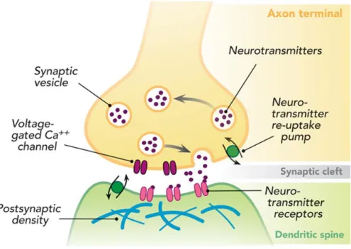
Figure 1 - Chemical Synapse, taken from ... 3
Figure 2 - Dendritic spine morphology ... 4
Figure 3 - PP1 docking motifs ... 11
Figure 4- Neurabin-1 domain structure ... 12
Figure 5 – Neurabin – 2 domain Structure ... 12
Figure 6 -Phactr3 domain structure ... 15
Figure 7- Interactome network, of PP1α and PP1γ... 40
Figure 8 – Merged Interactome Network of PP1α, PP1γ, Neurabin-1, Neurabin-2, Phactr3 and Cofilin. . 41
Figure 9 - Interactome Network of proteins directly binding to PP1α and PP1γ ... 42
Figure 10- Phase contrast photographs of SH-SY5Y cell line morphology during differentiation ... 45
Figure 11 - Neurabin-1 and Neurabin-2 expression in SH-SY5Y cells treated with Aβ1-42 ... 47
Figure 12- Phactr3 expression in SH-SY5Y cells treated with Aβ1-42 ... 48
Figure 13- Co-immunoprecipitation of PP1α binding proteins in SH-SY5Y ... 50
Figure 14 - Co-immunoprecipitation of PP1γ binding proteins in SH-SY5Y ... 50
Figure 15 - Interaction levels of PP1 complexes in co-immunoprecipitated lysates of SH-SY5Y treated with different concentrations of Aβ ... 51
Figure 16 - Proposed models for the effect of Aβ on the Neurabin/PP1 complex ... 57
II.
List of tables
Table 1 - List of Key Proteins retrieved from uniport and submitted to IntAct to obtain interactome
network ... 27
Table 2- Primary antibodies and dilutions used. ... 29
Table 3- Secondary antibodies used. ... 29
III.
Abbreviations
Aβ β Amyloid
AMPA α-amino-3-hydroxy-5-methyl-4-isoxazole propionic BCA Bicinchoninic acid
BSA Bovine serum albumin
CaMKII Calcium/calmodulin-dependent protein kinase II Cdk5 Cyclin-dependent-like kinase 5
Co-IP Co-immunoprecipitation
DARPP-32 Dopamine and cAMP-regulated neuronal phosphoprotein
ECL Enhanced luminescence
ERK2 Extracellular signal-regulated kinase 2
LB Loading Buffer
LTD Long-term depression LTP Long-term potentiation
MRTF Myocardin-related transcription factor MYPT1 Myosin phosphate-targeting subunit 1 NMDA N-methyl-D-aspartate
PDZ PSD-95/Dlg/ZO-1
PIPs PP1-interacting proteins
PKA Protein kinase catalytic subunit alpha PP1 Protein phosphatase 1
PP1α Protein phosphatase 1 catalytic subunit α PP1β Protein phosphatase 1 catalytic subunit β PP1γ Protein phosphatase 1 catalytic subunit γ PSD Postsynaptic density
RA Retinoic acid
SAM Sterile alpha motif SDS Sodium dodecyl sulfate
SDS-PAGE Sodium dodecyl sulfate polyacrylamide gel electrophoresis Ser/Thr Serine/threonine
TBS Tris Buffered Saline
1. Introduction
1.1. The human brain and neuronal cells
The human brain is a highly complex structure, which governs our lives. It dictates everything that we do, think and feel. It commands all our actions, thoughts, memories and emotions. To guarantee that its functions are properly executed, the brain needs a highly organized network.1–3
The brain network includes an elaborate neuronal matrix, capable of communication, through neuronal synapses.4,5 Most synapses are chemical, and rely
on transferring endogenous chemical compounds, called neurotransmitters, between neurons. The neurotransmitters are produced in the presynaptic neuron and encapsulated within synaptic vesicles, that are secreted to the synaptic cleft.6 After
diffusion through the synaptic cleft, neurotransmitters bind to specific receptors on the membrane of the postsynaptic neuron.7
1.2. Dendritic Spines
Dendritic spines are the morphologic correlates of excitatory post synapses8. They
are tiny, bulbous structures that protrude from the dendrites in neurons and receive fast excitatory synaptic input in the brain.9 This type of neuronal communication usually occurs
between an axon terminal (presynaptic component) and dendritic spines (post synaptic component). These structures, compartmentalize the postsynaptic machinery and biochemical signaling molecules needed to respond to input from single presynaptic terminals. It is now widely accepted that this compartmentalization serves a major function of spine structure: input specificity of synaptic plasticity10.
Dendritic spines are present on different populations of neurons in the brain and are preferentially located on the peripheral dendrites of neocortical and hippocampal pyramidal neurons, on the striatum and in cerebellar Purkinje cells. They are thought to be key structures in both learning and memory formation, as they receive the majority of excitatory and inhibitory outputs in the central nervous system.11
1.2.1. Structure and composition
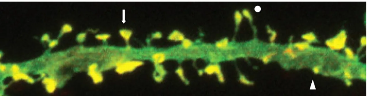
Morphologically, spines are specialized protrusions from a dendrite’s shaft, where neurons form synapses to receive and integrate information and can be classified into three major types: Mushroom spines, which have a large head and a thin neck; stubby spines, which have a large head but no discernible neck; and thin spines, which are slender without a discernible head8,9,11,12.
Figure 2 - Dendritic spine morphology. Circle, thin dendritic spine; Arrow, Mushroom dendritic spine; Triangle, Stubby dendritic spine. Taken from61.
However, dendritic spines are not static, i.e., they do not always have the same structure, since in developing neurons, the majority of dendritic spines change their shape over periods of minutes to hours. As for dendritic spines in mature neurons, they are not as motile as in developing neurons and thus, there are fewer changes in their shape. Large spines are functionally stronger in their response to glutamate, regulation of intracellular calcium, protein translation and degradation, and endosomal recycling than smaller spines, which are more flexible, rapidly enlarging or shrinking in response to subsequent activation. 9,11
The first structure of the dendritic spine, and probably the most complex spine organelle, is the postsynaptic density (PSD), an electron-dense thickening of the postsynaptic membrane that harbors hundreds of proteins involved in synaptic plasticity, including neurotransmitter receptors, such as N-methyl-D-aspartate (NMDA) receptors, α-amino-3-hydroxy-5-methyl-4-isoxazole propionic (AMPA) receptors, kainate and metabotropic glutamate receptors, along with numerous signaling and scaffolding proteins. 6,9,11,13
Below the PSD we can find both monomeric (globular, g-actin) and filamentous (F-actin) actin. The spines’ cytoskeleton is rich in f-actin, which modulates spine head structure in response to postsynaptic signaling and contributes to the overall structure of synapses. It organizes the PSD, anchoring and stabilizing postsynaptic receptors and localizing the translation machinery.11 The dendritic spine is rich in actin-binding proteins,
actin-associated proteins and some small GTPases that cooperate to regulate actin-based cellular events, such as the formation, elimination, motility, stability, size and shape of dendritic spines14. This actin cytoskeleton helps explain why a variety of dendritic spines
exist, since actin is very dynamic and allows for changes in size and shape of dendritic spines.9,14
Additionally, several other organelles can be found within dendritic spines, such as the smooth endoplasmic reticulum, a spinae apparatus, clusters of polyribosomes and proteasomes.
1.2.2. Dendritic Spine formation
There are three main views on the origin of dendritic spines, which imply different molecular mechanisms of spine origin. The first model states that once dendritic filopodia, predominant in younger neurons, establish synaptic contact they originate dendritic spines. This model is based on the fact that, as synapses form, the number of filopodia declines and the number of spine-like structures increase, which may suggest that filopodia are the precursors of dendritic spines. So, this model suggests that the highly motile filopodia act as a probe that search for appropriate contacts. The axon guidance (suggested by the fact that release of glutamate from sites of presynaptic vesicle release, promotes filopodia extension) and cell adhesion molecules present on developing dendrites and axons may contribute to directing the movements of filopodia and their selective adhesion to a compatible axonal partner.15
The second model proposes that dendritic spines arise from synapses that are initially formed on the dendritic shaft. This view is supported by the observation that most synapses in younger pyramidal neurons are located on the dendritic shaft rather than on filopodia.16
The third model states that spines can form even without synaptic contact. Spines of the distal dendritic branches of cerebellar Purkinje neurons form before the establishment of synaptic contact with the presynaptic parallel fibers. 16
Since these three models base their assumptions on different types of neurons, it is more likely that dendritic spines emerge through different mechanisms depending on the type of neuron. However, it has also been proposed that these three models might be part of the same process, which occur in specific temporal periods of a neuron’s life and may be dependent on the maturation of filopodia and dendritic spines. 15,16
Following genesis of dendritic spines, they pass through a process of maturation where there is an increase in spine density, a decrease in overall length and a decrease in
the number of dendritic filopodia with a simultaneous decrease in spine motility. This maturation process also results in synapse maturation, which involves the further recruitment of presynaptic and postsynaptic components (scaffolding proteins and neurotransmitters receptors, for example). 12
Finally, after maturation of dendritic spines and formed synapses, there is a retraction of some contacts and elimination of inappropriate synaptic proteins, which refine the neuronal circuitry, together with long-term potentiation (LTP) and long-term depression (LTD). 9,11
1.2.3. Synaptic plasticity
Synaptic plasticity is the ability that neurons have to positively or negatively change the efficacy of their connections in response to neuronal activity. This synaptic connectivity is dynamic, constantly changing in response to neural activity and other influences, and can vary in time from milliseconds to years. 17
Short-term synaptic plasticity includes facilitation, augmentation, potentiation and synaptic depression. Synaptic facilitation is a rapid increase in synaptic strength that occurs when two or more action potentials invade the presynaptic terminal within a few milliseconds of each other.5 Synaptic augmentation of potentiation is also elicited by
repeated synaptic activity and serves to increase the amount of neurotransmitters released from presynaptic to postsynaptic terminals. Synaptic depression opposes facilitation, causing a decline in neurotransmitter release during sustained synaptic activity. 17
Long-term synaptic plasticity includes LTP term potentiation) and LTD (long-term depression). LTP is a long-lasting increase in synaptic strength due to certain patterns of high frequency electrical stimulation, whereas LTD is a long-lasting decrease in synaptic strength due to certain patterns of low frequency electrical stimulation, both of which are thought to be important for memory formation and storage in the brain. LTP and LTD are intimately related with the neuronal F-actin cytoskeleton. LTP can result in spine head enlargement accompanied by an increase in F-actin levels, whereas LTD results in shrinkage of dendritic spine heads and even spine elimination, which is accompanied with actin
depolymerization. Thus, the g-actin/f-actin ratio and all the proteins that can interfere with the former affect the various aspects of dendritic spine morphology and consequently, synaptic plasticity.9
These enduring forms of synaptic plasticity lead to protein phosphorylation and changes in gene expression which greatly outlast the period of synaptic activity and can yield changes in synaptic strength that persist for hours, days, or even longer5. It is an
important neural mechanism, which modulates several forms of brain plasticity, such as learning new behaviors or acquiring new memories. 11
1.3. PP1
Protein phosphatase 1 (PP1) is a serine/threonine (Ser/Thr) phosphatase that belongs to the phosphoprotein phosphatase superfamily along with PP2A, PP2B, PP4, PP5, PP6 and PP7. These phosphatases catalyze over 90% of all eukaryotic protein dephosphorylation reactions and among them, PP1 is the most important one in terms of substrate diversity, with close to 400 PP1-interacting proteins (PIPs) already identified.18,19
PP1 is known to be involved in glycogen metabolism, transcription, protein synthesis, cellular division, meiosis and apoptosis. Additionally, through interaction with its regulatory proteins, PP1 can be involved in neurotransmission, neurite outgrowth and synapse formation20.
This versatility of PP1 is largely determined by the binding to different specific regulatory subunits, which can function as inhibitors of its activity, substrate-specifying subunits, targeting subunits or substrates. By interacting with its substrates, PP1 can dephosphorylate them at a single or multiple residue, activating or inactivating them18.
Some proteins can also mediate the targeting of PP1 to specific protein complexes, bringing PP1 near to specific substrates21,22. PP1 can also interact with proteins which enhance its
activity towards a specific substrate, such as myosin phosphate-targeting subunit 1 (MYPT1)23. Additionally, some proteins, such as dopamine and cAMP-regulated neuronal
phosphoprotein (DARPP-32) and inhibitors-1/2/3, can directly block PP1 activity, thus inhibiting the dephosphorylation of all PP1 substrates. 21
1.3.1. PP1 isoforms
The mammalian genome contains three different genes for PP1 (PP1CA, PP1CB and PP1CC) that encode four distinct catalytic subunits: PP1α, PP1β and the splice variants PP1γ1 and PP1γ2.18
PP1α, PP1β and PP1γ1 are ubiquitously expressed, while PP1γ2 isoform is testis-enriched and sperm-specific. Even though PP1α, PP1β and PP1γ1 are ubiquitously
expressed, their expression levels differ, depending on the cell type and tissue24. For
instance, PP1α is more enriched in the brain (especially in the striatum and hippocampus) and in the heart; PP1β is more enriched in the brain, small intestine, muscle and lung; and PP1γ1 is more enriched in the brain (especially in the striatum and hippocampus), heart and skeletal muscle. Additionally, even in the same cell type, isoforms have distinct localizations. In neuronal cells, PP1α is specially localized in dendritic spines, PP1β to the soma and dendritic shaft, and PP1γ1 to the dendritic spines and presynaptic terminals25.
1.3.2. PP1 Docking Motifs
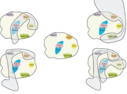
As previously mentioned, PP1 has nearly 200 validated PIPs and by using bioinformatic-assisted PIP identification screens it is estimated that hundreds more PIPs remain to be identified and validated. Moreover, PIPs interact with PP1 unique binding motifs, such as RVxF, SILK, MyPhoNE, SpiDoC, IDoHA and others.18
Among these binding motifs, the RVxF motif is the most common, being present in nearly 70-90% of all validated PIPs. This motif generally conforms to the consensus sequence [K/R] [K/R][V/I][x][F/W], with x being any reside other than Phe, Ile, Met, Tyr, Asp or Pro. Interaction between PIPs and PP1 through this motif does not change PP1 conformation and only serves to anchor the PIPs to PP1, bringing them closer to PP1 and promoting secondary interactions that determine the activity and substrate specificity of PP1. 18,26,27
The SILK motif, [GS]IL[KR], initially described as being essential to PP1 inhibition by inhibitor-2, is present in seven PIPs and, as the RVxF motif, seems essentially for PP1 anchoring18,28. The MyPhoNE motif, RxxQ[VIL][KR]x[YW], is present in seven PIPs and is
involved in substrate recognition. Both motifs are always N-terminal to the RVxF. 18,28,29
SpiDoC, is a domain in neurabin-2 which binds to the PP1 C-terminal groove, blocking access to substrates that require this groove for PP1 binding, thus directing the substrate specificity of PP1. 18,23
Aside from the RVxF and the SILK motif (Figure 3), inhibitor-2 has a third motif able to interact with PP1 – IDoHA. This motif binds PP1 in a manner that covers its active site, preventing PP1 activity. 18
1.4. Neurabins
1.4.1. Neurabin 1
Neurabin-1 is a multifunctional scaffolding protein first identified and characterized in 1997 from purified rat brain tissue. The human neurabin-1 is encoded by the PPP1R9A gene (protein phosphatase 1, regulatory subunit 9A) located on chromosome 7 and is comprised of 1098 amino acids (a.a.). It has an N-terminal F-actin binding domain (a.a. 1-144), followed by a PP1 binding domain (a.a. 425-502), a PDZ (PSD-95/Dlg/ZO-1) domain (a.a. 504-592), three C-terminal coiled-coil regions (a.a. 597-627, a.a. 670-824, 1033-1090) and a sterile alpha motif (SAM) domain (a.a. 988-1051). These two latter domains are known to mediate homo- and heterodimerization of other proteins. Within the PP1-binding domain it is possible to identify a KIKF motif, conserved in other PP1 regulatory subunits.
Figure 3 - PP1 docking motifs RVxF, SILK, MyPhoNE, SpiDoC and IDoHA. PIPs combine PP1 docking sites to form unique PP1 holoenzymes. Taken from18.
Neurabin-1 also has some consensus sequences for phosphorylation by several protein kinases. 30
Neurabin-1 is highly concentrated in the synapse and in the growth cone of developed and developing neurons, respectively31,32.
Figure 5 – Neurabin – 2 domain Structure. Boxes represent the several domains found in both Neurabin-1 and Neurabin-2. Actin BD, Actin Binding Domain; PP1 BD, PP1 Binding Domain; PDZ, PSD-95/Dlg/ZO-1 domain; CC, coiled-coil domain. Taken from42.
Figure 4- Neurabin-1 domain structure. Boxes represent the several domains found in Neurabin-1. Actin BD, Actin Binding Domain; PP1 BD, PP1-binding Domain; PDZ, PSD-95/DLG/ZO-1 domain; CC, coiled-coil domain; SAM, sterile alpha motif. Taken from33.
1.4.2. Neurabin 2
Neurabin-2, also known as Spinophilin, was initially identified and described in 1997 due to its ability to form a complex with the catalytic subunit of PP1 and due to its potent modulation of PP1 enzymatic activity in vitro. In humans it is encoded by the PPP1R9B gene located on the chromosome 17 and is comprised of 817 amino acids. It has two F-actin-, a receptor- and a PP1-binding domain, a LIZ and a PDZ domain, and three coiled-coil domains33.
The Neurabin-2 F-actin-binding domain (a.a. 1-154) is intrinsically unstructured, but upon binding to F-actin adopts a more ordered structure. Additionally, a second F-actin binding domain was described between a.a. 164-282. However, it is still unknown whether these two domains represent segments of a single domain or two independent domains33.
The receptor-binding domain, located between a.a. 154-444, interacts with the third intracellular loop of various seven transmembrane domain receptors, such as the dopamine D2 receptor and some subtypes of the alfa-adregenic and muscarinic-acetylcholine receptors33.
The PP1 binding domains located within residues 417-494 contains the pentapeptide RKIHF motif between 447-451, and is conserved in other PP1 regulatory subunits. Within this domain it is possible to find other regions able to bind to PP123,33. A
LIZ motif was described between a.a. 485-510, known to mediate protein-protein interactions and target protein kinases and protein phosphatases to membrane ion channels. The PDZ domain (a.a. 494-585) directly binds to C-terminal peptides derived from glutaminergic AMPA and NMDA receptors. The three-predicted coiled-coil domains between a.a. 664-814 allow for homo- hetero- dimerization between neurabin-2 and other proteins. Neurabin-2 also has some consensus phosphorylation sequences for several protein kinases. 23,33
Contrary to Neurabin-1, Neurabin-2 is ubiquitously expressed. It is highly concentrated in brain tissue, especially in neurons, where it is enriched in the PSD34.
1.5. Phactrs
1.5.1. Phactr 3
Protein phosphatase 1 (PP1) and actin regulatory (Phactr) proteins are a family that comprises four members in humans and other vertebrates; phactr1–4. Phactr-3 is diffusely expressed throughout the brain35.
Each Phactr protein contains four G-actin-binding RPEL motifs, including an N-terminal motif and a C-N-terminal triple RPEL repeat. The C-N-terminal triple RPEL repeat is adjacent to the PP1-binding domain. RPEL motifs are also found in the regulatory domains of myocardin-related transcription factor (MRTF) transcriptional coactivators where subcellular localization and activity is controlled by sensing signal-induced changes in the G-actin concentration36.
Phactr family proteins are involved in cell migration both in vitro and in vivo, and it is believed that they regulate cytoskeleton dynamics37–39. The Phactr protein family is
considered to be involved in cell migration and morphogenesis by modulating the actin cytoskeleton. Phactr3 is distributed to the plasma membrane in adherent cells, and it enhances cell migration. This indicates that phactr3 is a membrane-associated PP1 and an actin regulator36,40.
Phactr3 lacks fatty acid-attachment sites (e.g., palmitoylation, prenylation, and myristorylation sites) and lipid-binding domains with defined ternary structures (e.g., a PH domain)36. G-actin and PP1 competitively bind to the C-terminal regions of Phactr proteins
and the cytoplasmic G-actin concentration is determined by RPEL motifs, which control the formation of the phactr-PP1 complex. With an increase in cytoplasmic G-actin levels the Phactr-PP1 complex is inhibited. Previous studies suggest that the phactr-PP1complex modulates the phosphorylation status of cofilin or myosin; therefore, regulating actin cytoskeleton dynamics. Overall, these results suggest that Phactr protein is a membrane-associated PP1 regulator, which is itself regulated by signal-induced changes in the cytoplasmic G-actin levels. Therefore, it is likely that the Phactr-PP1 complex modulates
the phosphorylation status of actin cytoskeleton regulators such as cofilin and myosin, which associate with the plasma membrane36,40.
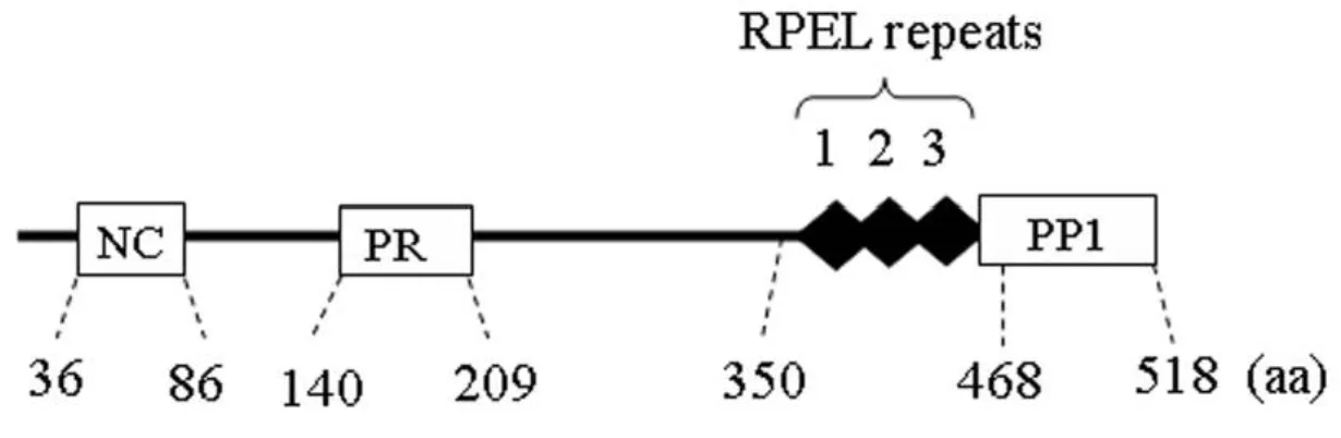
Figure 6 -Phactr3 domain structure. Boxes represent the several domains found in Phactr3. NC, N-terminal region that is conserved in the Phactr family; PR, the proline-rich region; RPEL, the three tandem repeats of PREL motifs; PP1, PP1-binding domains. Taken from37.
1.6. Protein/PP1 complex in dendritic spines
As aforementioned, PP1 is ubiquitously expressed41. However, in the brain, more
specifically in neurons, PP1α and PP1γ1 isoforms are highly localized to dendritic spines24.
PP1 has been identified as a key regulator in both LTP and LTD, with PP1 inhibiting LTP while promoting LTD. In fact, LTD-inducing stimuli promotes distribution of PP1 to dendritic spines, where it can dephosphorylate its substrates, such as calcium/calmodulin-dependent protein kinase II (CaMKII), AMPA and NMDA receptors.22
Neurabin-1 is highly concentrated in dendritic spines, even though it can be found in dendrites, axons, terminals and glia.42 Within dendritic spines, Neurabin-1 can be found
at high levels in the PSD and the 100nm subjacent to it. In fact, Neurabin-1 concentration falls with increasing distance from synapse. Neurabin-1 knockout mice exhibited a deficit in contextual fear memory and increased AMPA receptor synaptic transmission, suggesting that neurabin-1 regulates LTP. Additionally, neurabin-1 overexpression induced filopodia and dendritic spines, further suggesting that it has an important role in spine morphogenesis30.
Neurabin-2 is highly enriched at the synaptic membrane in dendritic spines, where it regulates the actin cytoskeleton42. Within dendritic spines, neurabin-2 localization is
similar to that neurabin-1. It is predominantly localized in the PSD and the subjacent 100nm of it, with its concentration decreasing with increasing distance from synapse. Knockout mice exhibited a marked increase in spine density during development and altered filopodia formation, suggesting it functions as a negative regulator of spine morphogenesis34.
Neurabin-1 and neurabin-2 can form hetero- and homodimers and rapidly shuttle on and off the actin cytoskeleton34. Through interaction with their respective partners, they
are able to regulate spine morphology and density, receptor function and synaptic plasticity. They bind an overlapping set of targets with some opposing effects42. Also, as
beforementioned, neurabin-1 overexpression induced filopodia and dendritic spines while neurabin-2 knockout mice exhibited a marked increase in spine density during development43,44. Additionally, neurabin-1 and neurabin-2 seem to have opposing effects
on the regulation of R4-RGS. Due to all these opposing effects, it has been proposed that neurabin-1 and neurabin-2 may act as negative regulators of each other, especially in dendritic spines, where they are highly concentrated32.
Both neurabins were initially identified as being PP1-targeting subunits. However, between PP1 isoforms, both neurabins showed a significant preference for PP1γ1 and PP1α over PP1β. Since neurabins showed enhanced localization to dendritic spines, this preference is to be expected, as both PP1 isoforms are enriched in dendritic spines, while PP1β is more enriched at the neuronal cell body25.
Disrupting the neurabins/PP1 complex prevents PP1 from reaching some of its targets, thus preventing PP1-mediated dephosphorylation of its substrates. The neurabin-1/PP1 complex is important in the formation of filopodia in young neurons and the transformation of neuronal filopodia into dendritic spines, since it has been shown that disrupting of its complex enhances filopodia and impairs surface GluR1 expression, hindering the morphological and functional maturation of dendritic spines43. Neurabin-2
targets PP1 to several substrates in dendritic spines (such as AMPA and NMDA receptors), controlling their phosphorylation of the AMPA receptors, decreasing the rundown of AMPA currents and increasing the latter’s activity.
The neurabins/PP1 complex can be disrupted by phosphorylation. It has been shown that cAMP-dependent protein kinase catalytic subunit alpha (PKA) phosphorylation of 1 at ser-461 results in the drastic reduction of PP1 activity, allowing neurabin-1 recruitment of other proteins. Even though the RVxF-flanking serine of neurabin-neurabin-1 is conserved in neurabin-2, PKA does not phosphorylate neurabin-2 at the same site. Instead, it phosphorylates a serine residue in the F-actin binding domain (Ser-94)44. This does not
result in reduced PP1 affinity for neurabin-2, but does reduce neurabin-2 interaction with F-actin, displacing neurabin-2 from the PSD to the cytosol, which may ultimately serve to control PP1-mediated changes in the actin cytoskeleton or PP1 anchoring to receptors45.
Neurabin-1 and 2 share a conserve site at Ser-17 for cyclin-dependent-like kinase 5 (Cdk5) phosphorylation44. However, different research groups reached two different
neurabin-1 can be phosphorylated by Cdk5 at ser-17 in vitro, while Causeret et al 2007 did not, reporting Cdk5 phosphorylation at Ser-95 instead. This latter research group found that Ser-95 phosphorylation of Neurabin-1 affects its ability to bind to F-actin, thus regulating neuronal morphology.30,46 As for Neurabin-2, Cdk5 phosphorylation at Ser-17 did
not affect its ability to bind to F-actin, while extracellular signal-regulated kinase 2 (ERK2) Ser-15 phosphorylation did. This latter kinase can also phosphorylate neurabin-1 at Ser-15, at least in vitro. However, the effect of this phosphorylation was not tested44,46.
Additionally, neurabin-2 can also be phosphorylated by CaMKII (100 and Ser-116), which, as with PKA phosphorylation at Ser-94, reduces neurabin-2 ability to bind to F-actin47. Neurabin-2 phosphorylated at Ser-100 showed enhanced concentration in
membrane fractions, including the synaptosomal membrane and synaptic plasma membrane. Both CaMKII and PP1 are enriched in the PSD (with the latter being bound to neurabin-2 which, in turn, is bound to F-actin)47.
CaMKII is auto phosphorylated at Thr-286, a critical process for the regulation of synaptic signaling, and PP1 selectively dephosphorylates phosphor-Thr-286 CaMKII, preventing its association with NMDA and AMPA receptors. So, CaMKII phosphorylation of neurabin-2 may reduce the interaction between the neurabin-2/PP1 complex with F-actin, thus preventing PP1 from dephosphorylating phospho-Thr-286 CaMKII45.
Moreover, neurabin-2 localization was shown to vary depending on the kinase that phosphorylates it. Thus, it has been suggested that phosphorylation of neurabin-2 regulates subcellular localization of the neurabin-2/PP1 complex, targeting this complex to specific locations within dendritic spines47. Finally, since neurabin-1 can also be
phosphorylated by PKA, ERK2 and Cdk5, but not CaMKII, it has been suggested that both Neurabin-1 and 2 scaffolding functions are differentially regulated by phosphorylation44.
To summarize, Neurabin-1 and 2 are scaffolding proteins, which can target PP1 (and also other proteins) to different location within dendritic spines. They seem to act as opposing regulators of each other since they have opposing effects on the actin dynamic of dendritic spines and R4-RGS regulation48. Additionally, their localization, and
not, which may explain how they can target PP1 to specific substrates within different cellular compartments.
Likewise relevant to cytoskeletal dynamics is the report that G-actin and PP1 competitively bind to the C-terminal region and the formation of the Phactr/PP1 complex is inhibited by an increase in the cytoplasmic G-actin concentration, which is induced by extracellular signals such as serum. The current hypothesis suggests that the Phactr/PP1 complex is controlled by the changes in the cytoplasmic G-actin concentration, which regulate the actin cytoskeleton dynamics by modulating the phosphorylation status of actin regulatory proteins. This suggests that Phactr proteins regulate both the PP1 activity and subcellular localization by sensing the cytoplasmic actin concentration through RPEL motifs49,50.
The catalytic subunit of PP1 forms many other complexes, for instance it interacts with noncatalytic subunits that determine the activity, substrate specificity, and subcellular localization of the phosphatase. PP1 can dephosphorylate cofilin and myosin. The actin filament-severing activity of cofilin, which stimulates the treadmill-like movement of the actin cytoskeleton in the lamellipodia and filopodia, is controlled by its phosphorylation status, and the force-generating activity of myosin is controlled by the phosphorylation status of myosin itself. In this context, several studies have shown that the Phactr/PP1complex modulates the phosphorylation status of cofilin or myosin, thereby regulating actin cytoskeleton dynamics49,50.
2. Objectives
Protein phosphorylation is a major regulatory mechanism in cellular processes, involving protein kinases and phosphatases. Of particular note, protein phosphatases exert their activity via the formation of protein complexes. The main aim of this thesis was to review PP1 complexes and to evaluate the effects of Aβ on three proteins implicated in dendritic spine morphogenesis and dynamics, Neurabin-1 (PPP1R9A), Neurabin-2 (PPP1R9B) and Phactr3, as well as their interaction with PP1.
The specific objectives were to:
- elucidate the network representing PP1 interactions with Neurabin-1,
Neurabin-2, Phactr3 and Cofilin 1;
- study the effect of Aβ on the expression levels of Neurabin-1, Neurabin-2 and
Phactr3;
- evaluate the effects of Aβ on the interaction of Neurabin-1, Neurabin-2 and
3. Materials and methods
3.1. Interactome Networks
To understand the relationship between different proteins we often resort to a systems biology approach to depict the known protein interactions, and these can be represented in networks. For this study key proteins were used for developing the
interactome networks; namely PP1, PP1, Neurabin 1, Neurabin 2, Phactr3 and Cofilin 1. The uniprot code for the Homo sapiens of each of the key proteins was retrieved, in order to eliminate interactions described in other species, and submitted to IntAct Molecular Interaction Database (see Table 1). Different databases (IntAct, String and BioGRID) were tested and IntAct demonstrated to be more complete and reliable, as well as being a curated source. For the PP1 networks two distinct sources were used. Given the expertise in our laboratory several PP1 interactors have been identified using the yeast two hybrid (YTH) system. There are over 400 known PP1 interactors19. Hence for the PP1
analyses the YTH data and the IntAct data were pooled.
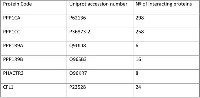
Table 1 - List of Key Proteins retrieved from uniport and submitted to IntAct to obtain interactome network. PPP1CA/PP1α; PPP1CC/PP1γ; PPP1R9A/Neurabin-1; PPP1R9B/Neurabin-2; PHACTR3/Phactr3; CFL1, Cofilin.
Protein Code Uniprot accession number Nº of interacting proteins
PPP1CA P62136 298 PPP1CC P36873-2 258 PPP1R9A Q9ULJ8 6 PPP1R9B Q96SB3 16 PHACTR3 Q96KR7 8 CFL1 P23528 24
All data recovered from IntAct were transferred to Cytoscape and the interactome networks were built for each protein and merged, so as to identify key proteins as presented in the results. The output from the databases is as the gene name, but given that were are dealing with protein:protein interactions, the term protein will be used.
3.2. Antibodies
Antibodies can be classified as monoclonal or polyclonal. Monoclonal antibodies contain a single antibody clone from B-Cells and recognize a single epitope from the antigen, while polyclonal antibodies contain several clones of antibodies recognizing different epitopes in the antigen. Monoclonal antibodies are highly specific, but less sensitive, while polyclonal antibodies are highly sensitive, but less specific.
Antibodies are central to the procedures carried out in the work here presented. The antibodies were used for Western blot analysis and immunoprecipitation procedures as indicated in table 2. Anti-PP1α and anti-PP1β were produced ‘in house’. The remaining antibodies, primary and secondary, were obtained from commercial sources.
The following tables (Table 2 and Table 3) summarize the antibodies used in this work.
Table 2- Primary antibodies and dilutions used.
Antibody Type Target Dilution
Anti-Neurabin I (D-4 Santa
Cruz Biotechnology, Inc.) Mouse, polyclonal Neurabin 1 WB: 1:1000 Anti-PPP1R9B
(Proteintech) Rabbit, polyclonal Neurabin 2 WB: 1:1000 Anti-Phactr3 (Proteintech) Rabbit, polyclonal Phactr3 WB: 1:1000
Anti-PP1α (CBC2C)51 Rabbit, polyclonal PP1α WB: 1:5000
Co-IP: 4µL/1000µg
Anti-PP1γ (CBC3C)51 Rabbit, polyclonal PP1β WB: 1:5000
Co-IP: 3µL/1000µg
Table 3- Secondary antibodies used.
Antibody Target Dilution
Horseradish peroxidase-conjugated
anti-mouse (Amershan Pharmacia) Anti-mouse primary antibodies 1:5000 Horseradish peroxidase-conjugated
anti-rabbit (Amershan Pharmacia Anti-rabbit primary antibodies 1:5000
3.3. Culture, growth and maintenance of SH-SY5Y cell
line
In neurosciences the use of mammalian neurons derived from embryonic the CNS tissue is very limited mainly because once these cells are terminally differentiated into mature neurons, they can no longer be propagated.
To overcome this limitation transformed neuron-like cell lines are commonly used. Among them, there is a very popular and well characterized cell line, the SH-SY5Y neuroblastoma cell line (ATCC® CRL-2266™).52
The SH-SY5Y cell line was originally derived from a metastatic bone tumor biopsy, and a subline of the parental line SK-N-SH, which were subcloned three times: first to SH-SY, them to SH-SY5, and finally to SH-SY5Y.52
In order to achieve the objectives proposed for the present study, the SH-SY5Y neuroblastoma cell line was chosen for the reasons described above, and because this cell line is a human cell line that can be easily differentiated into a more mature neuronal-like phenotype.
SH-SY5Y cells (ATCC CRL-2266) were grown and maintained in Minimal Essential Medium (MEM)/F12 (1:1) supplemented with 10% heat-inactivated fetal bovine serum (FBS), 0,5 mM L-Glutamine, 100 U/ml streptomycin. Cultures were maintained at 37ºC, 5% CO2. Cells were subcultured when 80-90% confluence was reached.
3.3.1. Differentiation of the SH-SY5Y neuroblastoma cell
line
SH-SY5Y cells can be differentiated into a neuronal-like phenotype through treatment with several compounds. Retinoic acid (RA), is one of the most commonly used compounds. It is a vitamin A derivative, with growth inhibiting and differentiation-promoting properties. Several variations of differentiation medium can be found in the literature, however it is usually administered at 10µM in free-serum or low serum medium.
SH-SY5Y underwent differentiation in 1%FBS Serum, with 10µM RA for 7 days, with medium renewal every 2 days. To confirm differentiation cells were cultured in the same medium without RA.
3.4. Aβ treatment
In order to evaluate the effects of Aβ on the expression and interaction of Neurabin 1, Neurabin 2 and Phactr3, with PP1α and PP1γ, SH-SY5Y cell line were incubated with different concentrations of Aβ peptide.
Synthetic Aβ 1-42 (GenicBio) was dissolved in water in order to prepare 1mM stock solution. Prior to cell exposure, an aggregation step was performed, by incubating the
peptides with phosphate buffered saline (PBS) 1x, 48h at 37ºC to a final concentration of 100µM.
SH-SY5Y cells were incubated for 7 days as described in Section 3.1.1. and then incubated in serum free MEM/F12 1:1 medium, supplemented with 0.5mM L-Glutamine, 100U/mL penicillin, 100U/mL Streptomycin and 10µM RA with 2µM and 10µM of Aβ 1-42. Prior to treatment cell were washed twice with PBS 1X.
In order to assure that the results obtained were not due to PBS1x, used to aggregate Aβ peptide, control cultures were incubated with PBS1x vehicle.
3.5. Lysates and protein quantification
Depending on the experimental procedure, cells were collected using different methods.
To analyze protein expression, cells were collected with 200µL of RIPA buffer. RIPA buffer, contains sodium dodecyl sulfate (SDS), which is a strong ionic detergent. It is able to solubilize lipids and proteins contained in cell membrane, creating pores leading to full cell lysis. The lysates were subjected to western blot analysis.
To analyze protein interactions through co-immunoprecipitation (Co-IP), cells were collected using lysis buffer (50mM Tris-HCL pH8.0, 120mM NaCl, 4%CHAPS) containing protease inhibitors (cOmpleteTM EDTA-free, Roche), to prevent proteolysis,
dephophorylation and denaturation of proteins. The lysis buffer was freshly prepared.
Following protein extraction, protein concentrations were measured through Pierce’s bicinchoninic acid (BCA) protein assay kit (Thermo Scientific) following the manufacturer’s instructions. In an alkaline medium, proteins reduce Cu2+ to Cu1+, which
links to bicinchoninic acid allowing a highly sensitive and selective colorimetric detection. 96-well Plates were used to perform BCA assay. Standard samples were prepared with known concentrations of bovine serum albumin (BSA)(see Table 3). Cell samples were prepared by mixing 5ul of lysate with 20uL of SDS1%, and incubated with 200µl working reagent. The working reagent was prepared by mixing BCA reagent A and B in 50:1 proportion. The 96well plate was incubated 30min, at 37ºC. After cooling to room
temperature, absorbance was measured at 562nm using a microplate reader (Infinite M200, TECAN). To determine protein concentration, a standard curve was calculated by plotting standard absorbance vs standard BSA concentration.
All measurements were performed in duplicate.
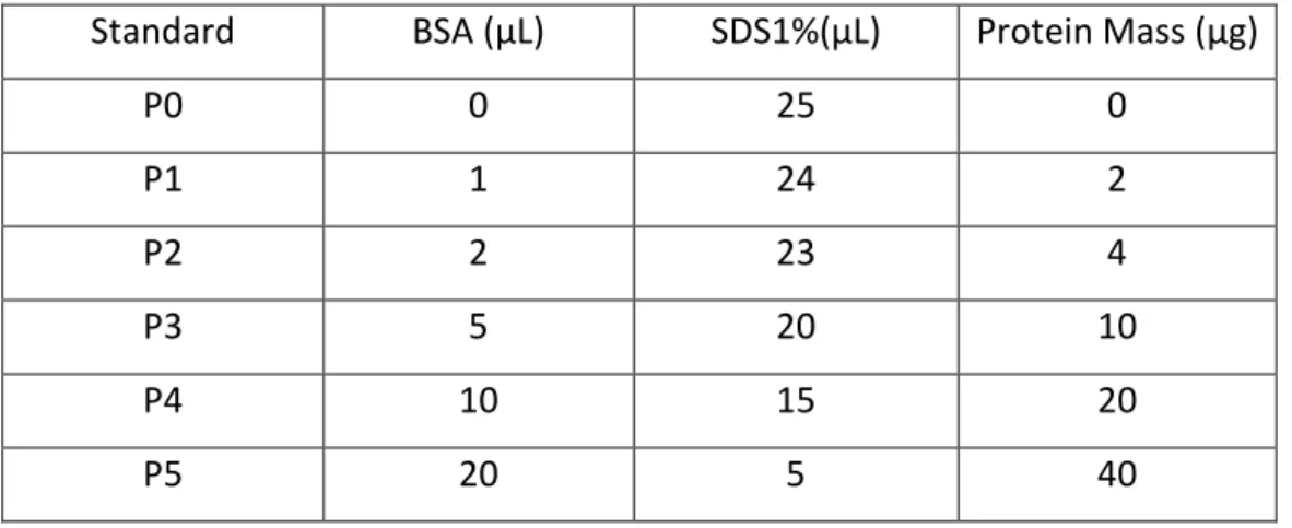
Table 4 - Protein standards used in BCA assay.
Standard BSA (µL) SDS1%(µL) Protein Mass (µg)
P0 0 25 0 P1 1 24 2 P2 2 23 4 P3 5 20 10 P4 10 15 20 P5 20 5 40
3.6. Co-Immunoprecipitation
To evaluate Aβ influence on PP1 complexes (both α and γ isoforms) with Neurabin 1, Neurabin 2 and Phactr3, a co-immunoprecipitation (Co-IP) was performed using lysates of SH-SY5Y cells. The Co-IP is a variation of immunoprecipitation, which relies in the immunodepletion of a protein of interest (PP1 isoforms), together with all interacting proteins (i.e. Neurabin 1, Neurabin 2 and Phactr3) in solution.
The Co-IP assay was performed using Dynabeads Protein G (Invitrogen), due to rapid and easy magnetic bead separation. This method preserves most of protein-protein interactions, and reduces background caused by non-specific binding. The procedure is based in the capacity of Dynabeads protein G binding to the Fc region of antibodies. The antibody-bound beads are placed in a Dynal magnet, which allows the beads to migrate to the magnet side, allowing easy supernatant removal. Sample proteins are loaded onto the
beads, permitting the binding of protein of interest and all interacting proteins consequently. By placing the complex in the Dynal magnet, and removing the supernatant, only the protein complex of interest remains. In this experimental procedure, the complex was analyzed through SDS-PAGE.
Cell cultures were performed as described in Section 3.1.1., at a 1x10^5 cell density. Following Aβ treatment for 24h, cells were washed with cold PBS1x and gently scrapped off the culture plate with Lysis buffer described in Section 3.5. Lysates were sonicated twice for 10seconds.
Each sample had its mass normalized by BCA assay and 1000ug of each sample were precleared using 15µl of Dynabeads for 1h, 4ºC with agitation. Simultaneously, the primary antibody (PP1α and γ as depicted in Table 2) was incubated with 40µL of Dynabeads in the same conditions. Prior to use, Dynabeads were washed thrice with washing solution (3%BSA/PBS1x).
After 1h, each sample was transferred to the antibody-bound beads, and incubated overnight, 4ºC with agitation. Following incubation, supernatant was removed, and the beads washed thrice with 500µL of PBS1x, 10min at 4ºC with agitation. After the last wash, supernatant was fully discarded and 100µL of Loading Buffer (LB) was added to the beads, boiled for 10min at 90ºC to disrupt beads-proteins complex. Dynabeads were removed and samples stored at -20ºC until needed.
Co-IP controls were performed by incubated cell lysates with either Dynabeads without antibody, or Dynabeads with IgG controls.
3.7. Western Blot
Western Blot or Immunoblot, is a widely used technique for detection and analysis of proteins. It comprises several steps. Initially prepared samples are separated by electrophoresis and lastly transferred to a nitrocellulose membrane. The membranes are incubated with antibodies to allow detection and further analysis.
In this experimental procedure, electrophoresis gels in 5-20% gradient SDS-PAGE were used, allowing protein separation only by molecular weight. It consists of 2 two gels, a resolving gel (bottom), with higher polyacrylamide concentration to separate proteins, and a stacking gel (top) with a lower polyacrylamide concentration.
Prior to loading, the samples are incubated with Loading Buffer (LB). The loading buffer consists of glycerol, to increase sample density, SDS, to mask any inherent charge of proteins, β-mercaptoethanol (reducing agent), breaking disulfide bonds and thus disrupting quaternary and tertiary structures, and bromophenol blue (dye), which allows sample tracking and progression in the gel.
Electrical current is applied, enabling protein separation. After separation, proteins are transferred by electrophoresis onto a nitro-cellulose membrane. This immobilizes all proteins at their relatively migration positions.
The membranes can be stained with Ponceau S, to confirm protein transfer and assess equal gel loading. Membranes are then blocked with either non-fat milk or BSA, in order to block non-specific sites of the primary anti-body. Following blocking, membranes are incubated with specific primary antibody and appropriate secondary antibody. After antibody biding, proteins can be detected by chemiluminescence. The horseradish peroxidase conjugated onto secondary antibody, catalyze the oxidation of luminol, emitting light. This light signal can be detected with appropriated equipment, Chemidoc XR, BioRad, and the resulting images further analyzed with Image Lab, BioRad.
In the present work, samples were collected, and protein concentration determined as described in Section 3.5. The samples were separated in a 5-20% gradient SDS-PAGE in a Hoefer electrophoresis system. The gradient gels were prepared and allowed to
polymerize for at least 45minutes at room temperature. The stacking gels were prepared and polymerized on the top of the gradient gel. A comb was inserted before polymerization of the stacking gel, and removed only after polymerization was ensured. The samples were incubated with 4X LB for 10min at 90ºC.
Samples were loaded onto the gel alongside a molecular weight marker (Precision Plus Protein Dual Color Standards, BioRad). The gels were run at 90mA for approximately 3hours. Proteins were then transferred onto nitro-cellulose membranes for 18hours at 200mA.
Membranes were incubated with Ponceau S for 5 minutes and photographed in Chemidoc (BioRad) to ensure equal gel loading. Following washing TBS-T1x to remove Ponceau S, membranes were blocked with 5% BSA in Tris Buffered Saline Tween (TBS-T) 1x for 4hours. Membranes were then washed in TBS-T 1x, 10min, and incubated with a specific primary antibody overnight (Anti-Neurabin-1, Anti-PPP1R9B, Anti-PHACTR3) or for 2 hours at room temperature (Anti-PP1α and Anti-PP1γ), washed thrice with TBS-T1x, 10min, incubated with appropriated secondary antibody for 2hours and washed thrice with TBS-T1x, 10min. Following washing, membranes were incubated with enhanced luminescence (ECL) detection kit for 1minute. After incubation with ECL membranes were exposed in Chemidoc XR, BioRad. Band intensity was determined and used to correlate with intracellular protein levels.
4. Results and discussion
4.1. PP1 Interactome
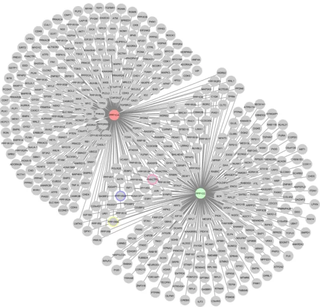
In order to select potential candidates to study the effects of Aβ in Protein/PP1 complexes, the interactome of PP1 was identified using a systems approach. Firstly, PP1α and PP1γ interactors were retrieved from IntAct, as well additional PP1 interactors identified previously by our laboratory by yeast to hybrid (YTH) system, but not present in the database (Figure 7)19. Analysis of the merged network of the two
isoforms reveals that each central node has a significant number of interactors (as indicated in table 1), but there is also a significant number of overlapping nodes. In fact, 82 interactors are shared by PP1α and PP1γ.
Upon analyzing the GeneOntology (GO) of the PP1 merged interactome the most represented Molecular Functions (MF) were protein binding/binding, Catalytic activity, Kinases and phosphatase activity (supplemental figure 1). It is also worth noting that cytoskeletal protein binding and actin binding are also well represented as well as receptor binding activity. Taken together the major nodes in the MF point to important roles for PP1 as important signal transduction mediators, this is highly consistent with all the available information on PP1.
Upon analyzing the PP1 merged interactomes (Figure 7), proteins implicated in synapse plasticity and neuronal morphology were also present and these were selected for further analysis. The candidate proteins were Neurabin-1, Neurabin-2, Cofilin 1 and Phactr3. All these interact directly with PP1 and all have important roles in synapse plasticity and morphology. Consequently, the interacting proteins for each were retrieved from IntAct. The interactomes of the above mentioned proteins were merged with those of PP1α and PP1γ and are shown in figure 8. It was very striking that the
Figure 7- Interactome network, of PP1α and PP1γ. PPP1CA, PP1α; PPP1CC, PP1γ; Highlighted border for key proteins; Pink, Phactr3; Blue, Neurabin-1, Yellow, Neurabin-2.
merged interactome of these 6 central nodes had only an additional 10 interactors when compared to the merged interactome of PP1α and PP1γ alone. This represents a strong overlap of interactors and is suggestive of very close functional relationships. Also of note PP1α and PP1γ remain as central nodes (Figure 8). It is also noteworthy that a central hub emerged around Cofilin 1 (CFL1). In the experimental design CF1 was not highlighted for subsequent studies, but given the data emerging from this systems approach it will have to be included in follow up analysis.
The GO for MF of the 6 merged interactome networks was also investigated (supplemental figure 2). Not surprisingly the most represented MFs remained the same when compared to the merged PP1 network, that is: catalytic activity, Kinases and phosphatase activity, cytoskeletal protein binding and actin binding. This is so because the total number of nodes for the two merged networks is almost the same. (see if there are any differences)
Subsequently only proteins which interact with PP1 isoforms and another interactor were selected (Figure 9). This was important to simplify the network and home in on key nodes. By doing so we obtained a list of candidate proteins that interact with PP1. Given the central role of PP1 in AD and the fact that Aβ is a model to study AD in cell cultures 14,53,54,
Figure 9 - Interactome Network of proteins directly binding to PP1α and PP1γ. Colored nodes represent most relevant proteins. PPP1CA, PP1α; PPP11CC, PP1γ; PPP1R9A, Neurabin – 1; PPP1R9B, Neurabin – 2; PHACTR3, Phactr3; CFL1, Cofilin.
the nodes identified in Figure 9 are potential targets to study the effect of Aβ at a molecular level.
The final sub-network of common interactors yielded a total of 103 nodes (Figure 9). It is noteworthy that this came from an original pool of around 500 proteins. Thus one can deduce that this was important to simplify the network and home in on key nodes for future studies. Emerging central nodes from this work that deserve to be further addressed in future studies include MYO 19, GRB2, CAPZA2, MYO1C, SYNPO and PHACTR4. These are important because they have several interactors and can potentially be central hubs.
4.2. Differentiation of the SH-SY5Y cell line
In order to use a cell line closer to characteristics of a neuron, SH-SY5Y cell line, was differentiated for 7days, with media renewal every 2 days. Photographs were taken every two days to access correct differentiation.
To evaluate cell differentiation morphological changes were analyzed, such as neurite extension. Non-differentiated cells, were used as a control to access morphological changes.
4.2.1. Morphology evaluation
To evaluate the morphological changes induced by differentiation media, photographs were taken prior to media change, every 2 days, as depicted in the following images (Figure 10).
It was possible to observe that cells in media with 10% FBS and 1%FBS, the proliferative rate was higher than media with retinoic acid. However cells in 1%FBS media, show some neurite extention compared to cells in 10% FBS media. This can be explained by depletion of serum, essential for cell proliferation, which may induce some cells to differentiate. Cells in differentiation media (1% FBS and 10uM RA) show higher neurite extention, as expected. With this data it is possible to access a proper differentiation of SH-SY5Y cell line, and conclude that the characteristics of the cells were as close as possible to a neuronal cell, and the requirements to study Protein/PP1 complexes were met.
Day FBS 10% FBS 1% FBS1% + 10µM RA
0
3
5
Figure 10- Phase contrast photographs of SH-SY5Y cell line morphology during differentiation. White arrows represent extended neurites. White triangles represent “S” population.
4.3. Aβ effect on Neurabin expression
Aβ is a neurotoxic peptide, which is reported to cause spine and synapse degeneration. Even when applied extracellularly it causes neurite damage, leading to spine degeneration. Several studies, including from our own laboratory, have been carried out using cell exposure to Aβ as an AD model system55,56.
As discussed in the introduction synaptic dysfunction is an AD hallmark. Further, since neurabins are concentrated in the spine it is possible that in the presence of Aβ, their expression could be decreased.
In order to evaluate this hypothesis, differentiated SH-SY5Y cells were treated with two different concentrations of the toxic peptide (previously aggregated), 2µM and 10µM. The sample lysates of the cells collected were analyzed by western blot in order to evaluate changes in intracellular protein levels (Figure 11).
The results obtained, show a significant decrease in intracellular Neurabin levels. It is observed for both neurabins, and may be due dendritic spine degeneration caused by Aβ. This suggest that in AD, the decrease in this protein may lead do dysregulation of the actin dynamics, leading to dendritic spine a synapse loss. Whether this is the synaptic loss so well described for AD remains to be directly demonstrated, but it is nonetheless a very attractive hypothesis.
A
120kD 200kD 180kDB
C
Neurabin-2Control Aβ 2µM Aβ 10µM
Neurabin-1
D
D
Figure 11 - Neurabin-1 and Neurabin-2 expression in SH-SY5Y cells treated with Aβ1-42. A. Western blot analysis of SH-SY5Y lysates treated with different concentrations of Aβ1-42. B. Loading Control, Ponceau Staining. C. Comparison of Neurabin-2 expression levels in cells for different concentrations of Aβ1-42. D. Comparison of Neurabin-2 expression levels in cells for different concentrations of Aβ1-42. All data was normalized to Ponceau S levels prior to analysis. * P < 0,05; n.s, no significant difference, P>0,05.
* *
* *
4.4. Aβ effect on Phactr3 expression
As already discussed, Aβ is a neurotoxic peptide. It was reported that Aβ interferes with cytoskeleton dynamics, thus interfering with actin levels and interfering with polymerization/depolymerization of F-actin, increasing its polymerization57. It is also
known that Phactr3 is regulated by the levels of G-actin and regulates several proteins involved in cytoskeleton and actin dynamics. This leads to an indirect effect of Aβ on Phactr3 expression37.
Figure 12- Phactr3 expression in SH-SY5Y cells treated with Aβ1-42. A. western Blot analysis of SH-SY5Y lysates treated with different concentrations of Aβ1-42. B. Loading Control, Ponceau Staining C. Comparison of Phactr3 expression levels in cells for different concentrations of Aβ1-42. All data was normalized to Ponceau S levels prior to analysis. n.s, no significant difference, P>0,05. 75kD Phactr3 Control Aβ 2µM