UNIVERSIDADE DE LISBOA
FACULDADE DE CIÊNCIAS
DEPARTAMENTO DE BIOLOGIA VEGETAL
The Effect of G418 and PTC124 as Suppression Therapy for
Beta Thalassemia
Cláudia Filipa de Noronha Estima
Mestrado em Biologia Molecular e Genética
Dissertação orientada por:
Doutora Luísa Romão
Doutora Juliane Menezes
ii “Take up one idea. Make that one idea your life. Think of it, dream of it, live on that idea. Let the brain, muscles, nerves, every part of your body, be full of that idea, and just leave every other idea alone. This is the way to success."
iii INDEX FIGURES ... iv TABLES ... iv ACKNOWLEDGMENTS ... v RESUMO ... vii PALAVRAS CHAVE ... ix ABSTRACT ... x KEYWORDS ... xi ABBREVIATIONS... xii 1. INTRODUCTION ... 1 1.1. Beta thalassemia ... 1
1.2. Nonsense-mediated mRNA decay ... 2
1.3. Translation ... 6
1.4. Suppression therapy ... 7
2. AIMS ... 10
3. MATERIALS AND METHODS ... 11
3.1. Plasmid construction... 11
3.2. Cell culture and plasmid transfection ... 11
3.3. Drug treatment and cell lysis ... 12
3.4. RNA extraction ... 12
3.5. Reverse transcription-coupled quantitative PCR (RT-qPCR) ... 12
3.6. Semiquantitative PCR ... 12
3.7. SDS-PAGE and Western blot ... 13
3.8. Statistical analysis ... 13
4. RESULTS ... 14
4.1. Beta globin detection By Western blot ... 15
4.2. Analysis of BN and B39 mRNA levels when transfected HeLa cells are exposed to G418 or PTC124 ... 18
5. DISCUSSION AND FUTURE DIRECTION ... 22
6. REFERENCES ... 25
APPENDIX 1 ... 1
APPENDIX 2 ... 2
APPENDIX 3 ... 3
iv
FIGURES
Figure 1: Nonsense-mediated mRNA decay ... 5
Figure 2: The effect of readthrough compounds on protein translation. ... .9
Figure 3: Western blot of cell lysates using an anti-beta globin antibody ... 15
Figure 4: Insertion of a c-myc tag in the restriction site of BstXI ... 16
Figure 5: Western blot of cell lysates using anti-c-myc and anti-H2B antibodies ... 17
Figure 6: Western blot of cell lysates using anti-c-myc and anti-beta globin antibodies. ... 18
Figure 7: RT-qPCR analysis performed on RNA isolated from HeLa cells containing the BN, B15 and B39 transcripts without any drug treatment ... 18
Figure 8: RT-qPCR analysis performed on total mRNA isolated from HeLa cells containing the BN and B39 transcripts after treatment with G418 aminoglycoside. ... 19
Figure 9: RT-qPCR and semiquantitative PCR performed on RNA isolated from HeLa cells containing the BN, B15 and B39 transcripts after treatment with PTC124 ... 20
Figure 10: RT-qPCR analysis performed on RNA isolated from HeLa cells containing the BN and B39 transcripts after treatment with 3µM, 30µM and 100µM of PTC124 ... 21
Figure 11: RT-qPCR analysis performed on RNA isolated from HeLa cells containing the BN and B39 transcripts after treatment with 50µM, 100µM and 150µM of PTC124 ... 21
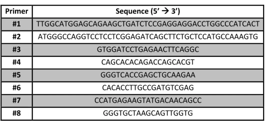
TABLES Table 1: Sequences of the primers used in the current work ... 13
v
ACKNOWLEDGMENTS
O meu pai sempre me diz “Se queres ir rápido vai sozinha, mas se queres chegar longe vai acompanhada”, portanto não posso deixar de agradecer a todas as pessoas que fizeram com que esta etapa extraordinária fosse possível.
Em primeiro lugar gostaria de agradecer à Doutora Luísa Romão por ter permitido que eu participasse neste projecto, que foi uma das experiências mais desafiantes e enriquecedoras da minha vida. Agradeço também a liberdade que sempre me deu quando eu sugeria alguma experiência que não estava inicialmente nos planos e por ter confiado em mim. Espero ter estado sempre à altura do desafio e que não a tenha desiludido.
Não posso deixar de exprimir a minha gratidão ao Doutor João Lavinha, assim como à Doutora Glória Isidro por me permitirem aprender tanto neste Mui Nobre Departamento de Genética Humana do Instituto Nacional de Saúde Doutor Ricardo Jorge.
Acima de tudo quero demonstrar o meu profundo agradecimento à Doutora Juliane Menezes: uma excelente pessoa que nunca irei esquecer, por me ter ensinado tanto não só no laboratório mas também como pessoa. Espero um dia ser tão meiga e compreensiva a ensinar como tu. As palavras não são suficientes para exprimir a gratidão e respeito que tenho por ti e espero que também guardes um cantinho para mim no teu coração. Obrigada!
Ao Paulo, por todo o tempo e paciência que dedicou a ajudar-me a delinear estratégias para o meu projecto. Por todos aqueles “e então?”, quando voltava da câmara escura, que me mostravam que não estava sozinha nesta luta. Por todos os preciosismos que te caracterizam e que me ajudaram a ser ainda mais exigente comigo própria. E como a vida não é só trabalho, obrigada por todas as futeboladas depois dos dias difíceis.
De tudo o que experienciei durante este ano, o que mais gostei, para além do que aprendi, foi das pessoas que conheci e com quem partilhei o meu dia-a-dia. Quero agradecer, do fundo do coração, aos meus companheiros de laboratório, que me ajudaram incondicionalmente e pelos quais guardo grande carinho e respeito. À Joana, Raquel, Rafaela, Rafael, André e Andreia, que me proporcionaram muitos bons momentos de aprendizagem, mas também de diversão.
Para além dos meus colegas de laboratório quero agradecer a todo o grupo de Oncobiologia e, em especial, à Doutora Patrícia Barros, que nunca recusa ajuda a quem quer que seja. Quero também agradecer ao José Ferrão, um exemplo de que não precisamos de estar sérios para sermos competentes no nosso trabalho, obrigada pela alegria e boa disposição.
À minha família, por me ter acompanhado e apoiado sempre nos desafios da minha vida. Obrigada por plantarem em mim a vontade de ser melhor e de vencer. Orgulho-me de ter tido uns
vi Pais que nunca me castigaram por uma nota menos boa, mas que me alertavam quando sabiam que não tinha dado o meu melhor.
À minha Mãe, apesar de ser da área de Letras, por ter lido a minha tese do início ao fim, deve ter sido bastante aborrecido. Obrigada por me teres ajudado todas as vezes que eu ia ao teu quarto e perguntava “Ó mãe, diz aí um sinónimo de (…)”.
Ao meu Pai, por me perguntar todos os dias como iam as minhas filhas (células) e se já tinha resultados que fizessem sentido. Por todas as vezes que insististe comigo para ir ao ginásio converter a energia má do dia em energia boa. Acima de tudo, obrigada por acreditares sempre em mim, seja qual for o desafio a enfrentar: “Eu não quero saber se tu és a melhor, apenas quero saber que deste o teu melhor”.
Ao meu irmão Diogo, o meu geniozinho, obrigada por me fazeres sentir que sou um bom modelo de irmã mais velha (não adianta desmentires porque me imitas em tudo). Apesar de estudares há menos anos que eu, inspiras-me com a tua capacidade de concentração e foco num objectivo e isso motiva-me a trabalhar mais. Quero que tenhas sempre em mente que se eu consegui, tu também consegues, tenho a certeza.
Aos meus Avós Maria Trindade e Aluíno Noronha que, pelo seu exemplo de vida, me mostraram que tudo é possível, basta sonhar, acreditar e trabalhar. Foram das pessoas que mais me apoiaram no meu percurso académico, desde o primeiro dia… com dezassete aninhos e cheia de medo, em que me disseram “Claudinha, tens de te mentalizar que tens de estudar”. Em breve espero que estejamos todos à beira-mar a almoçar e que já seja eu a pagar, como prometido.
Aos meus amigos pelos finais de dia no jardim, cada um a queixar-se para seu lado e a gozar com quem entra mais cedo no dia seguinte: “mañana es mañana”. Estes anos juntos fizeram-me perceber que, se não fossem estes pequenos momentos, a vida tornar-se-ia muito mais difícil. Quero salientar a Bruna, o Félix, a Joana e o Dário, por todo o apoio e por me terem feito acreditar que conseguia fazer um bom trabalho. Ao meu amigo de longa data, João Pereira, com o qual sei que posso desabafar sobre tudo, espero que estejam muitos mais anos para vir.
Por fim, ao meu diamante, o meu namorado Marcos. Mesmo que passasse o resto da vida a agradecer-te, por tudo o que já fizeste e fazes por mim, não seria suficiente. As palavras não chegam para te dizer quão grata estou, por isso espero demostrá-lo ao longo da nossa vida. Obrigada por todos os conselhos e ralhetes que me dás, sei que posso contar contigo para tudo. Espero que estejas sempre lá, no final do dia, tanto quando venho eufórica, como quando venho triste e tens de aguentar uma choradeira de uma hora (desculpa). Ensinaste-me tanto… sinto que és meu pai, meu irmão, meu melhor amigo e, por vezes, até, a minha consciência. Espero que estejas orgulhoso de mim, pelo que te dedico esta tese.
vii
RESUMO
A beta talassémia é uma doença genética que se caracteriza pela redução ou ausência de síntese de beta globina, um dos constituintes da hemoglobina, resultando no aumento de glóbulos vermelhos e, consequentemente, em anemia. Uma das causas desta doença tem origem na alteração de um nucleótido, levando à introdução de um codão de terminação prematura (na sigla inglesa PTC ou codão nonsense) nos transcritos de beta globina. A presença de um PTC não permite que a tradução progrida normalmente, ocorrendo, na maior parte dos casos, a formação de péptidos truncados que são, possivelmente, não funcionais ou, até, tóxicos para a célula.
No sentido de recuperar a homeostase, a célula desenvolveu mecanismos de controlo de qualidade para assegurar a fidelidade da expressão genética. Entre estes mecanismos está o decaimento do RNA mensageiro (mRNA) mediado por mutações nonsense (na sigla inglesa NMD – nonsense mediated mRNA decay) que permite identificar e degradar transcritos que contenham um PTC, reduzindo a sua abundância. Deste modo, a eliminação destes transcritos anómalos evita a formação e acumulação de proteínas truncadas que, caso contrário, danificariam a célula. Assim, o NMD tem, na maioria dos casos, um papel protetor contra erros genéticos. A degradação do transcrito que contém o PTC inicia-se logo após a exportação da ribonucleoproteína mensageira (na sigla inglesa mRNP) para o citoplasma. Durante a ronda pioneira de tradução, se o transcrito não possuir um PTC, o ribossoma remove da mRNP todos os complexos de junção exão-exão (na sigla inglesa EJC) até chegar ao codão de terminação. Os EJC são complexos multiproteicos, depositados 20 a 24 nucleótidos (nt) de distância a montante das junções exão-exão (local de excisão dos intrões) durante o splicing. Se, por ventura, o transcrito possuir um PTC e este se localizar a, pelo menos, 50 a 55 nt de distância a montante da última junção exão-exão, a tradução é terminada prematuramente e pelo menos um EJC fica associado ao mRNA. Nestas condições, o EJC facilita o recrutamento e a interacção dos factores do NMD com o complexo de terminação da tradução, activando o NMD. Este é o caso mais comum, no qual o transcrito é degradado, reduzindo os seus níveis drasticamente (entre 0 a 20% dos níveis normais). Como exemplo para esta situação temos o caso do transcrito de beta globina com uma mutação nonsense no codão 39. Contudo, esta regra posicional dos EJC não é tão linear como aparenta, dado que existem transcritos que, mesmo estando nas condições previstas pelo modelo, conseguem resistir à acção do NMD, como é o caso de mRNAs que contenham PTCs próximos do codão de iniciação. Como exemplo de um mRNA que escapa à degradação pelo NMD, temos o transcrito de beta globina que possui uma mutação nonsense no codão 15, sendo que os seus níveis de expressão são semelhantes aos do gene da beta globina normal. De facto, verificou-se que existem outras características do mRNA, para além dos EJC, que influenciam a activação do
viii NMD, tais como distância física entre um PTC e a proteína citoplasmática de ligação à cauda poli-A do mRNA (na sigla inglesa PABPC1). Para além disso, o tamanho da região não traduzida a 3’ (na sigla inglesa 3’UTR) é outro dos fatores que influencia a ativação do NMD. Apesar de, na maioria dos casos, o NMD assumir um papel protector, este pode também agravar o fenótipo da doença pelo facto de eliminar mRNAs que originam proteínas parcialmente funcionais ou com actividade residual. O processo de terminação da tradução ocorre quando o codão stop é reconhecido por proteínas que formam o complexo de terminação, promovendo a libertação da cadeia polipeptídica formada até então. Em certos casos, ao invés de ser reconhecido pelo complexo de terminação, o codão stop é identificado por um aminoacil tRNA, complementar a duas das três bases (na terminologia inglesa near cognate aminoacyl tRNA). Ocorre, assim, a recodificação do codão de terminação prematura, permitindo que a elongação prossiga em fase, até que o ribossoma atinja um codão stop normal, possibilitando a formação da proteína completa e potencialmente funcional. Este processo de readthrough de um PTC ocorre espontaneamente na célula, embora não seja frequente (cerca de 1%). No entanto, certas moléculas de baixo peso molecular demonstram elevar a frequência destes acontecimentos, uma vez que favorecem a competição entre os near cognate aminoacyl tRNAs e o complexo de terminação. Desta forma, nas últimas décadas tem sido desenvolvida uma terapia, designada como terapia de supressão, que utiliza estes compostos com vista a aliviar os sintomas de diversas doenças causadas por mutações nonsense, devido ao aumento da quantidade de proteína funcional disponível. Os compostos mais estudados e utilizados na terapia de supressão são os aminoglicosídeos, um grupo de antibióticos cujo potencial de supressão provém da sua capacidade de reduzir a fidelidade na incorporação dos aminoácidos (na terminologia inglesa misincorporation), promovendo a introdução de near cognate aminoacyl tRNAs. À parte dos aminoglicosídeos, existe um composto, o PTC124, que tem vindo a ser alvo de grande interesse devido ao facto de não apresentar actividade antibacteriana nem toxicidade, assumindo um papel promissor no desenvolvimento da terapia de supressão.
Este trabalho teve como objectivo investigar se os dois compostos a testar, o aminoglicosídeo G418 (geneticina) e o não-aminoglicosídeo PTC124 (atalureno), possuem a capacidade de aumentar os níveis de beta globina normal para, possivelmente, atenuar as manifestações da beta talassémia devido a mutações nonsense. Para tal, foi delineada uma estratégia experimental, que consiste em transfectar células HeLa com plasmídeos que contêm o gene da beta globina humana normal (BN) ou o equivalente com uma mutação nonsense no codão 39 (B39), expondo-as aos dois compostos. No final, foram analisadas as diferenças nos níveis de mRNA, consoante as concentrações utlizadas. Os nossos resultados mostraram que o G418 produz diferenças subtis entre as amostras tratadas e não tratadas, não sendo claro se o composto é capaz de aumentar os níveis de mRNA da beta globina, no
ix modelo experimental usado. Os resultados relativamente ao composto PTC124 foram, de certa forma, inconsistentes, uma vez que o aumento dos níveis de mRNA não foi idêntico ao longo das experiências, embora seja bastante normal aquando de uma fase de testes. Ainda assim, o PTC124 induziu o aumentou de 4 vezes os níveis de mRNA do transcrito B39 na concentração de 100µM, quando comparamos com o B39 sem tratamento, o que pode indicar um possível efeito da droga, embora as diferenças observadas não sejam estatisticamente significativas.
De modo a obter resultados mais esclarecedores, é aconselhável testar diferentes concentrações para ambas as drogas. Devido ao facto de não ter sido possível testar o efeito das drogas relativamente à sua expressão proteica, no futuro seria de grande interesse avaliar o efeito destes dois compostos a este nível. Para além disso, a utilização de outro modelo celular, como as células MEL (murine erythroleukemia), seria de grande interesse, devido ao facto de se tratar de células eritróides. Uma vez que a terapia de supressão é uma abordagem na qual é extremamente difícil alcançar resultados consistentes, reprodutíveis e satisfatórios, futuramente é recomendável acoplar esta terapia com outras metodologias, por exemplo, a inibição do NMD.
Apesar da investigação no ramo da terapia de supressão ser bastante complexa e exigente, é de recordar que este tratamento não se foca nos sintomas, mas sim na origem molecular de muitas doenças genéticas.
PALAVRAS CHAVE
x
ABSTRACT
Nonsense mutations are alterations that introduce, prematurely, in the coding region of the messenger RNA (mRNA), a translational termination codon. Nonsense mutations or premature termination codons (PTCs) can arise from various types of mutations in germ or somatic cells. PTCs promote premature translational termination and, in most cases, the induction of nonsense-mediated mRNA decay (NMD). NMD is a quality control pathway that recognizes and rapidly degrades mRNAs that contain PTCs, preventing the synthesis of C-terminally truncated proteins, possibly toxic for the cell. In recent years, a therapeutic approach called suppression therapy is being developed using low molecular weight compounds to induce the translation machinery to recode a PTC into a sense codon. Beta thalassemia is one of the most common genetic diseases worldwide, being characterized by reduced or absent beta globin chain synthesis, resulting in diminished hemoglobin in red blood cells, increased red blood cells production and anemia. Some studies have shown that aminoglycosides and non-aminoglycosides can suppress PTCs in cystic fibrosis and Duchenne’s muscular dystrophy, but it remains unclear whether beta thalassemia would also be responsive to a similar drug treatment. Preliminary results obtained in our lab have shown that the aminoglycoside G418 (geneticin) can suppress a nonsense mutation at codon 39 of the human beta globin mRNA, although at low levels in cultured erythroid cells. To investigate if suppression therapy can restore enough beta globin protein to possibly correct the disease manifestations of beta thalassemia, HeLa cells were transfected with plasmids containing the human beta globin wild type gene (BN) or the counterpart carrying a nonsense mutation at codon 39 (B39). HeLa cells were then treated with G418 or PTC124 (ataluren) to ascertain if these compounds are able to induce efficient levels of suppression, in a dose-dependent manner. Our results showed that G418 produces slight differences between the treated and untreated samples, and so it is not clear if the compound is capable of restoring beta globin mRNA levels. Results from PTC124 treatment were somehow inconsistent because the increase of the mRNA levels was not identical throughout the experiments, although it is quite normal considering a testing phase. On the other hand, PTC124 increased nearly 4-fold the mRNA levels of the B39 transcript when comparing with the B39 without treatment, which may indicate a possible effect of the drug, although the observed differences are not statistically significant. To obtain more clarifying results we suggest testing more drug concentration ranges. Due to the fact that we were unable to test the effect of the drug on the protein level, in the future it would be of great interest to evaluate the effect of these two compounds on protein expression levels. Additionally, since suppression therapy is an extremely difficult approach to achieve efficient results, future methodologies should combine this therapy with others, for example, NMD inhibition.
xi
KEYWORDS
xii
ABBREVIATIONS
A-site aminoacyl-site
B15 human beta globin transcript with a PTC at codon 15
B39 human beta globin transcript with a PTC at codon 39
BN normal/wild type human beta globin transcript
CBC cap binding complex
CBP cap binding protein
cDNA complementary deoxyribonucleic acid
C-terminal carboxyl-terminal
DECID decay inducing complex
DMEM Dulbecco's modified Eagle medium
DNA deoxyribonucleic acid
eEF eukaryotic translation elongation factor
eIF eukaryotic initiation factor
EJC exon junction complex
eRF eukaryotic translation release factor
G418 geneticin
GAPDH glyceraldehyde-3-phosphate dehydrogenase
GTP guanosine triphosphate
h hour(s)
HeLa human cervical cancer cell line
Met metionin
Met-tRNAi methionine-loaded initiator tRNA
min minute(s)
mRNA messenger ribonucleic acid
mRNP messenger ribonucleoprotein particle
NMD nonsense-mediated mRNA decay
NP40 nonidet-P40
nt nucleotide
ORF open reading frame
PABP poly(A)-binding protein
PABPC1 cytoplasmic poly(A)-binding protein 1
PBS phosphate buffered saline
PCR polymerase chain reaction
Poly(A) poly-adenilate
P-site peptidyl-site
PTC premature translation termination codon
PTC124 ataluren
Puro puromycin
RNA ribonucleic acid
RT reverse transcription
RT-qPCR reverse transcription quantitative polymerase chain reaction
SDS sodium dodecyl sulphate
SMG suppressor of morphological defects on genitalia
SURF SMG1-UPF1-eRFs complex
tRNA transfer ribonucleic acid
UPF up-frameshift protein
UTR untranslated region
WT wild type
1
1. INTRODUCTION 1.1. Beta thalassemia
Beta thalassemia is one of the most common autosomal recessive disorders worldwide1, being the total annual incidence of symptomatic individuals estimated at 1 in 100,000 throughout the world and 1 in 10,000 people in the European Union2. This disease is more prevalent in Mediterranean countries, the middle east, central Asia, India, southern China, north coast of Africa and in south America. The highest carrier frequency is reported in Cyprus (14%), Sardinia (10.3%), and southeast Asia (3-5%)3. The high frequency of beta thalassemia in these regions is most likely related to the selective pressure from Plasmodium falciparum, which causes malaria3.
Beta thalassemia is a genetic disease caused by mutations in the beta globin (HBB) gene4. The HBB gene, which spans 1.6 Kb, contains three exons and two introns, is located in the short arm of chromosome 11 in a cluster also containing the delta globin gene, the embryonic epsilon gene, the fetal A-gamma and G-gamma genes, and a pseudogene1. More than 200 mutations have been identified to cause beta thalassemia. The majority are nonsense mutations, defined as an alteration in a single nucleotide base, which results in an in-frame premature termination codon (PTC) in the coding region5. The introduction of a PTC originates a smaller truncated protein, which can lead to a complete loss of protein function and a reduction in mRNA levels due to nonsense-mediated mRNA decay (NMD)5. PTCs can occur for numerous reasons, namely point or frameshift mutations, programmed DNA rearrangements and errors in RNA splicing6. Rarely beta thalassemias result from gross gene deletion1. As a consequence, this disease is characterized by reduced (beta+) or absent (beta0) beta globin chain synthesis, resulting in diminished hemoglobin (Hb) in red blood cells (RBC), increased RBC production and anemia2.
This disease can exhibit variable phenotypes extending from severe anemia to clinically asymptomatic individuals. There are three forms of beta thalassemia: thalassemia major, thalassemia intermedia and thalassemia minor. Individuals with thalassemia major usually present, within the first two years of life, severe anemia requiring regular RBC transfusions. Thalassemia intermedia reveals its symptoms later in life with moderate anemia and do not require regular transfusions. Thalassemia minor is clinically asymptomatic but some patients may have moderate anemia2. The clinical severity of beta thalassemia is related to the extent of imbalance between the alpha and non-alpha globin chains1.
As far as the control of the disease is concerned, RBC transfusions are required to increase the oxygen-carrying capacity of the blood through raising the hemoglobin concentration of patients with acute or chronic anemia7. Thus, in patients with thalassemia major, regular transfusions correct the anemia, suppress erythropoiesis, and inhibit increased gastrointestinal absorption of iron. The
2 transfusion procedure is designed to obtain an Hb concentration of 95–100 g/L, given every 2–3 weeks1. Splenectomy and folic acid supplementation is a characteristic treatment for patients with thalassemia intermedia, being the RBC transfusions sporadic. Treatment by means of transfusion has many disadvantages being the most common iron overload. This problem can be surpassed with chelation therapy using desferrioxamine B or, less often, deferiprone and deferasirox. Chelation therapy using these agents has many side effects due to their toxicity at diverse levels (ocular and auditory toxicity, growth retardation and several others), requiring close monitoring1.
Owing to the fact that beta thalassemia is a disease with high prevalence, its symptoms can be very severe and the associated therapy is complex and entails many collateral issues, it is a need to develop a treatment being the suppression therapy an alternative.
1.2. Nonsense-mediated mRNA decay
Cells produce a large number of damaged mRNAs, which harbour premature termination codons (PTCs) that are eliminated by nonsense-mediated mRNA decay (NMD). NMD is a post-transcriptional quality control mechanism that exists in all eukaryotes8. It acts by identifying and inducing the degradation of newly synthesized transcripts that harbour PTCs, preventing, in this manner, the production of truncated proteins that may be harmful for the cell9. Besides having its role on the degradation of aberrant transcripts, NMD also participate in the downregulation of many physiological mRNAs acting as a controller of gene expression in response to cellular needs8–10. The biological significance of NMD is evidenced by the fact that, as mentioned before, approximately 30% of all inherited genetic disorders are due to PTCs and NMD is capable of influencing the severity of the clinical phenotype11. In most cases, NMD has a beneficial effect by eliminating transcripts harbouring PTCs that would give rise to truncated proteins either with a complete loss of function or with a dominant-negative function leading to toxicity. Besides, NMD can also aggravate the disease phenotype by eliminating mRNAs that would originate partially functional proteins12.
It is important to underline that NMD strictly depends on reading frame recognition and consequently on translation9. The degradation of mRNAs that contain PTCs is initiated with the detection of the NMD target and it takes place during the pioneer round of translation, where the newly synthesized messenger ribonucleoprotein complexes (mRNP) are exported to the cytoplasm and then remodeled13,14. At this stage the 5’ end of the transcript is bound to the cap-binding protein heterodimer CBP80-CBP20, which constitutes the cap-binding complex (CBC) (Figure 1a). Exon-junction complexes (EJCs) are also bound to the mRNP, deposited 20-24 nucleotides (nts) upstream of exon-exon junctions15, being a hallmark from splicing8. At the 3’ end, poly(A)-binding protein nuclear 1 (PABPN1) and poly(A)-binding protein cytoplasmic 1 (PABPC1) bind to the poly(A) tail.
3 Remodeling of mRNP includes replacement of CBC for eukaryotic translation initiation factor 4E (eIF4E); removal of EJCs and replacement of PABPN1 for PABPC113. During remodelling the ribosome displaces from the open reading frame (ORF) any EJC deposited in the mRNA, until it reaches a stop codon. If the transcript possesses a PTC localized at more than 50-55 nts upstream of the last exon-exon junction16, the ribosome is incapable to physically remove the EJC. In contrast, if the PTC is located at less than 50-55 nucleotides upstream of the last exon-exon junction of a transcript, all EJCs are removed and the NMD is not activated. However, the “50-55nt rule” is not the only determinant on the NMD activation. For example, the beta globin transcripts possessing a PTC near the AUG fail to trigger NMD, despite the existence of downstream EJCs17. In fact, it has been suggested that the decision of whether NMD is triggered or not relies on the competition between UPF1 and PABPC1 for binding to eRF3 on the terminating ribosome. If PABPC1 is in close proximity to a stop codon, it interacts with the termination complex, stimulating proper translation termination, and represses NMD. On the other hand, if the distance between the terminating ribosome and the poly(A) tail is large, the interaction of PABPC1 with the termination complex is reduced and UPF1 interacts with eRF3 triggering NMD18. Thus, the presence of an EJC downstream of a stop codon is characteristic of PTC-containing mRNAs and not of most normal endogenous mRNAs, being this situation considered aberrant and consequently NMD is activated13,15.
In human cells, the surveillance complex comprises the factors up-frameshift 1 (UPF1), UPF2 and UPF3 and the suppressor with morphogenetic effects on genitalia 1 (SMG1), SMG5, SMG6 and SMG7. UPF1 is a highly conserved ATP-dependent RNA helicase and is the central NMD factor. UPF1 associates with the terminating ribosome through interaction with the eukaryotic release factor 1 (eRF1) - eRF3 heterodimer, forming a complex with SMG1, called the SURF complex (SMG1-UPF1-eRFs complex)19 (Figure 1a). UPF1 also binds to UPF2 and UPF3 forming the decay-inducing complex (DECID), which promotes UPF1 phosphorylation by the protein kinase SMG1 (Figure 1b and c). Hyperphosphorylation of UPF1 is a prerequisite for degradation of PTC-bearing mRNAs because it contributes to the recruitment of the additional SMG factors13 (Figure 1d). Depending on the recruited SMG factor two different strategies of degradation can be performed. The recruitment of SMG6 causes an endonucleolytic cleavage between the PTC and the EJC, resulting two fragments that will be degraded by exonucleases. On the other hand, when SMG5-SMG7 or SMG5-PNRC2 (proline-rich nuclear receptor 2) are involved, they further recruit decapping and/or deadenylating enzymes, promoting the exonucleolytic degradation of the mRNA8,9,13,15 (Figure 1e).
4
a)
SURF formationb)
UPF1-SMG1 joins EJC leading to DECID formationc)
SMG1-mediated UPF1 phosphorylation5
e)
Two different pathways of degradation:Endonucleolytic cleavage by SMG6 followed by exonucleolytic degradation
Decapping and/or deadenylation followed by exonucleolytic degradation
Figure 1: Nonsense-mediated mRNA decay (Adapted from Popp & Maquat, 2014)13. Immediately after nuclear export, mRNP is subject to pioneer round(s) of translation. a) If a premature termination codon (PTC) resides ≥ 50-55 nucleotides (nts) upstream of an exon-exon junction that is bound by an EJC, then CBC escorts UPF1−SMG1 to the eRF3 constituent of the eRF1−eRF3 heterodimer in the context of the terminating ribosome, forming the SURF complex. b) CBC also escorts UPF1−SMG1 to the EJC, to which UPF2 is bound via UPF3 or UPF3X to form the DECID complex. c) In this configuration, SMG1 phosphorylates various serines and threonines near the N- and C-termini of UPF1, producing hyperphosphorylated UPF1. d) UPF1 activation via phosphorylation has several functions namely the induction of translational repression and the recruitment of several enzymes that, depending on which are involved, will culminate in two different pathways of degradation (e). Upper panel: Recruitment of SMG6 will produce one or more endonucleolytic cleavages between the PTC and EJC, leaving bare extremities, exposed to the action of exonucleases. Bottom panel: Recruitment of SMG5−SMG7 or SMG5−PNRC2, which further recruit decapping and/or deadenylating enzymes facilitating exonucleolytic degradation of the mRNA.
6
1.3. Translation
Translation of mRNA into protein represents the final step in the gene expression pathway, which mediates the formation of the proteome from genomic information20. The translation process can be divided into four stages - initiation, elongation, termination and ribosome recycling - each of which requires a particular set of conditions and factors21.
Translation initiation requires the function of several eukaryotic translation initiation factors (eIFs) as well as the assembly of elongation-competent 80S ribosomes at the initiation codon (generally AUG). Commonly, the start codon is identified by a scanning mechanism, where the 43S pre-initiation complex binds to the mRNA at the 5’ untranslated region (5’UTR) scanning for an AUG codon. The 43S pre-initiation complex comprises the small (40S) ribosomal subunit, the eIFs 3, 1, 1A, 5 and a ternary complex, which consists of the methionine-loaded initiator tRNA (Met-tRNAiMet) and eIF2 coupled to GTP20,22,23. Binding of the 43S pre-initiation complex to the mRNA requires the cooperative action of eIF4G and eIF4E subunits of the cap-binding complex (CBC). In addition, the CBC contains an RNA helicase - eIF4A - which unwinds the 5’UTR of the mRNA allowing ribosomal attachment. After a stable binding of the 43S pre-initiation complex to the AUG codon, the large (60S) ribosomal subunit joins, resulting in the formation of the 80S initiation complex which is competent to catalyze the formation of the first peptide bond and start elongation24,25.
Translation elongation represents the ordered addition of amino acids to the growing polypeptide chain by ribosomes. Ribosomes have three tRNA-binding sites: the A-site, which accepts the incoming aminoacyl-tRNA, the P- (peptidyl) site, which holds the tRNA with the nascent peptide chain and the E- (exit) site that holds the deacylated tRNA before it leaves the ribosome26. Various aminoacyl-tRNAs successively enter the A site as ternary complexes with the translation elongation factor eEF1A and GTP until a cognate tRNA (carrying an anticodon with a correct match for the codon) is selected. This process is monitored at two distinct steps separated by the irreversible step of GTP hydrolysis by eEF1A, thus ensuring that aminoacyltRNA selection is achieved with a high level of accuracy5,27.
The process of adding amino acids is repeated numerous times, until a stop codon is reached. Two translation termination factors are required to mediate eukaryotic translation termination. eRF1 directly recognizes any of the three stop codons (UAA, UAG, and UGA). Notably, codon recognition during termination is the only step in translation where a protein factor—rather than a nucleic acid adaptor (the tRNA)—serves as the adaptor that decodes a codon. The second termination factor, eRF3, is a GTPase that binds eRF1 and assists in the termination process28,29. When a stop codon enters the ribosomal A site to form the pre-termination complex, the process of sampling to identify the appropriate binding partner occurs again. This includes both aminoacyl-tRNAs (in a ternary
7 complex with eEF1A and GTP) and the release factor eRF1 (in a ternary complex with eRF3 and GTP). Upon initial stop codon recognition by eRF1, GTP hydrolysis by eRF3 induces conformational changes in eRF1 that finalize stop codon recognition. This allows eRF1 to stimulate hydrolysis of the ester bond of the peptidyl-tRNA, thus facilitating the release of the completed polypeptide chain5,28.
1.4. Suppression therapy
Nonsense mutations are associated with one third of all genetic disorders, comprising many types of cancer4. Several of these diseases do not have an efficient treatment and hence the relevance to discover a cure. PTC suppression was first described in 1996 as a potential therapy for diseases caused by nonsense mutations in the CFTR gene (mutations in which result in the common genetic disease Cystic Fibrosis). That study demonstrated that the aminoglycoside geneticin (G418) suppresses nonsense mutations in the CFTR gene and restores significant levels of both CFTR protein and function in cultured cells30.
The introduction of a PTC in the ORF will terminate mRNA translation prior to the completion of a full length polypeptide, leading to the formation of a truncated protein that can be non-functional or unstable. mRNAs containing PTCs are, most of the time, degraded by surveillance mechanisms namely NMD (previously addressed), being the available functional protein scarce or inexistent, originating the symptoms of the disease. In the last decades a therapeutic approach that utilizes low molecular weight compounds has been developed, which induce the translation machinery to recode a PTC into a sense codon31. By these means, the translation elongation will continue in the correct reading frame until it reaches the normal stop codon, allowing the formation of the full-length polypeptide.
During translation elongation, the recognition of a sense codon differs from the recognition of a stop codon since those sense codons are identified by aminoacyl tRNAs but stop codons are recognized by proteins called eukaryotic release factors (eRF). Thus, when a stop codon enters the A site of a ribosome the sampling of aminoacyl tRNAs is initiated as it does on the sense codons but since there are no aminoacyl tRNAs that are completely matching, an eRF binds inducing the releasing of the polypeptide chain (Figure 2a and b). There are some aminoacyl tRNAs, near-cognate aminoacyl tRNAs, that are complementary to two of the three nucleotides of a stop codon and can compete with the release factors for the A site binding32. In the presence of the compounds that promote the suppression of PTCs the capacity of the near-cognate aminoacyl tRNAs to bind the A site is enhanced5,32,33. The process that recodes a stop codon into a sense codon is referred to as a “readthrough” event. The amino acid inserted during the readthrough event may not be the one normally encoded, however, as long as the substituted amino acid does not carry out an essential
8 function (for example, as a critical active site residue), the resulting protein may have normal or, at least, partial activity5 (Figure 2c). Increasing the frequency that PTCs are recoded into sense codons, enough full-length functional protein can be restored and consequently provide a therapeutic effect to patients that carry PTCs. The basal level of termination suppression at naturally occurring stop codons occurs at a frequency of <0.1%34. In contrast, the basal level of termination suppression at PTCs occurs at higher frequencies (<1%)35.
PABP plays an important role in translation termination. Due to the fact that it is bound to the poly(A) tail, PABP is normally close to the termination complex at normal stop codons, allowing it to interact with eRF3 during translation termination and stimulate polypeptide chain release36. In contrast, termination at a PTC usually does not occur in proximity to the poly(A) tail, which limits the interaction between eRF3 and PABP and leads to prolonged ribossomal pausing at PTCs37. This has important implications for nonsense suppression, because it increases the extent of aminoacyl-tRNA sampling and makes PTCs more susceptible to readthrough5.
Focusing on the compounds that increase the frequency that PTCs are recoded into sense codons, aminoglycosides are the most studied. Aminoglycosides are a class of antibiotics consisting of a 2-deoxystreptamine ring linked to multiple amino sugars. This group of antibiotics acts by binding to the decoding center of the bacterial ribosome. The decoding center carries out a proofreading function that monitors codon–anticodon interactions to ensure that only cognate aminoacyl-tRNAs are correctly accommodated into the peptidyltransferase center where peptide bond formation can occur. This characteristic of aminoglycosides is the key factor in suppression therapy because it allows the reduction of ribosome’s proofreading activity, enhancing the misincorporation of near-cognate aminoacyl tRNAs into the ribosomal A site5. Even though aminoglycosides lead to an extensive translational misincorporation in bacteria, the same is not verified in eukaryotes. Various aminoglycosides, including gentamicin, amikacin, paromomycin, G418 (geneticin), lividomycin, tobramycin, and streptomycin have been shown to suppress disease-causing PTCs in mammalian cells and partially restore protein function38,39. Although these results are promising, there are several obstacles that must be overcome before aminoglycosides can be used long-term in the suppression of nonsense mutations. On the one hand, the efficiency of suppressing PTCs is greatly influenced by the sequence of the stop codon and the context of the surrounding mRNA sequence. On the other hand, the use of aminoglycosides long-term is limited due to toxic side effects, which include nephrotoxicity and ototoxicity. The toxicity associated with aminoglycosides does not appear to be attributable to their ability to suppress PTCs but due to their association with off-target sites such as lysosomal membranes. Apart from aminoglycosides, one of the compounds that have shown good results in PTC readthrough and protein function restore is PTC124, also known as Ataluren. This
9 compound is an oxadiazole discovered by PTC Therapeutics® that suppresses termination at PTCs in mammalian cells without affecting translation termination at natural stop codons40. Preclinical studies showed that PTC124 is safe, has minimal off-target side effects, has no antibacterial activity, and is orally bioavailable5.
Figure 2: The effect of readthrough compounds on protein translation (Adapted from Carriço et al, 2012)28. (a) During normal translation, a tRNA carrying the appropriate amino acid enters the A site of the rRNA, upon recognition of the mRNA codon by the tRNA anticodon. The peptidyl tRNA, with the nascent polypeptide, is located in the P site. The amino acid in the A site binds to the nascent polypeptide, and the ribosome moves along the mRNA three nucleotides, with transfer of the tRNA from the A site to the P site. (b) In the presence of a PTC, there is no tRNA matching the stop codon. Instead, the release factors eRF1 and eRF3 bind and terminate translation by releasing the polypeptide, which is a truncated protein. (c) When aminoglycosides or PTC124 (readthrough compounds – RTC) bind to the rRNA there is no premature termination of translation, despite the presence of a PTC. An alteration of rRNA conformation is induced upon binding of the small molecule, reducing the accuracy of the codon–anticodon interaction. This enables incorporation of an aminoacylated tRNA, moving translation towards the canonical stop codon and originating a full-length protein. The proteins produced may be functional, or non-functional, depending on whether the introduced amino acid affects conformation and/or binding to other proteins.
10
2. AIMS
In recent years, a new therapeutic approach called suppression therapy has been developed that utilizes low molecular weight compounds to induce the translation machinery to recode a PTC into a sense codon31,41. Suppression of translation termination at a PTC allows translation elongation to continue in the correct reading frame so that synthesis of a full-length polypeptide can be completed and protein function can be restored31. The goal of suppression therapy is to enhance the ability of near-cognate aminoacyl tRNAs to compete with the release factor complex for binding PTCs in the ribosomal A site in a specific way for suppression termination at PTCs and not at natural stop codons32.
Although there have been several studies showing that aminoglycosides and non-aminoglycosides can suppress PTCs in disorders such as cystic fibrosis and Duchenne muscular dystrophy, it remains unclear whether different nonsense mutations responsible for beta thalassemia would be equally responsive to aminoglycoside or other drugs treatment. In this project we aim to test this hypothesis. Our preliminary results show that the aminoglycoside G418 can suppress a nonsense mutation at codon 39 of the beta globin mRNA, although at low levels in cultured erythroid cells. To further support the proof of principle that the suppression therapy can restore enough beta globin protein to correct the disease manifestations of beta thalassemia, we proposed to address:
(i) How the beta thalassemic nonsense mutation at codon 39 will respond to nonsense suppression therapy with different compounds;
(ii) Which compounds (aminoglycoside G418 and non-aminoglycoside PTC124) will have efficient levels of suppression, in a dose-dependent manner.
11
3. MATERIALS AND METHODS 3.1. Plasmid construction
The wild-type (wt) HBB gene (BN), and the two beta globin variants (with mutation at codon 15 and 39, respectively B15 and B39) were already available in the laboratory, cloned into the pTRE2pur vector, as previously described in Inácio et al, 200417. The mutation at codon (CD) 15 comprises the alteration from TGG to TGA and the mutation at CD 39 from CAG to TAG. In order to more easily detect beta globin protein, c-myc tag was cloned by using an annealing reaction, where two oligonucleotides (Table 1), one forward #1 and another reverse #2, were designed to contain the sequence of the c-myc tag and the BstXI restriction sites in the extremities. (The annealing was performed following the temperature programme: 5 min at 95°C, 10 min at 85°C, 10 min at 75°C and 10 min at 65°C). After annealing, this fragment was then ligated to the pTRE2pur vector, previously digested with the same enzyme. With this approach, only the B39_c-myc variant was obtained. The other two variants (BN and B15) were created through another strategy, by using two restriction enzymes: NotI and BsrGI, which digest, respectively, on the plasmid and on the HBB gene, as illustrated in figure 4 (Results section). The B39_c-myc variant was digested with these two restriction enzymes, resulting two fragments: one (smaller) containing the nonsense mutation, and another one (larger), comprising the plasmid with the c-myc tag (Appendix figure 2). In parallel, the BN and B15 variants were digested with the same two enzymes and the small fragment, containing the nonsense mutation (in the case of B15), was ligated to the large fragment, containing the c-myc tag, from the B39_c-myc digestion.
3.2. Cell culture and plasmid transfection
HeLa cells were grown in Dulbecco’s modified Eagle’s medium supplemented with 10% (v/v) fetal bovine serum (DMEM 1X + GlutaMAXTM-I; Gibco® by Life Technologies™, USA), incubated at 37°C in an atmosphere containing 5% CO2. HeLa cells were plated in 35-mm plates and transfected with 3 µg of plasmid DNA of each variant (BN, B15 and B39). Transfection of cells was performed using 1mg/mL Lipofectamine® 2000 Transfection Reagent (Invitrogen® by Life Technologies™, USA) and Reduced Serum Medium (Opti-MEM® I (1X); Gibco® by Life Technologies™, USA), following the manufacturer’s instructions. In parallel, non-transfected cells were plated at the same conditions and used as a control.
12
3.3. Drug treatment and cell lysis
Twenty-four hours post-transfection, cells were either treated or untreated with G418 (Sigma-Aldrich®, USA) or with PTC124 (Selleckchem, USA). The culture medium was removed and new medium supplemented with 0 µg/mL, 200 µg/mL, 300 µg/mL or 500 µg/mL of G418 or 0 µM, 50 µM, 100 µM or 150 µM of PTC124 was added. Cells were harvested 24h post treatment with the above mentioned drugs by rinsing with 1mL of Phosphate Bufferd Saline (PBS) and lysed through solubilisation and scrapping in 100 µL of Nonidet P40 1% (NP40) (Roche, Germany). Lysates were then stored at -80°C until they were used. Once defrosted, lysates were centrifuged for 2 minutes at full speed and then 40 µL of the supernatant was collected for further RNA extraction, being the remaining 60 µL used for protein detection by Western blot (WB).
3.4. RNA extraction
Total RNA was extracted from cultured HeLa cells using the RNA extraction kit NucleoSpin® RNA II (Macherey-Nagel, Germany) according to the manufacturer’s instructions.
3.5. Reverse transcription-coupled quantitative PCR (Real Time PCR)
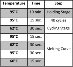
cDNA synthesis was achieved using 1 μg of total RNA and Reverse Transcriptase (RT; NZYTech, Portugal), according to the manufacturer’s instructions. Real-Time PCR was performed in ABI Prism 7500 Sequence Detection System, using SybrGreen Master Mix (Applied Biosystems® by Life Technologies™, USA) and specific primers for the gene of interest (HBB: primer #3 and #4; puromycin resistance gene-internal control: primer #5 and #6) (Table 1). Quantification was performed using the relative standard curve method (ΔΔCt, Applied Biosystems® by Life Technologies™, USA), following the temperature programme on Table 2. Technical triplicates from each experiment, as well as each sample, were assessed in all cases.
3.6. Semiquantitative PCR
Three dilutions (1:2, 1:4 and 1:8) were prepared using the RT product from the BN sample. Three µL of each of the diluted samples were amplified with 0.3 µL of Taq DNA polymerase (5 U/µL; Ambion), 5 µL of 10X PCR buffer (with 15 mM MgCl2; Ambion), 0.5 µL of dNTPs (10 mM) and 1 µL of each corresponding forward #3 and reverse primer #4 (Table 1) for HBB gene, in a total volume of 50 µL. Thermocycler conditions were 95°C for 5 min, followed by 30 cycles of 95°C for 1 min, 60°C for 1 min, and 72°C for 1 min, followed by a final extension of 72°C for 5 min. The same procedure was carried out in parallel using GADPH specific primers (#7 and #8 – Table 1) as the internal control. Thermocycler conditions were 95°C for 10 min, followed by 24 cycles of 95°C for 45 sec, 60°C for 30
13 sec, and 72°C for 45 sec, followed by a final extension of 72°C for 10 min. Aliquots from each sample were analysed by electrophoresis on 2% agarose gels.
3.7. SDS-PAGE and Western Blot
Cell lysates were denatured for 10 minutes at 95°C. Twelve µL of SDS sample buffer 5x was added to 60 µL of lysate and these were loaded into a 14% polyacrylamide gel and resolved at 20mA/gel. Then proteins were transferred to a PVDF membrane (Bio-Rad, USA) for 1 hour at 100V. The membrane was blocked in 5% (w/v) skimmed milk + TBS-Tween 20 (Sigma-Aldrich®, USA) 0,1% (v/v) for 1 hour and probed using mouse anti-α-tubulin antibody (loading control; Roche, Switzerland) at 1:10 000 dilution and mouse monoclonal anti-beta globin (Sigma-Aldrich®, USA, USA) at 1:250 or rabbit monoclonal anti-c-myc (Santa Cruz Biotechnology Inc., USA) at 1:250 overnight. After incubation with the primary antibody, membranes were washed 3 times in TBS-Tween 20 (1X) (Sigma-Aldrich®, USA) 0.1% (v/v). Detection was carried out by incubating the membranes for 1 hour with the secondary antibodies, peroxidase conjugated anti-mouse IgG (Bio-Rad, USA), anti-rabbit IgG (Bio-Rad, USA) antibodies, followed by enhanced chemiluminescence reaction.
3.8. Statistical analysis
Results are expressed as mean ± standard deviation of at least 3 experiments in which the mRNA levels expressed from B15 and B39-containing plasmids are normalized to the BN mRNA levels arbitrarily set to 1. Student’s t test was used for estimation of statistical significance (unpaired, two tails). Significance for statistical analysis was defined as p< 0.05.
Table 1: Sequences of the primers used in the current work
Primer Sequence (5’ 3’) #1 TTGGCATGGAGCAGAAGCTGATCTCCGAGGAGGACCTGGCCCATCACT #2 ATGGGCCAGGTCCTCCTCGGAGATCAGCTTCTGCTCCATGCCAAAGTG #3 GTGGATCCTGAGAACTTCAGGC #4 CAGCACACAGACCAGCACGT #5 GGGTCACCGAGCTGCAAGAA #6 CACACCTTGCCGATGTCGAG #7 CCATGAGAAGTATGACAACAGCC #8 GGGTGCTAAGCAGTTGGTG
14
Temperature Time Step
95°C 10 min. Holding Stage
95°C 15 sec. 40 cycles Cycling Stage 62°C 30 sec. 95°C 15 sec. Melting Curve 62°C 30 sec. 95°C 30 sec. 60°C 15 sec.
15
4. RESULTS
4.1. Beta globin detection by Western blot
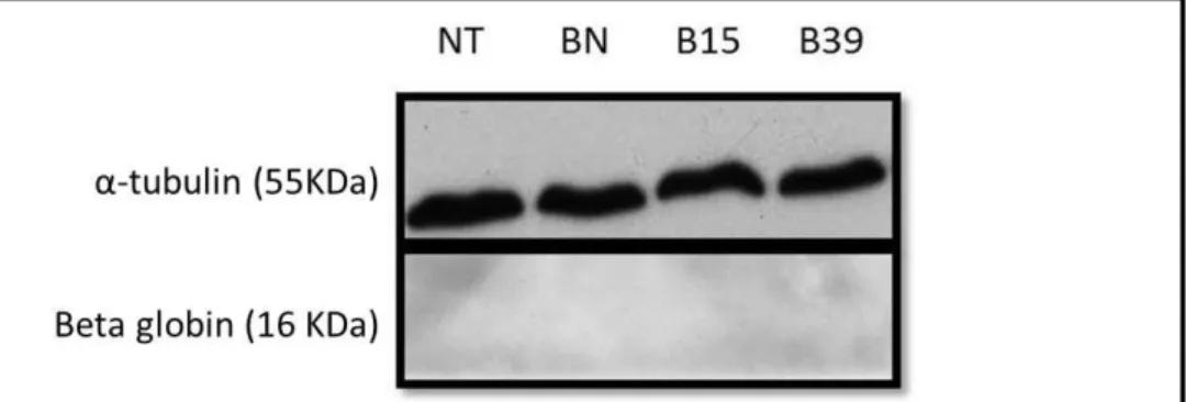
The first experiment had the purpose of evaluating beta globin protein levels, produced by each transcript (BN, B15 and B39). For that, HeLa cells, transiently transfected with the plasmids containing the three beta globin variants, were lysed and denatured leaving the protein in solution and available to be detected by WB. As observed in Figure 3, it was not possible to detect beta globin with the tested conditions. However, we observed α-tubulin with a specific antibody, meaning that the WB was correctly carried out. Thus, this negative result may be due to the incapacity of the antibody to detect the protein.
To overcome this obstacle, we decided to clone a c-myc tag in the human HBB gene exon 3, specifically in the restriction site of BstXI, using annealing reaction (Figure 4). The two oligonucleotides were annealed, following the protocol on Materials and Methods section, forming a double stranded oligo (Figure 4a) that was ligated to the plasmid, containing the HBB gene, previously digested by BstXI (Appendix Figure 1). With this approach only B39_c-myc variant was created (Figure 4b) since for BN and B15 we did not get any transformant colonies, possibly due to ligation inefficiency. Because of this problem, a new strategy based on restriction enzymes was chosen. For that, NotI and BsrGI enzymes were used to digest the B39_c-myc variant, as well as BN and B15, which do not contain the c-myc tag yet. Eventually, the resulting fragments (Appendix Figure 2) were ligated. Particularly, the large fragment from the B39_c-myc variant was ligated with the small fragments of each BN and B15 variant (Figure 4c). The beta globin gene was fully sequenced after the cloning strategy.
Figure 3: Western blot of cell lysates using an anti-beta globin antibody (Sigma-Aldrich®). The molecular
weights of the α-tubulin (55 kDa) and beta globin (16 kDa) proteins are indicated on the left side of the autoradiography. The first lane corresponds to a sample with non-transfected cells (NT).
16 After obtaining the three variants containing the c-myc tag, we tried to detect beta globin protein once again (data not shown), before testing any of the readthrough drugs. The cell culture, transfection, cell lysis and WB were performed at the conditions previously used, but this time with an anti-c-myc antibody. Unfortunately, it was not possible to detect beta globin once again even though the c-myc tag was correctly inserted. This lack of results may be due to a bad transfer of the small proteins to the PVDF membrane. To ascertain whether the small proteins are being transferred, the membrane was stained with Comassie Blue (Sigma-Aldrich®) which showed that the transfer stopped around the 25kDa (Appendix Figure 3a). This problem may be due to the blot buffer or the voltage/amperage that were used in the transfer. In order to solve this problem, the blot buffer recipe was replaced for the Towbin buffer which is the most suitable for small proteins like beta globin (recipe available in Appendix 4). Since there were no noticeable differences in the transfer using the Towbin buffer, the original blot buffer was used. It was suggested that blot buffer’s recipe might not be correct and, therefore, a blot buffer with a new recipe was made (Appendix 4). By using this blot buffer the transfer was much more efficient (Appendix Figure 3b). However, as observed in Figure 5, beta globin was not able to be detected, despite all the adjustments to the original protocol. In parallel with the two tested samples (BN and B39) it was decided to run, as a Figure 4: Insertion of a c-myc tag in exon 3 of HBB gene. a) Agarose gel of the annealing reaction, from left to
right: oligonucleotide forward (F), oligonucleotide reverse (R), double stranded oligo (DS), DNA marker – NZY DNA ladder VI (M). b) Comparison of the three matching sequences: reference sequence (top), c-myc sequence (middle) and B39_c-myc (bottom). c) Cloning strategy to create BN_c-myc and B15_c-myc variants using NotI and BsrGI enzymes.
c-myc tag
48bp
17 control, four other samples (C1, C2, C3 and C4) that do not contain beta globin. These four controls (gift from Oncobiologia lab, INSA) are lysates which are supposed to contain a small protein - H2B histone – with approximately 15 kDa. The purpose of these four samples is to verify if the absence of signal in the BN sample is due to a transfer problem during WB. Although α-tubulin is detectable, the small proteins could be over transferred, owing to their small molecular weight, passing through the PVDF membrane. As we can observe in Figure 5, the small protein used as a control was detected, indicating that the absence of beta globin is not due to transfer inefficiency.
At this point it was suggested that the production of beta globin by the cells was very low due to the weak promoter (SV40) present in the pTRE2pur plasmid. To solve this problem, for each beta globin variant, two 35-mm plates containing HeLa cells were plated instead of one, concentrating the sample at the cell lysis step. The experiment was repeated considering this new alteration and using a new control (CP), a lysate in which the protein of interest contains a c-myc tag. The use of this control makes it possible to know if the anti-c-myc antibody is working. As we can observe in figure 6a, the antibody is working since the CP sample shows a clear band, unlike the remaining samples. Because there is no simple explanation to the absence of band in the BN sample, as was expected, it was decided to test the anti-beta globin one last time (Sigma-Aldrich®) and this time it was possible to obtain a band (Figure 6b). Due to the fact that this strategy was not viable owing to time and money constrictions, it was decided to continue the work by studying the effect of the G418 and PTC124 only at the mRNA level.
Figure 5: Western blot of cell lysates using anti-c-myc antibody (Santa Cruz biotechnology) and anti-H2B
antibody (Santa Cruz biotechnology). Only the first two lanes are samples related to the beta globin analysis. The other four (C1, C2, C3 and C4) are controls supposed to contain a small protein (H2B histone). (M) Molecular weight marker.
18
4.2. Analysis of BN and B39 mRNA levels when transfected HeLa cells are exposed to G418 or PTC124
Our first approach was to evaluate the mRNA levels produced by each transcript before testing any of the readthrough drugs. For that, HeLa cells were transfected with a plasmid containing the three beta globin gene variants: BN, B15 and B39 and 24 hours post transfection, the cells were lysed followed by RNA extraction and cDNA synthesis. The beta globin mRNA levels were quantified by Real Time PCR. As it is observed in Figure 7, B39 is 21% and B15 is 83% of the BN, which is arbitrarily set to 1, as expected.
Once optimal conditions were verified, it was possible to start the experiments using the readthrough compounds. Twenty-four hours after transfection, cell cultures were either treated or Figure 6: Western blot of cell lysates using c-myc antibody (Santa Cruz Biotechnology) on (a) and an anti-beta globin antibody (Sigma-Aldrich®) on (b). These experiments revealed that c-myc and anti-beta globin
antibodies are working properly, however the first was not able to detect the c-myc tag in our samples.
Figure 7: RT-qPCR analysis performed on mRNA isolated from HeLa cells expressing the BN, B15 and B39 transcripts without any drug treatment. Statistical analysis of the three independent experiments compares
B15 and B39 with the wild-type (BN). p<0,001
(c-myc Ab)
19 untreated with increasing concentrations (150 µg/ml, 300 µg/ml or 500 µg/ml) of G418 and harvested 24 hours later. The RNA was extracted, followed by cDNA synthesis and lastly analysed by Real Time PCR (Figure 8). As it is possible to observe in figure 8, G418 did not have a significant readthrough effect since there is not a clear increase in the beta globin mRNA levels. The highest effect is noticeable in the 500 µg/ml drug concentration, which rose from 12% (B39 0µg/mL) to 21% (B39 500µg/mL).
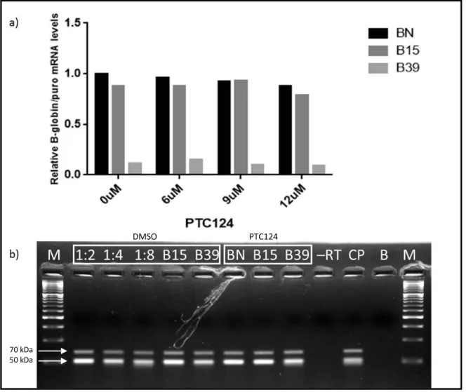
The second drug tested was PTC124 at 6, 9 and 12 µM (Figure 9), using the same experimental approach used for G418. Beta globin mRNA levels did not increase in any of the tested concentrations excepting at 6 µM that rose slightly from 11% to 16%, when compared to B39 0 µM (Figure 9a). In parallel, it was done a semiquantitative PCR to ascertain if it was possible to see any clear alterations and, when observing the two lanes that concern the B39 sample, it is noticeable that the sample containing the drug is slightly brighter, indicating a higher beta globin expression, although the RT-qPCR does not validate that information (Figure 9b). Looking for a more stimulating result, we decided to change the drug concentrations. In order to reach that goal, three concentrations of PTC124: 3 µM, 30 µM and 100 µM were used, maintaining the remaining conditions. This concentration range was chosen based on Welch et al. (2007)40, that uses this drug in a different working model.
Figure 8: RT-qPCR analysis performed on total mRNA isolated from HeLa cells containing the BN and B39 transcripts after treatment with G418 aminoglycoside - Relative beta globin mRNA levels are normalized to
the expression of the wild-type mRNA of each condition (BN). Statistical analysis of the three independent experiments compares B39 0µg/mL with the B39 of each drug concentration. p>0,05
NS NS NS
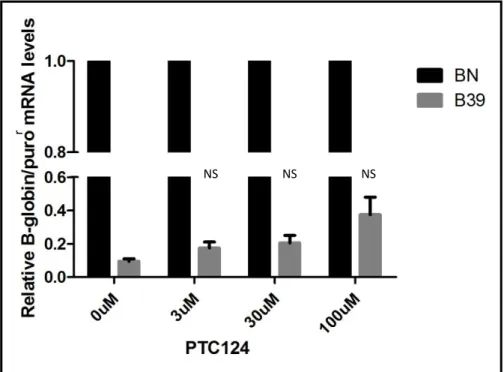
20 After the RT-qPCR analysis, the results were very promising (Figure 10) since all samples containing PTC124 exhibited an increase in the beta globin mRNA levels. The most efficient concentration was 100 µM, which showed an increase of almost 4-fold (38%) when compared to the B39 0 µM (10%). Finally, one last range of concentrations was tested, comprising 50 µM, 100 µM and 150 µM (Figure 11). 150 µM was the most efficient concentration when the readthrough is concerned, since the beta globin mRNA levels duplicated (18%) when compared to the B39 0 µM (9%).
Figure 9: RT-qPCR (a) and semiquantitative PCR (b) performed on RNA isolated from HeLa cells containing the BN, B15 and B39 transcripts after treatment with PTC124 – (a) Relative mRNA levels are normalized to
the expression of the BN mRNA that do not contain drug (first bar). (b) The top bands are referred to the
GAPDH internal control, being the bottom ones referred to the beta globin. From left to right: DNA marker –
NZY DNA ladder VI (M); BN 0 µM 1:2; BN 0 µM 1:4; BN 0 µM 1:8; B15 0 µM; B39 0 µM; BN 9 µM; B15 9 µM; B39 9 µM; sample that do not contain reverse transcriptase (-RT); positive control (CP); sample that only contains the PCR mix (B).
DMSO PTC124
70 kDa 50 kDa
21 Figure 10: RT-qPCR analysis performed on RNA isolated from HeLa cells expressing the BN and B39 transcripts after treatment with PTC124 - Relative mRNA levels are normalized to the expression level of the
BN mRNA of each condition. Statistical analysis of the two independent experiments compares B39 0µM with the B39 of each drug concentration. p>0,05
Figure 11: RT-qPCR analysis performed on RNA isolated from HeLa cells expressing the BN and B39 transcripts after treatment with PTC124 - Relative beta globin mRNA levels are normalized to the expression
of the BN mRNA of each condition. Statistical analysis of the three independent experiments compares B39 0µM with the B39 of each drug concentration. p>0,05
NS NS NS
r
NS NS NS
22
5. DISCUSSION AND FUTURE DIRECTION
The introduction of a PTC in the ORF does not allow the translation to progress until the full-length protein is synthesized. For this reason, nonsense mutations are the origin of a large proportion of all genetic diseases, being imperative the development of a therapy. Suppression therapy utilizes low molecular weight compounds that induce the translation machinery to recode a PTC into a sense codon, allowing the translation elongation to continue in the correct reading frame until it reaches a normal stop codon, culminating with the formation of a full-length polypeptide31. Although the translation termination process is generally accurate, the readthrough process can naturally occur if a near-cognate aminoacyl-tRNA (whose anticodon is complementary to two of the three nucleotides of the codon) binds the stop codon32. The error rate for suppression of normal stop codons at the end of genes is generally less than at premature termination codons. Suppression generally occurs at a rate of 0.001–0.1% at normal stop codons42 and 0.01–1% at PTCs35.
In the present thesis the aim was to ascertain how beta thalassemic nonsense mutation at codon 39 will respond to nonsense suppression therapy with an aminoglycoside, G418, or a non-aminoglycoside, PTC124. The final purpose is to discover which compounds will have efficient levels of suppression, in a dose-dependent manner.
The intention was to analyze the effect of these two compounds in the mRNA as well as in protein levels to obtain more complete and accurate information about the readthrough ability of each drug. Concerning the mRNA analysis (Figure 7), the results were in line with literature. B39 transcript exhibited a level of expression of 21% when compared to the wild type, indicating that these nonsense-mutated mRNAs undergo rapid decay, as demonstrated by Romão et al., 200043. Statistical analysis indicated that there is a significant difference between the mRNA levels of BN and B39 (p<0,001). The low levels of the B39 transcript is explained by the fact that it possesses a PTC localized at more than 50-55 nts upstream of the last exon-exon junction and so the ribosome is incapable to physically remove downstream EJCs, activating NMD16. On the other hand, B15 levels are identical to the wild type transcript as it was expected. Although this transcript has a nonsense mutation on codon 15 and, for this reason, has downstream EJCs, it fails to trigger NMD, due to the fact that it is in close proximity to the translation initiation codon (AUG-proximal PTC)17. The mechanism for this NMD resistance was related to the fact that the PABPC1 is capable of inhibiting NMD when placed close to a PTC, even in the presence of EJCs44. Taking this into account and that the mRNA acquires a circular conformation owing to the interactions of PABPC1 with the scaffold protein eIF4G at the 5’-Cap, eIF3 (eukaryotic initiation factor 3) and the 40S ribosome, it is supposed