UNIVERSIDADE DE LISBOA
Faculdade de Medicina Veterinária
THE USE OF GASTROSCOPY IN THE EVALUATION OF TREATMENT RESPONSE OF EQUINE GASTRIC ULCER SYNDROME (EGUS)
FILIPA GRAÇA PAIT CORTEZ
CONSTITUIÇÃO DO JÚRI ORIENTADORA
Doutora Paula Alexandra Botelho Garcia de Andrade Pimenta Tilley
2019 LISBOA Doutora Berta Maria Fernandes
Ferreira São Braz
Doutora Paula Alexandra Botelho Garcia de Andrade Pimenta Tilley Doutora Maria Rita Martins Garcia da Fonseca Pequito
UNIVERSIDADE DE LISBOA
Faculdade de Medicina Veterinária
THE USE OF GASTROSCOPY IN THE EVALUATION OF TREATMENT RESPONSE OF EQUINE GASTRIC ULCER SYNDROME (EGUS)
FILIPA GRAÇA PAIT CORTEZ
DISSERTAÇÃO DE MESTRADO INTEGRADO EM MEDICINA VETERINÁRIA
CONSTITUIÇÃO DO JÚRI ORIENTADOR
Doutora Paula Alexandra Botelho Garcia de Andrade Pimenta Tilley
2019 LISBOA Doutora Berta Maria Fernandes
Ferreira São Braz
Doutora Paula Alexandra Botelho Garcia de Andrade Pimenta Tilley Doutora Maria Rita Martins Garcia da Fonseca Pequito
ii
“My mother, is the bone of my spine, keeping me straight and true. She is my blood, making sure it runs rich and strong. She is the beating of my heart. I cannot imagine a life without here.”
Kristin Hannah
I wish to dedicate this thesis to my late mother, for always nursing me with love and strength while I pursue my purpose in life.
iii
Acknowledgements
First and foremost, I must thank my adviser, Prof. Dr Paula Tilley, for allowing me to work on this project under her guidance, for all the years of knowledge shared, her patience and kindness that she has always shown throughout these years of schooling. I am truly grateful.
To Dr Joana Simões, for all her advice, counselling and mentoring.
To Professor Rita Garcia da Fonseca and Professor Luis Lamas, for their guidance and knowledge transmitted.
Moreover, to my fellow interns Xico, Dianna, Constance and Bruno, for sharing this experience with me and always adding some fun to our time together.
Also, to Bruno and João, for all their help, outstanding care for the horses at FMV-ULisboa and their excellent Horsemanship.
To Dr Telmo Nunes for his help and direction with the statistical analysis of the results, without which I would be clueless. Also, to Professor Rute Roda, whose guidance led me to my results. To my beautiful, loving family whom I adore, admire and cherish, thank you for all your support. No matter what I choose to do in life, you keep on cheering me on.
To my Mother, my role model, the strongest and the most beautiful person I have ever known. Your strength was such a blessing, and I am so proud of you. Thank you seems very small to say for all the sacrifices you made for me, so I could always pursue my dreams and reach my goals.
To my “sestra” Anica, who has always been by my side, from giggles to tears, smiles and frowns, everything from dead serious, to goofing around, life’s ups and downs, tornados and twisters.
To Carlitos, whom I can thank for all his love, support, friendship and guidance.
To my Father, to whom I have inherited the love of animals, who introduced me to the real meaning of Horsemanship, and always pushed me to pursue my dream.
To my special friend and roommate, Sara Bernardino, for sharing the ups and downs of this long journey since the beginning. I am so lucky to say that I have gained a friend for life.
To “my Sheldon”, for all his support, friendship, love and understanding. Thank you for helping me get by the roughest experience in life and for always pushing me to progress and succeed. Lastly, to all my little furry angels for their unconditional love and devotion. I thank them for all the lessons they taught me, and which made me a better person.
iv
Abstract
Title: The use of gastroscopy in the evaluation of treatment response of Equine Gastric Ulcer
Syndrome (EGUS)
Despite the existence of effective treatment (Omeprazole) for EGUS, given the high cost and recurrence rate, it is essential to take preventive management and dietetic measures.
The purpose of this retrospective study was to evaluate the treatment response with omeprazole using gastroscopy, identify risk factors associated with the appearance of gastric lesions and evaluate the effect of management changes in the prevention of recurrence, between the years of 2010 and 2015.
69 horses with suspected EGUS were evaluated by physical examination, gastroscopic examination and measurement of gastric fluid pH values. The final sample group were the 22 horses with confirmed lesion grade≥1 in the first gastroscopy, then treated with omeprazole 4mg/kg bwt, PO, SID, for 28 days, followed by 2mg/kg bwt, SID, PO, for another 28 days, and recommended management and nutritional measures. After this period, all horses were re-evaluated. Information was gathered from the horse’s owners using two questionnaires regarding clinical complaints at both examinations and regarding treatment follow-up.
The occurrence of EGUS was confirmed in 100% (n=69). Clinically relevant symptomatology was identified, were signs of colic in the last year was the most frequent clinical complaint (59,10%) and was significantly associated with higher numeric scores (p=0,029) whereas the coat condition was significantly associated with severity score (p=0,038). After treatment, there was a general improvement of the lesions and clinical signs, with total remission of the lesions in 4,5% (n=2/22). Owner’s responses on signs of colic (p=0,031) and body weight (p=0,008) and body weight registered at physical examination (p=0,011) were statistically different, along with an increase in mean pH values show evidence of a certain level of efficacy of treatment with omeprazole. None of the presumable risk factors were found significantly associated with the presence or degree of gastric ulceration (p>0,05). Still, exercise and administration of NSAIDs may have determined a lower treatment response. No significant changes in ulcer scores were identified for any levels of implementation of management measures. Nevertheless, decreased ulcer scores were identified in 45,5% (n=10/22) in those who implemented the management changes in some way.
v
Resumo
Título: Utilização de gastroscopia na avaliação da evolução do tratamento de Síndrome de
Úlcera Gástrica Equina (SUGE)
Apesar da existência de tratamento eficaz (Omeprazol) para SUGE, dado o elevado custo e taxa de recorrência, é essencial tomar medidas preventivas de maneio ambiental e dietética. O objetivo deste estudo retrospectivo foi avaliar a resposta ao tratamento com Omeprazol utilizando gastroscopia, identificar fatores de risco associados ao aparecimento de lesões gástricas e avaliar o efeito das alterações de maneio na prevenção de recorrência, entre os anos de 2010 e 2015.
Foram avaliados 69 equinos com suspeita de SUGE por exame físico, exame gastroscópico e medição dos valores de pH gástrico. A amostra final foi de 22 equinos com lesões grau≥1 confirmados na primeira gastroscopia, tratados com Omeprazol 4 mg/kg PV, PO, SID, por 28 dias, seguido de 2mg/kg BWT, SID, PO, por mais 28 dias, e recomendado alterações de maneio ambiental e dietético. Após esse período, todos os equinos foram reavaliados. Informação a respeito das queixas clínicas em ambos os exames e do seguimento do tratamento foi recolhida dos proprietários dos cavalos usando dois questionários.
A ocorrência de EGUS foi confirmada em 100% (n=69). Foi identificado sintomatologia clinicamente relevante, tendo sido a queixa clínica mais frequente sinais de cólica no último ano (59,10%, n=13/22) estando associada significativamente com o grau numérico (p=0029), enquanto a condição de pelagem foi significativamente associada ao grau de gravidade (p=0038). Após o tratamento, verificou-se uma melhoria geral das lesões e dos sinais clínicos, com remissão total das lesões em 4,5% (n=2/22). As respostas dos proprietários sobre sinais de cólica (p=0,031) e peso corporal (p=0,008) e peso corporal registados no exame físico (p=0,011) foram estatisticamente diferentes, juntamente com um aumento nos valores médios de pH mostram evidências de um certo nível de eficácia do tratamento com Omeprazol. Nenhum dos fatores de risco foram encontrados significativamente associados à presença ou grau de ulceração gástrica (p > 0,05). No entanto, o exercício e a administração de AINEs podem ter determinado uma resposta de tratamento mais baixa. Não foram identificadas alterações significativas nos graus de úlcera tendo em conta níveis de implementação das medidas de maneio diferentes. No entanto, foi identificada a diminuição do grau de ulceração em 45,5% (n=10/22) naqueles que implementaram as mudanças de maneio de alguma forma.
Palavras-chave: Síndrome de Úlcera Gástrica Equina, gastroscopia, omeprazol, maneio,
vi Table of Contents Dedication ... ii Acknowledgements ... iii Abstract ... iv Resumo ... v Table of Contents... vi
List of Figures ... viii
List of Tables ... ix
List of Graphics ... x
List of Abbreviations and Symbols ... xi
I. Report on the activities developed during the curricular internship ... 1
II. Literature review ... 3
1. Introduction ... 3
2. Terminology ... 3
3. Anatomy of the equine stomach and gastric acid secretion ... 4
4. Gastric mucosal defence mechanisms ... 8
5. Pathophysiology of ESGD and EGGD ... 10
5.1. Equine Squamous Gastric Disease (ESGD) ... 10
5.2. Equine Glandular Gastric Disease (EGGD) ... 11
6. Prevalence ... 12
7. Epidemiology ... 12
7.1. Intrinsic factors: age, gender, temperament and breed ... 13
7.2. Intense exercise ... 14
7.3. Feeding routine (intermittent versus continuous feeding, feed deprivation), water deprivation and stall confinement ... 15
7.4. Diet type – Starch content and forage type ... 16
7.5. Stress ………..17
7.6. Transportation ... 18
7.7. Nonsteroidal anti-inflammatory agents ... 18
7.8. Parasites ... 20
7.9. Bacteria ... 21
8. Clinical signs of EGUS ... 22
9. Diagnosis of EGUS ... 24
9.1. Blood analysis - haematological and biochemical changes ... 24
9.2. Faecal occult blood test ... 25
9.3. Endoscopic examination ... 25
9.3.1. Scoring system for gastric ulcers in the horse ... 26
9.3.2. Complications and disadvantages ... 27
9.4. Other diagnostic methods ... 27
10. Management of EGUS ... 27
10.1. Pharmaceutical treatment ... 27
10.1.1. Proton pump inhibitors ... 28
10.1.1.1. Omeprazole ... 28 ➢ Pharmacokinetics ... 28 ➢ Presentation ... 28 ➢ Dosage ... 29 ➢ Efficacy ... 30 10.1.1.2. Esomeprazole ... 31 10.1.1.3. Pantoprazole ... 32
10.1.2. Histamine type-2 receptor antagonists ... 32
10.1.3. Coating and binding agents ... 32
vii
10.1.5. Antimicrobial agents ... 33
10.2. Pharmaceutical prevention ... 33
10.3. Environmental management of EGUS ... 34
10.3.1. Modification of exercise intensity and duration ... 34
10.3.2. Pasture turnout ... 34
10.4. Dietary management of EGUS ... 35
10.4.1. Eliminate bolus feeding and increase forage intake ... 35
10.4.2. Decrease the size and increase the frequency of concentrate feeding ... 36
10.4.3. Oils ... 36
10.4.4. Nutraceuticals, nutritional supplements and other alternatives ………37
III. Retrospective study ... 39
1. Objectives ... 39
2. Material and Methods ... 39
2.1. Sample selection ... 39
2.2. Epidemiological questionnaire ... 40
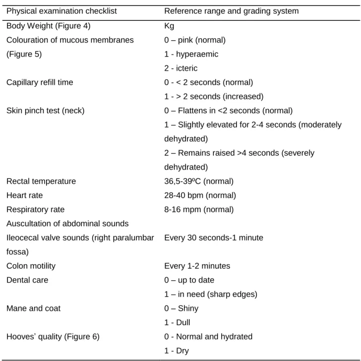
2.3. Physical examination ... 40
2.4. Gastroscopy examination ... 43
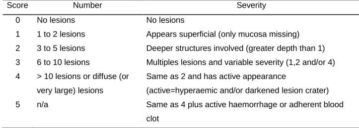
2.5. Gastric ulcer lesions scoring system ... 45
2.6. Measurement of gastric pH value ... 47
2.7. Questionnaire on treatment follow up... 47
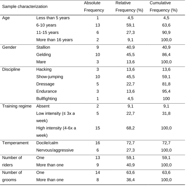
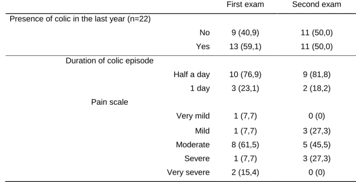
2.8. Statistical analysis ... 47 3. Results ... 48 3.1. Sample selection ... 48 3.2. Epidemiological questionnaire ... 49 3.2.1. Sample characterization ... 49 3.2.2. Clinical complaints ... 51 3.3. Physical examination ... 53 3.4. Gastroscopy examination ... 55
3.4.1. Measurement of gastric pH value ... 58
3.5. Identification of associations between independent variables and ulcer scores. ... 60
3.6. Effect of screening interval on treatment outcome ... 62
3.7. Influence of implementation of recommended management changes over the treatment outcome ………..63
4. Discussion ... 67
5. Conclusion ... 76
IV. Further studies ... 78
V. References ... 79
VI. Annexes ... 96
Annexe 1a. Submitted and accepted abstract for poster presentation for the IV Jornadas do GTIE……….96
Annexe 1b. Poster presented at the IV Jornadas do GTIE ... 97
Annexe 2. Epidemiological questionnaire ... 98
Annexe 3. Gastroscopy report form ... 99
viii
List of Figures
Figure 1 – An overview of the recommended terminology for describing equine gastric erosive
and ulcerative diseases (Adapted from Sykes et al., 2015). ... 4
Figure 2 - A post mortem specimen of the equine stomach illustrating the anatomical regions of the stomach (Hepburn, 2011)... 5
Figure 3 - Schematic illustration of the parietal cell transport mechanism (Adapted from Hepburn, 2004). ... 7
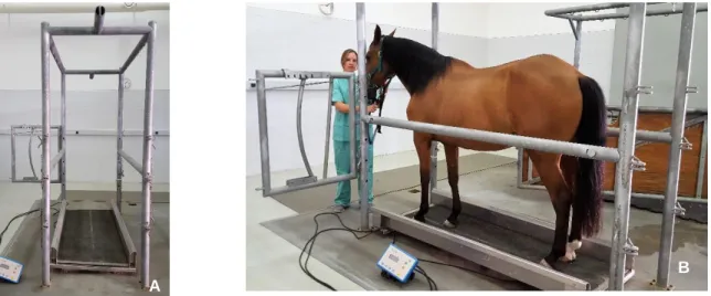
Figure 4 - Illustrative image of weighing. ... 42
Figure 5 - Oral mucosa classification ... 42
Figure 6 - Classification of the hooves’ quality. ... 42
Figure 7 - Sedation protocol. ... 44
Figure 8 - Image of the Storz® equine-gastroscope used for this study . ... 44
Figure 9 - Gastroscopy procedure. ... 44
Figure 10 - Illustrative image of the horse immobilised for the gastroscopy procedure. ... 44
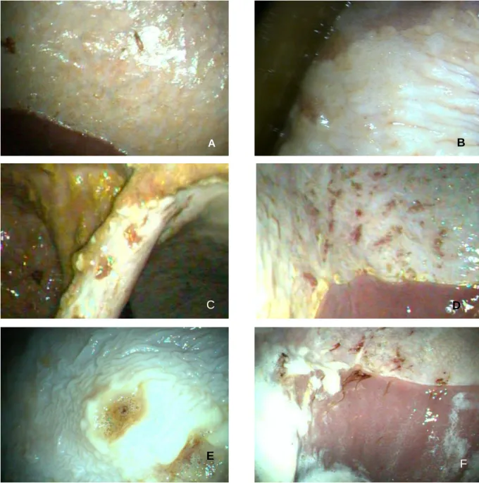
Figure 11 - Exemplification of characteristic lesions of each severity score according to the adapted scoring system of MacAllister et al. (1997). ... 46
Figure 12 - Collection of gastric fluid and measurement of gastric pH value ... 47
Figure 13 - Number of gastroscopies performed on each horse (n=69) ……….47
ix
List of Tables
Table 1 - Parameters included in the physical examination, reference range and grading system (Rose & Hodgson, 2000). ... 41 Table 2 – Number/Severity scoring system for grading of EGUS lesions in horses (Adapted by MacAllister et al. (1997)). ... 45 Table 3 - Distribution regarding sample characterization according to age, gender, equestrian discipline, training regime, temperament, also number of riders and grooms (n=22). ... 50 Table 4 - Distribution of presence of colic in the last year and characterisation of the colic episodes. ... 51 Table 5 - Minimum, maximum, mean, median and standard deviation regarding the number of colic episodes accounted in the last year, determined for both moments. ... 52 Table 6 - Distribution of number of colic episodes accounted in the last year at both examination moments (n=13)... 52 Table 7 - Comparison between results of the epidemiological questionnaires (n=22) ... 53 Table 8 - Descriptive statistics regarding weight, respiratory and heart rates (n=22). ... 54 Table 9 - Distribution of qualitative analysis for physical examination findings in both examinations (n=22). ... 55 Table 10 - Distribution regarding lesion location (n=22). ... 56 Table 11 - Distribution regarding gastric lesion scores at both gastroscopic examinations (n=22). ... 57 Table 12 - Number of horses per numeric score in each gastroscopy (n=22). ... 57 Table 13 - Number of horses per severity score in each gastroscopy (n=22). ... 58 Table 14 – Distributions of the levels of implementation of the recommended management changes according to score evolution. ... 59 Table 15 - Chi-square tests to determine association between level of implementation of recommended changes and score evolution. ... 59 Table 16 - Testing of changes in the number and the severity of lesions before and after treatmet. ... 60 Table 17 - Descriptive statistics regarding the gastric pH value (n=22). ... 62 Table 18 – Comparison of lesion number score and lesion severity score with independent variables (P-values resulting from non-parametric tests of Mann-Whitney and Kruskal-Wallis). 62 Table 19 - Descriptive statistics regarding the number of days between examinations. ... 62 Table 20 - Distribution regarding endoscopic screening interval. ... 63 Table 21 - Distributions regarding score evolutions and endoscopic screening interval. ... 66 Table 22 - Chi- square tests to determine association between endoscopic interval and score evolution (n=22). ... 63
x
List of Graphics
Graphic 1 - Distribution regarding the level of implementation of environmental management changes (n=22). ... 64 Graphic 2 - Distribution regarding the level of implementation of dietary and nutritional management changes (n=22). ... 64 Graphic 3 - Level of implementation of recommended management changes established (n=22). ... 65
xi
List of Abbreviations and Symbols
% - Percentage & - And + - Plus < - Less than > - More than = – Equal to ± – Plus or minus ® – Registered trademark
AINEs – Anti-inflamatórios não esteróides BID - bis in die
bwt - Body weight
CI95% - Confidence Interval of 95 per cent Cl- - Chloride ion
cm – Centimetre
CRT - Capillary refill time DM – Dry matter
DGEE – Doença Gástrica Escamosa Equina DGGE – Doença Gástrica Glandular Equina e.g. – exempli gratia
ECL - Enterochromaffin-like cells
EGUS – Equine Gastric Ulcer Syndrome EGF - Epithelial growth factor
EGFr - Epithelial growth factor receptor ESGD - Equine Squamous Gastric Disease EGGD - Equine Glandular Gastric Disease EP3 - Prostaglandin E2 receptor 3
FOBT – Faecal Occult Blood Test
FVM-ULisboa - Faculdade de Medicina Veterinária – Universidade de Lisboa g - Gram
h - Hour
H+ - Hydrogen ion
i.v - Intravenous
mg/kg - Milligram per kilogram ml - Millilitre
xii mmol/l - Millimoles per litre
MR – Mean rank
n - Total sample number n/a – Not applicable
NSAIDs - Nonsteroidal anti-inflammatory drugs NSC - Nonstructural carbohydrates
NTA - Nothing to add p - p-value PGE2 - Prostaglandin E2 PO – per os SB - Standardbred SC - Subcutaneous SD – Standard deviation SID - semel in die
SUGE – Síndrome de Úlcera Gástrica TB – Thoroughbred
TM - Trademark
χ2 - Pearson's chi-squared test kg - Kilogram
1
I. Report on the activities developed during the curricular internship
This report concerns the curricular internship from the integrated master’s degree in veterinary medicine. The internship took place at the Equine Unit of the Teaching Hospital, Faculdade de Medicina Veterinária, Universidade de Lisboa (FMV-ULisboa).
In the last two years of her integrated Masters, the author took an on and off training period starting in September 2013, while finishing her studies, working under the guidance of Professor Paula Tilley, and under the supervision of Dr Joana Simões, Professor Rita Garcia da Fonseca, and Professor Luis Lamas. Following this period, she started the curricular internship per se in 2016 for seven months, between March and September. Overall, the author summed a total of 1500 hours of training.
During this time, she collaborated in several services of the hospital, such as internal medicine, imaging services, surgical services and postoperative care, as well as accompanying Professor Tilley on house calls.
The Equine Unit of the FMV-ULisboa is considered a referral hospital, but also provides primary and preventive health care programs, as well as a wide range of medical, surgical and dental services, as well as an emergency service 24hours a day, 365 days a year.
In the internal medicine service, she observed several cases of prophylactic care, namely vaccination and wellness care, preventive dental care, equine identification, blood sampling for identification purposes, microchipping, deworming, and prepurchase examination. Besides prophylactic care, there were other consultations with appointments of different departments such as dermatology, gastroenterology, odontology, orthopaedics, trauma, neurology, ophthalmology, oncology, cardiology, pneumology, andrology, gynaecology and obstetrics, neonatology, intensive care and parasitology.
While in the internal medicine service, a variety of cases were observed, from physical examinations of patients to assisting the responsible physician with any medical acts and diagnostic techniques. These included a wide range of minimally-invasive procedures, in particular, digital radiology, ultrasound (musculoskeletal, cardiac, pulmonary, abdominal and obstetric) and endoscopy (gastroscopy, vaginoscopy, rectoscopy, static and dynamic upper and lower respiratory endoscopy). The author assisted the physician in charge by preparing the patient for the exam, preparing and placing of the necessary equipment, as well as the cleaning and disinfection of the equipment (ultrasound probe and endoscope).
While in the surgery service she was involved in several procedures, starting with the preparation of the operating room, next in the preoperative care of the surgical patient, performing a physical examination of the surgical patient, administering the proper medication prescribed by the surgeon, as well as the anaesthesiologist, and postoperative care.
2
The author had the opportunity to assist in small procedures such as skin suturing, drainage and removal of abcesses, as well as large procedures such as castration, tenotomy, exerisis of tumours, rectum and perineum reconstruction.
The surgeries that were observed took place in the operating room of the hospital or in the field. During postoperative care, the author was responsible for monitoring vital signs of recovering patients and administering the medication prescribed by the supervising physicians.
During this period the author also collaborated in a few courses that took place at the teaching hospital of FMV-ULisboa, such as the XXXIX Jornadas Médico-Veterinárias (Dynamic Endoscopy Workshop) which took place in October of 2015, and the “Avaliação cardio-respiratória, endoscopia no cavalo e ecografia do aparelho musculo-esquelético” in November of 2015.
The necessary information was gathered for this study during this time frame, namely the chart information of suspected patients of Equine Gastric Ulcer Syndrome (EGUS), such as their physical examination, weight, gastroscopy examination, gastric pH level, and epidemiological questionnaire. The development of this work was accepted to be presented as a poster at the IV Jornadas do Grupo de Trabalho de Investigação em Equídeos, Golegã (November 2015) (available in Annexe 1).
3
II. Literature review 1. Introduction
Equine gastric ulcer syndrome (EGUS) is a wide-reaching and common disorder in the horse regardless of age, gender, or breed. It has been considered a conundrum for several decades by practitioners, horse owners, riders and trainers for its economic impact and clinical importance, due to its high prevalence, unspecific clinical signs and negative impact on performance (Nadeau & Andrews, 2009).
Many risk factors have been implicated in the aetiology of this syndrome, such as stress, diet and feeding routine, type and intensity of exercise, stall-confinement and the administration of non-steroidal anti-inflammatory drugs (NSAIDs) (Sykes & Jokisalo, 2015). Despite many efforts, due to the nonspecific nature of the symptomology and the absence of specific biochemical and haematological parameters, endoscopic examination remains the only reliable method to determine a definite diagnosis (Sykes, Hewetsen, Hepburn, Luthersson & Tamzali, 2015).
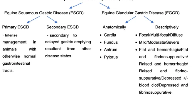
2. Terminology
The term EGUS was first recognised in a consensus compiled by the Council in 1999 to describe gastric ulceration in the horse (Andrews, Bernard,et al., 1999). However, since then, the term has been misused, and therefore, an effort has been made to define and elucidate the terminology appropriately (Merritt, 2009). Similarly, in Human Medicine, the term peptic ulcer disease (PUD) is considered a hypernym to define ulcerative lesions in the oesophagus, stomach and duodenal mucosa (Malfertheiner, Chan, & McColl, 2009).
Recently the terminology has been polished, recognising EGUS as the blanket term to describe gastric ulceration, while distinguishing Equine Squamous Gastric Disease (ESGD) and Equine Gastric Disease (EGGD) separately according to the affected anatomic region (Sykes, Hewetson, et al., 2015). Therefore, it is unfitting to extrapolate the knowledge of one syndrome to the other. Furthermore, collection of information is required for each condition due to the stated differences between the squamous and glandular mucosae, which include differences in prevalence within a population (Begg & O’sullivan, 2003; Habershon-Butcher, Hallowell, Bowen & Sykes, 2012; Luthersson, Hou Nielsen, Harris & Parkin, 2009b; Murray Nout & Ward, 2001b; Tamzali, Marguet, Priymenko & Lyazrhi, 2011), risk factors (Habershon-Butcher et al., 2012), response to treatment with omeprazole and prevention (Sykes, Sykes, & Hallowell, 2014d, 2014a, 2015). However, the presence of squamous and glandular ulceration within an individual was found unrelated (Begg & O’sullivan, 2003; Luthersson et al., 2009b; Murray, Nout, et al.,
4
2001b). A synopsis of the reinforced and recommended terminology of EGUS is displayed in Figure 1.
Figure 1 – An overview of the recommended terminology for describing equine gastric erosive and ulcerative diseases (Adapted from Sykes et al., 2015).
3. Anatomy of the equine stomach and gastric acid secretion
The horse’s stomach consists of a single composite large chamber interposed between the oesophagus and small intestine (König & Liebich, 2004). The oesophagus joins the stomach through the cardia in the right median plane of the abdomen, while the pylorus continues into the duodenum more to the left, both of which are controlled by sphincters (König & Liebich, 2004). The cardiac sphincter in the horse is very well developed, and this, together with the oblique entrance of the oesophagus is believed to be responsible for the presumed inability of the horse to vomit. Although vomiting and regurgitation are considered rare, it may occur (König & Liebich, 2004).
It possesses visceral and parietal surfaces and greater and lesser curvatures (König & Liebich, 2004). The lesser curvature is very short, compared to the greater curvature, causing the cardia and pylorus to have a nearby location (Sisson, 1986).
The stomach’s position is closely related to the development of the greater omentum and the lesser omentum (König & Liebich, 2004). Also, the exact shape and position depend on the degree of filling (König & Liebich, 2004). Still, the physiological capacity varies between 5 and 15 litres, which is smaller compared to the size of the animal and the volume of forage it consumes
5
(Dyce, Sack & Wensing, 2004). This fact should be considered when administering fluids via a nasogastric tube to avoid overdistension (König & Liebich, 2004).
The structure of the gastric wall is the same as that of the oesophagus and consists of the following layers, inward to outward: mucosa, submucosa, muscular layer and serosa (König & Liebich, 2004).
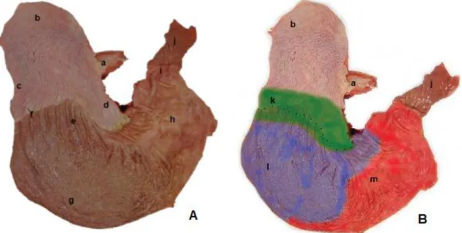
The mucosa can be divided into two core regions: the non-glandular region and the glandular region (Figure 2). The squamous non-glandular region includes the dorsal third of the stomach (orad), which is covered with stratified squamous epithelium, identical to the one found lining the oesophagus, as well as the entrance to the stomach via the cardia (Hepburn, 2011). Histologically, no glandular structures of any kind are found within it that could establish assumed digestive or mucosal protective mechanisms. Further, it was found that in an Ussing chamber it maintains no short circuit current, demonstrating the absence of active transport processes (Merritt, 1999). The functional role of this region is yet to be determined (Hepburn, 2011). However, it is believed that since it is not generally exposed to acid, it allows fermentative digestion processes to occur (Klein, 2013).
Figure 2 - A post mortem specimen of the equine stomach illustrating the anatomical regions of the stomach (Addapted from Hepburn (2011)).
Legend: A–Equine gastric mucosal anatomy; B–Illustration of gastric pH and content stratification.
a-Oesophagus; b-Squamous fundus; c-Greater curvature; d-Lesser curvature; e-Cardia; f-Margo plicatus; g-Glandular fundus; h-Pylorus region; i-Pylorus; j-Duodenum; k-Low density matter (pH 5-7); l-Medium density matter (pH 4-5); m-High density matter (pH 1-2).
6
The margo plicatus is the delineation region between the squamous and glandular regions of the stomach that lines the distal half of the equine stomach (Hepburn, 2011). Some authors have considered the margo plicatus as part of the non-glandular mucosa, while others found that, due to its distinct and complex capillary network and susceptibility to ischemic lesions, it should be seen as an independent region in the transition between the two mucous membranes (Staszyk, Jackowiak, Godynicki, & Gasse, 2001).
The distal two-thirds of the stomach is covered with glandular mucosa, which can then be divided into three regions: glandular fundus (body), pyloric antrum and cardiac glandular region. The glandular fundus (body) occupies the mid third of the stomach, the pyloric antrum occupies the most distal third, and the cardiac glandular region is the narrow strip of glandular mucosa adjacent to the margo plicatus (Hepburn, 2011). All these areas contain similarly structured glands, but each has particular cell types with different functions (Klein, 2013; König & Liebich, 2004).
The glandular mucosa of the stomach is comprised in part of simple columnar epithelium, which contains gastric pits, forming the entrance into the gastric glands (König & Liebich, 2004). These epithelial cells, known as surface mucous cells, excrete a thick alkaline mucus that protects the mucosa from the highly acidic gastric fluid (Klein, 2013).
Within the parietal region of the gastric fundus are parietal cells, zymogen (or chief) cells and enterochromaffin-like cells (ECL) that make up the gastric glands (Merritt, 1999). The parietal cells are clustered in the neck of the gland and have the primary function of secreting hydrochloric acid, under the stimulation of histamine, which is produced in a paracrine mechanism by the ECL cells. These cells also modulate gastric mucosal blood flow via the action of serotonin (5-HT) (Klein, 2013). While, in the base of the gland, the chief cells secrete pepsinogen, the precursor to the digestive enzyme pepsin (Klein, 2013). Also, among the parietal cells are neck mucus cells that secrete a thin mucus, less viscous than that of the surface mucous cells. Besides their secretory function, they secondarily function as replacement epithelial cells (Klein, 2013).
The cardiac and pylorus region resemble parietal region in structure, however, contain different cell types (Klein, 2013). The gastric antrum and pylorus have no parietal cells, but their ramified glands contain gastrin-producing G-cells (Klein, 2013). Also, a large number of somatostatin-immunoreactive cells have been shown in this region of the equine stomach. Along with other functions, somatostatin has been shown to be involved in the endogenous control of gastric acid secretion, while sampling intragastric pH and modulating G-cell gastrin release (Merritt, 1999; Hepburn, 2004). These regions can be incidentally parasitised with larvae of gastrophilus intestinalis and may be marked by scars left after their removal (Dyce et al., 2004).
7
The cardiac mucosa, in the horse and most species, forms a narrow band around the gastric opening of the oesophagus, while in swine it covers a more substantial portion of the proximal stomach (Klein, 2013). Little is known about its function in the horse, but it is thought that the primary secretion is sodium bicarbonate, which may protect the adjacent oesophageal mucosa from the stomach’s acid secretion (Klein, 2013).
The horse secretes gastric acid in a continuous, variable manner (Murray, 1994), and the median pH of the ventral stomach of 3.0 over a normal 24h period reflects this (Husted, Sanchez, Olsen, Baptiste, & Merritt, 2008). The acid secretion is predominantly triggered by paracrine, endocrine, and neural stimuli (Yao & Forte, 2003), stimulated by the gastrin, histamine and acetylcholine, a neurotransmitter from the Vagus nerve (Campbell-Thompson & Merritt, 1990).
The hydrogen and chloride ions are secreted by different cellular mechanisms (Klein, 2013). However, the ability of the parietal cell to secrete acid is dependent on active transport, through the H+/K+ ATPase pump (Figure 3), otherwise known as proton pump enzyme, in the cell membrane (Yao & Forte, 2003). This acid secretion occurs against both a concentration and electric gradient, higher than 3 million-fold (Yao & Forte, 2003). Within the parietal cell, H+ ions
are formed from the dissociation of water, then the hydration of CO2 leads to the formation of bicarbonate ion (HCO3-), a reaction catalysed by carbonic anhydrase. The bicarbonate is then
transported out of the basolateral membrane in exchange for chloride (Yao & Forte, 2003). The discharge of bicarbonate into the blood leads to a small elevation of blood pH (alkaline tide), which serves to maintain intracellular pH in the parietal cell (Yao & Forte, 2003). Hydrogen ions are driven out of the cell, into the lumen, in exchange for potassium through the action of the proton pump (Forte & Zhu, 2010). The bicarbonate trapped in the mucous barrier adhering to the stomach wall forms a pH gradient that allows a physiological pH at the mucosal surface and a pH similar to that of stomach acid at the luminal surface (Andrews et al., 1999).
The horse’s stomach presents a dorsoventral gradient (Figure 2B), starting with the squamous mucosa of the fundus (pH 5.46±1.82), followed by the squamous mucosa of the margo plicatus Figure 3 - Schematic illustration of the parietal
cell transport mechanism (Adapted from Hepburn (2004)).
8
(pH 4.12±1.62), then the glandular mucosa of the pylorus (pH3.09±1.9), being the gastric fluid the most acidic (2.72±1.86) (Murray, Grodinsky, Anderson, Rague, & Schmidt, 1989).
The gastric contents of an adult equid with a regular hay/grain diet, where the roughage is available on a free-choice basis, also have a similar dorsoventral layering. The roughage component of the diet determines this fact seeing that the dense acid liquid is trapped ventrally by the lower density/larger particle size components found dorsally, which is vital to normal gastric function (Hepburn, 2011; Merritt, 2013). Gastric emptying is continuous at a speed that according to the type of content, where liquid contents take only 30 minutes to reduce in half the volume, while solid contents take about 1·5 hours (Hepburn, 2011).
Adult horses may secrete up to 35-40 litres of saliva per day, the majority of which is parotid in origin, with a pH of approximately 7.4 (Eckersall, Aitchison, & Colquhoun, 1985). Saliva secretion has the core function of buffering gastric pH (Bell, Mogg, & Kingston, 2007), consequently, feeding has the buffering effect on gastric pH through effects of increased production of saliva with an increase of mean intragastric pH by one to two units (Murray, Nout, et al., 2001a). The fasting state was shown to increase the acidity of the gastric fluid from a median pH of 3.1 to 1.55 (Murray, Nout, et al., 2001a). Still, evidence was found that the greater the dry matter content of the feed, the greater the amount of saliva secreted due in part to the physical composition of the meal and in part to the time needed for adequate mastication (Merritt, 2013).
Hourly recordings regarding proximal gastric pH displayed a clear circadian pattern, and that the pH decreased during the night. This pattern was thought to be involved with the horses feeding behaviour during the night, associated to the fact that the horse consumes most of the daily food intake during the day (Husted et al., 2008).
4. Gastric mucosal defence mechanisms
The mucosal defence system is a complex and dynamic process. It is composed of numerous factors that permit the mucosa to remain intact despite its frequent exposure to substances with a wide range of temperature, pH, and osmolarity, as well as to cytotoxic substances (acids, bile salts, pepsin and digestive enzymes), and bacterial products capable of causing local and systemic inflammatory reactions (Andrews, Bernard, et al., 1999).
The squamous nonglandular mucosa is limited in defence mechanisms, relying mainly on acid repulsion and intracellular buffering (Murray, 1994). Mucopolysaccharides and intercellular tight junctions in the superficial epithelium are the only defences against H+ entry, however minimal
buffering capacity if still offered by the intercellular secretion of HCO3- and by intracellular
9
mucosal thickening, whereas prolonged exposure to increased acidity is likely to cause hyperkeratosis, erosion, and ulceration (Murray, 1994).
Gastric glandular epithelium, on the other hand, has several mechanisms to prevent injury by acid secretion, which include secretion of mucus, EGF, bicarbonate buffering, mucosal blood flow, cellular repair, and prostaglandins (Andrews, Bernard, et al., 1999).
Of these, mucosal blood flow is considered to be the most important as it provides the mucosa with the oxygen and nutrients necessary to produce the mucus-bicarbonate layer and allow rapid turnover of epithelial cells (Wallace, 2008). It is also required for removal of VFAs produced by intragastric carbohydrate fermentation, and to prevent the epithelial damage to progress to necrosis of deeper layers of the mucosa (Wallace, 2008).
Epidermal growth factors, which are found in salivary gland secretions, are known to promote mucosal protection, by inhibition of gastric acid secretion, the proliferation of gastric mucosal cells, and also interfere in prostaglandin synthesis process (Andrews, Bernard, et al., 1999). Although earlier studies regarding observation of the EGF receptor (EGFr) in gastric mucosa have focused on the glandular stomach of laboratory animals as an extrapolation for the human glandular stomach, Jeffrey, Murray and Eichorn (2001) found significant evidence that EGFr is also induced in peptic-injured equine gastric squamous epithelium and its involvement in the healing of gastric squamous mucosal ulcers in horses.
The mucus secreted by neck cells acts as both a protective physical barrier and lubricant against physical damage from ingesta, maintaining a neutral pH environment for the mucosal cells, besides being implicated in resisting the diffusion of protons (Andrews, Bernard, et al., 1999). Although the importance of the mucus layer thickness remains unknown, there is evidence that a change in the mucus layer may cause damage to the mucosa, and eventually ulceration (Wallace, 2008). In response, epithelial cells, with the help of bicarbonate secreted by the mucosal cells at the luminal surface, form a mucoid cap in order to maintain a high pH environment. The resulting inflammatory response includes an increase in mucosal blood flow, which helps with the mucoid cap and facilitates leukocyte recruitment and plasma exudation. If this response is weakened, then mucosal repair is slowed or halted, resulting in gastritis or ulceration (Wallace, 2008).
The prostaglandins (PGE1 and PGE2) promote numerous protective functions within the gastric mucosa (Murray, 2010). These substances promote gastric mucosal blood flow, maintain intercellular tight junctions, stimulate bicarbonate and mucus secretion leading to suppression of HCl secretion and gastric acid production. They also facilitate basal cell migration towards the lumen for repairing the mucosa and maintaining the integrity of nonglandular and glandular
10
mucosa; this takes place by stimulation of active surface-protecting phospholipid production (Andrews & Nadeau, 1999; Murray, 2010; Murray, Schusser, Pipers, & Gross, 1996).
5. Pathophysiology of ESGD and EGGD
Gastric ulceration occurs whenever there is an increase in acid exposure (pH <4). Squamous ulceration occurs rapidly due to minimal defences and reflects increased mobility of acidic gastric fluid, while glandular ulceration forms more slowly after the breakdown of the mucous and bicarbonate barrier (Hepburn, 2011).
In short, as discussed previously, there are three types of EGUS: (1) primary squamous ulceration, (2) primary glandular ulceration, and (3) secondary squamous ulceration (Sykes & Jokisalo, 2014). Primary squamous ulceration occurs when increased acid exposure arises due to a change in the normal gastric pH gradient and content stratification. The primary glandular ulceration is defined when acid exposure occurs due to a failure of the mucosal defences. Finally, the secondary squamous ulceration, in which delayed gastric emptying (typically pyloric disease) increases the residual gastric fluid volume and, later, results in dorsal movement of acid (Sykes, Hewetson, et al., 2015).
5.1. Equine Squamous Gastric Disease (ESGD)
The pathogenesis of ESGD is well described as a result of increased exposure of tissue with limited defence mechanisms to highly acidic gastric contents (pH of <4) (Lorenzo-Figueras & Merritt, 2002). Any disruption of the normal stratification of gastric pH results in an increased risk of ESGD (Lorenzo-Figueras & Merritt, 2002). Mucosal injury was identified within 30 minutes of exposure to solutions of hydrochloric acid using in vitro models (Nadeau et al., 2003; Widenhouse, Lester & Merritt, 2002). Other known factors considered to contribute to the development of the disease are volatile fatty acids (acetic, butyric and propionic acids), the by-products of carbohydrate fermentation by gastric bacteria (Andrews, Buchanan, Smith, Elliott & Saxton, 2006; Nadeau et al., 2003), associated with grain feeding (Luthersson, Hou Nielsen, Harris & Parkin, 2009a). Also, a longer carbon chained fatty acid (5 carbons), known as valeric acid, was found to have a more profound effect on function of the nonglandular mucosal barrier and is thought to be the reason that some cases of ESGD persist despite adequate acid control (Andrews, Buchanan, et al., 2006).
Concentrations of duodenal bile salts may also play a part and can be found in the gastric contents of horses deprived of feed for as little as 14 h (Berschneider, Blikslager, & Roberts, 2010).
11
Numerous studies (MacAllister et al., 1999; Sykes, 2019; Sykes, Sykes, et al., 2014d; Sykes, Underwood, Greer, McGowan, & Mills, 2017; Sykes, Underwood, McGowan, & Mills, 2015) demonstrated evidence of ESGD healing with effective antacid omeprazole therapy, in the absence of risk factor reduction. Therefore, there is a clear indication that gastric acid is the foremost erosive agent and that the role of short-chain fatty acids and duodenal bile salts are likely to be less critical (Sykes and Jokisalo, 2015).
Exercise and the increase in intra-abdominal pressure are considered an essential factor in the disruption of the pH gradient (Lorenzo-Figueras & Merritt, 2002).
Feed deprivation has also been shown to cause ulcers in the squamous mucosa of horses (Murray, 1994), which is due to repeated exposure of the squamous mucosa to high acidity (Murray, 2010).
Various experimental and epidemiological studies have identified risk factors, while focusing primarily on feeding and exercise practices, for ESGD which influence the management of clinical cases (Bell, Kingston, Mogg, & Perkins, 2007; Dionne, Vrins, Doucet, & Paré, 2003; Husted, Sanchez, Baptiste, & Olsen, 2009; Husted et al., 2008; Luthersson et al., 2009a; Pedersen, 2017).
5.2. Equine Glandular Gastric Disease (EGGD)
The pathogenesis of EGGD has not yet been identified (Sykes, Hewetson, et al., 2015). The normal physiological conditions of glandular mucosa are to be exposed to extremely acidic gastric contents with the pH in the ventral portion of the stomach fairly stable at around 3 (Husted et al., 2008). Thus, it is believed that EGGD results from a failure of the normal defence mechanisms that protect the mucosa from acidic gastric contents (Sykes and Jokisalo 2015). Moreover, the glandular mucosae anatomy and physiology differs according to the different regions (pyloric, fundic and cardiac regions) and the response to treatment is likely to be different between these regions (Rendle et al., 2018).
The factors believed to have a role in the disruption of the defence mechanisms in horses are bacterial agents and nonsteroidal anti-inflammatory drugs (NSAIDs), which are the predominant causes of gastric ulceration in man (Malfertheiner et al, 2009), and will be discussed further in more detail.
12
6. Prevalence
The prevalence of gastric ulceration varies between ESGD and EGGD and according to breed, purpose or level of training (Sykes, Hewetson, et al., 2015).
The high-risk horse populations have been the main focus of many authors over the last years, such as racehorses in training, which revealed the highest prevalence of between 80 and 100% within the squamous mucosa (Bell, Kingston, Mogg, et al., 2007; Habershon-Butcher et al., 2012; Hammond, Mason, & Watkins, 1986; Rabuffo et al., 2002; Roy, Vrins, Beauchamp, & Doucet, 2005; Vatistas, Snyder, Carlson, et al., 1999). Other horse populations were shown to also be at risk, reaching between 40 and 58%, such as endurance horses (Nieto et al., 2004; Tamzali et al., 2011), show horses (McClure, Glickman, & Glickman, 1999; Murray et al., 1989) and elite western performance horses (Bertone, 2000).
The prevalence of EGGD is less well described than ESGD (Sykes and Jokisalo, 2015), but it has been reported in several studies, ranging from 8% (Sykes and Jokisalo 2015b) to 70% in various populations (Sykes, Bowen, Habershon-Butcher, Green, & Hallowell, 2019; Ward, Sykes, Brown, Bishop, & Penaluna, 2015).
7. Epidemiology
Despite the many factors implicated in the cause of EGUS discussed above, several risk factors for its development have been identified (Videla & Andrews, 2009).
While the notion of distinguishing EGGD and ESGD is a somewhat a recent one, the few studies that have considered these two syndromes independently found that the risk factors have been mostly different. However, the information on risk factors specifically associated with EGGD is still lacking (Sykes, Hewetson, et al., 2015).
Nevertheless, the generally known risk factors for EGUS (not differentiating EGGD and ESGD) are intense exercise (Lorenzo-Figueras & Merritt, 2002; Nieto et al. 2004), a high grain-low roughage diet (Luthersson et al., 2009; Nadeau et al., 2000), water deprivation (Luthersson et al., 2009a), feed deprivation (Husted et al., 2009; Murray & Eichorn, 1996), hospitalization, and use of NSAIDs (MacAllister, Morgan, Borne, & Pollet, 1993; Rabuffo, Hackett, Grenager, Boston, & Orsini, 2009). Other risk factors include stress, transportation (McClure, White, et al., 2005), stall confinement (Bell, Mogg, et al., 2007; Murray & Eichorn, 1996) and the administration of hypertonic electrolytes (Holbrook, Simmons, Payton, & MacAllister, 2005).
Development of EGUS can be as brisk as arising within seven days in some studies (MacAllister et al., 1999; McClure, White, et al., 2005) and the risk of disease increases with time in work (Habershon-Butcher et al., 2012).
13
7.1. Intrinsic factors: age, gender, temperament and breed
Results suggesting a higher susceptibility towards developing EGUS due to intrinsic factors such as gender, breed, age, and temperament remains debatable (Martínez & Silveira, 2013).
Different associations have been suggested between the age of the horse and the prevalence of gastric ulceration. Vatistas et al. (1999) found that the incidence of gastric ulceration was lowest in 2-year-old horses (70%) and highest in 6-year-old horses (100%). In other studies (Murray et al., 1996; Orsini, Hackett, & Grenager, 2009; Rabuffo et al., 2002), despite little association between age and prevalence of ulcers, there was a significant association between age and severity of ulcers, where young horses (2-year-olds) had the lowest mean ulcer scores, compared with older horses. Several authors (Bell, Kingston, Mogg, et al., 2007; Dionne et al., 2003; Roy et al., 2005) stated that the presence or severity of ulceration did not vary significantly with the horse’s age or location within the stomach. On the other hand, Luthersson et al. (2009b) found evidence of older horses being more prone to have lesions in both the glandular and nonglandular regions despite not presenting a higher risk of developing clinically significant EGUS.
In various published studies (Bell, Kingston, Mogg, et al., 2007; Cate, Nielsen, Spooner, O’Connor-Robison, & Schott II, 2012; Dionne et al., 2003; Marqués et al., 2011), sex did not arise as a significant risk factor for gastric ulceration in active racehorses. A few studies (Rabuffo et al., 2002; Vatistas, Snyder, Carlson, et al., 1999) distinguish between castrated and sexually intact males, rather than grouping stallions, colts, and geldings together as males, describing a higher prevalence in geldings. The cause of these findings is thought to associated with a decrease in salivary epidermal growth factor concentration, stimulated by reproductive hormones (Rabuffo et al., 2002). Another study (Chameroy et al., 2006) reported the opposite, where the majority of horses were mare.
McClure et al. (1999) conducted a prevalence study on show-horses which identified the horses' temperament as a possible risk factor, where horses with a nervous character were more likely to have ulceration than quiet or behaviourally normal horses whereas Jonsson and Egenvall (2006) found no influence of temperament over gastric ulceration scores. In a more recent study (Malmkvist et al., 2012), horses with severe glandular ulcers were found to have a higher stress hormone response to novelty, hence more stress sensitive.
Regarding the importance of breed, as previously mentioned, most prevalence studies of gastric ulceration in adult horses have been conducted on Thoroughbreds with the highest incidence, and less on Standardbreds (Begg & O’sullivan, 2003; Dionne et al. 2003; Jonsson & Egenvall, 2006; Luthersson et al., 2009b, 2009a). Both breeds of racehorses are submitted to demanding training requirements and management; however, genetic, behavioural, and gait differences
14
should be considered (Dionne et al., 2003). Warmbloods were also found to be predisposed to EGGD with a moderate prevalence, along with increased risk associated with multiple handlers/riders (Mönki, Hewetson, & Virtala, 2016).
Prevalence studies on endurance horses are less frequent then racehorses, nevertheless no relationship has been found to date between age, breed or gender on gastric ulcer scores (Tamzali et al., 2011).
7.2. Intense exercise
Once initiating training, several management changes are enforced upon horses, many of which have been proven to increase the risk of EGUS, one of which includes exercise (Vatistas, Snyder, Carlson, et al., 1999).
There are still many contradictory results regarding exercise as a possible risk factor of gastric ulceration. Luthersson et al. (2009a) found no relationship between the level of exercise and EGUS. Numerous other studies conducted in racehorses (Bezděková, Jahn, Vyskočil, & Plachý, 2005; Murray et al., 1996; Orsini et al., 2009; Roy et al., 2005; Sykes et al., 2019) have been comparable with results reported by Hammond et al. (1986), which show that an increase of prevalence and lesion grades of specific anatomical lesion sites within the squamous mucosa were significantly associated with the increasing intensity of long-duration training. Evidence showed that horses in light to heavy training for as short as eight days were at risk of developing gastric ulcers (White et al., 2007).
However, Roy et al. (2005) suggested that seasonality may also have been an influencing factor on EGUS prevalence results because horses did not have access to pasture nor free access to pasture throughout the study period, which has been reported to decrease the prevalence of squamous ulcers in horses. On the other hand, another publication reports a high prevalence of EGUS in reproductive mares kept in pasture (Le Jeune, Nieto, Dechant, & Snyder, 2009).
There are several proposed mechanisms in order to explain the influence of exercise in the development of EGUS. The best described is the previously mentioned relationship between exposure of squamous mucosa to acidic content and training, demonstrated by a study which used an intermittent feed deprivation model (Murray, Eichorn, & Jeffrey, 2001). The acidic gastric contents are pushed dorsally into the squamous portion of the stomach by increased intra-abdominal pressure and gastric compression associated with gaits faster than walk (Lorenzo-Figueras & Merritt, 2002). McClure et al. (2005) suggested that the tensing of abdominal muscles may also occur during any other activity that is typical in recreational use of horses, such as trailer transport, and ESGD can develop within five days. This process is heightened by the fact that physical stress, hence intensive training, has been found associated
15
with an increase in gastrin plasmatic concentration, which promotes glandular secretion of HCl production within the gastric lumen (Furr, Taylor, & Kronfeld, 1994; Mills, 1996), thus predisposing the mucosa to acid damage (Mönki et al., 2016).
Authors (Vatistas, Snyder, Carlson, et al., 1999) have also proposed that horses may become excited before a race, which could yet again decrease the gastric pH. Another possible cause is that before any event horses are feed deprived, which could induce a loss of buffering effect by reducing feed content and therefore increase acidity in the stomach (Vatistas, Snyder, Carlson, et al., 1999). Squamous ulceration may occur secondary to delayed gastric emptying, caused by lesions within the distal stomach and/or duodenum, which cause abnormal reflux of acidic contents into the proximal stomach (Merritt, 2012). Lorenzo-Figueras and Merritt (2002) also described a decrease in saliva secretion during exercise, which usually buffers gastric fluid, thus contributing to more prolonged acid exposure.
7.3. Feeding routine (intermittent versus continuous feeding, feed deprivation), water deprivation and stall confinement
Feeding practices, along with stall confinement, have been identified as significant risk factors for developing EGUS (Murray & Eichorn, 1996).
When horses initiate training, they are stabled, fed intermittently, and many times have no access to grazing. Grazing causes a constant secretion of saliva and flow of feed material into the stomach, having a buffering effect against the constant gastric acid secretion in the horse (Murray, Nout, et al., 2001a).
Vatistas, Snyder, Carlson, et al. (1999) were able to show evidence of the development of EGUS in all the horses within 14 days of being stabled and initiating a training regimen.
It has been suggested that horses in training fed ad libitum may have a lower risk of developing gastric ulcers (Husted et al., 2009; Murray, 1994). Though, even when offered with feed ad libitum, they may spend less time eating when stabled, which may decrease this important salivary buffering mechanism (Buchanan & Andrews, 2003).
Horses grazing at pasture, in most cases, have shown a decreased prevalence of ESGD (Murray & Eichorn, 1996). However, other studies presented conflicting results regarding intermittent feed deprivation. Bell, Kingston, Mogg et al. (2007) reported that pasture turnout did not show evidence of having a protective effect. Husted et al. (2008) described a decreased proximal gastric pH in horses apparent after as little as four hours of feed deprivation. This result was only significant during the day, describing the presence of a nocturnal pH gradient in the fed horses. Feeding routines with intervals longer than 6 hours were found to increase the risk of nonglandular lesions (grade ≥2/5) by 3.9 times (95% confidence interval = 1.5 – 10.4) in Danish
16
pleasure horses (Luthersson et al., 2009a). Pedersen et al. (2015) showed that horses fed forage once daily were at a higher risk of developing ESGD (grade ≥2/4) than horses fed twice daily or more repeatedly.
Conversely, a prevalence study showed that both pastured pregnant and non-pregnant mares had a high prevalence of ESGD (Le Jeune et al., 2009). The authors acknowledged the hypotheses of grain and hay supplementation of these mares as a possible cause of increased gastric VFA concentration and therefore squamous ulceration (Andrews et al., 2017; Nadeau et al., 2000). Another assumption to explain the high prevalence of squamous ulcers in these pastured mares might have been due to the horses consuming less forage during evening hours than during daytime hours, which may result in less saliva production and a low pH environment in the proximal stomach (Husted et al., 2008).
Water deprivation in pasture turnout has also been associated with both ESGD ≥2 and EGGD ≥2. Despite the number of hours not being a significant factor, the risk increased when a horse is deprived of water for 4 hours, without increasing past this period (Luthersson et al., 2009a). Water intake is thought to cause dilution of gastric fluid (Andrews, Frank, et al. 2006) and thus pH, although the passage of water through the stomach may depend on the nature of the gastric contents (Luthersson et al., 2009a).
7.4. Diet type – Starch content and forage type
Size and composition of complementary grain meal are believed to be a critical factor associated with developing ESGD (Andrews et al., 2017; Reese & Andrews, 2009).
High starch diets are rich in digestible carbohydrates, resulting in higher production of VFA in the upper part of the stomach (Nadeau et al., 2000). The extensive fermentation in this area, due to a higher dry matter content and a slower mixture of stomach matter and gastric fluid, associated to higher lactic acid production in the lower gastric layers, causes a rapid decrease in gastric fluid pH (Harris, Coenen, Frape, Jeffcott, & Meyer, 2006). This low pH (<4) of gastric matter causes acid damage to the nonglandular squamous mucosa (Andrews, Buchanan, et al., 2006; Nadeau et al., 2000, 2003), similar to gastroesophageal reflux disease (GERD) in Humans (Lorenzo-Figueras & Merritt, 2002).
Feeding practices of 0,5 kg of grain-based concentrates (40% NSC) per 100 kg bwt have been proven to produce in general 20 mmol/l of VFAs concentration in the stomach up to 5 h after feeding (Nadeau et al., 2000). The high VFAs concentration leads to a decrease in sodium transport accompanied by a loss of tissue resistance and an increase in tissue permeability in the squamous mucosa in an in vitro Ussing system (Andrews, Buchanan, et al., 2006). Andrews, Buchanan, et al. (2006) reported valeric acid to have the most profound impact on mucosal
17
barrier function in comparison with acetic, propionic, or butyric acids while Nadeau et al. (2000) reported 78% of acetic acid out of the VFA in the gastric contents of the horses.
Luthersson et al. (2009a) emphasised the fact that the grain (starch) volume of starch fed per day or per meal, over a certain amount, increased the risk of EGUS ≥2 or NG ≥2.
Moreover, a high-grain diet resulted in a higher serum gastrin concentration, hormone known to stimulate secretion of HCl (Sandin, Skidell, Häggström, & Nilsson, 2000) and, is generally ingested quicker in horses with squamous (Malmkvist et al., 2012). Additionally, it may reduce hay uptake, restricting the production of buffering saliva (Nadeau et al., 2000, 2003).
Cereals deliver low levels of calcium (Harris et al., 2006) and, perhaps, other buffering agents, therefore, contributing to increased risk (Nadeau et al., 2000). Contrary to feeding alfalfa hay with grain, which provides a high calcium content (14.4 mg/g of dry weight) and high concentrations of crude protein, contributing with buffering effects on gastric contents (Nadeau et al., 2000).
Another study in horses showed evidence that a high starch-low forage diet increased squamous gastric ulcer scores in the horses compared to horses on a low starch diet (Al Jassim, McGowan, Andrews, & McGowan, 2008).
Horses that were fed straw as the only forage also increased the risk of EGUS ≥2 or NG ≥2, presumably due to the low protein and calcium content and, thus, lacking the buffering aid (Luthersson et al., 2009a).
7.5. Stress
Stress may be the primary cause of glandular gastric ulcers in foals and horses, and stress minimisation could be beneficial in reducing the risk of EGGD in some animals (Sykes et al., 2019). Authors identified trainer as a risk factor for EGGD (Sykes et al. 2019), which aligns with results of a previous Finnish study that identified an increased number of caretakers and riders as possible risk factors (Mönki et al., 2016).
It has been suggested that stress-induced release of endogenous cortisol increases the risk of developing gastric ulceration; e.g. stress of parturition in foals and stress of training and confinement in horses, by inhibition of prostaglandin synthesis, and therefore a failure in mucosal protective factors (Andrews & Nadeau, 1999). Horses with EGGD have augmented cortisol response to exogenous ACTH administration (Scheidegger et al., 2017) and were found to be more stress sensitive presenting in average a 26% higher cortisol concentration in response to novel stimuli (Malmkvist et al., 2012). Still, the authors reported a possible paternal influence (three stallions fathering 89%) on the risk of having gastric ulceration, justifying specific characteristics such as fearfulness and stress-sensitivity as having a genetic component.
18
In foals, stress was found to be implicated in the development of gastric glandular lesions but not in squamous lesions (Furr, Murray, & Ferguson, 1992).
The stress of hospitalisation for horses with the complaint of colic and those with non-colic complaints may also induce gastric mucosal lesions (Rabuffo et al., 2009). Additionally, as previously mentioned, the physical stress of high performance in thoroughbred and standardbred racehorses may also play a role in the higher occurrence of gastric ulcers in the non-colic patients (Orsini et al., 2009).
7.6. Transportation
The increase in equestrian sports recently has led to a rapid increase in the transportation of horses (Friend, 2001). Activities that are typical in recreational use of horses, besides transportation, but also housing off-site, were found to be ulcerogenic, with development of squamous lesions within five days under these conditions (McClure, Carithers, et al., 2005). The stressful impact of transportation on the horse has been proven by repeated increases in plasma cortisol concentrations (Möstl et al., 2009). Additionally, the cortisol concentration during transport has been reported to be positively correlated with transport time (Fazio, Medica, Aronica, Grasso, & Ferlazzo, 2008).
The increased risk of EGUS associated with transportation may be associated with temporary disturbances in the feed as well as water intake (Luthersson et al., 2009a; Möstl et al., 2009).
7.7. Nonsteroidal anti-inflammatory agents
The role of NSAIDs in the development of EGGD is also debatable. The NSAIDs are commonly used in equine clinical practice to treat acute abdominal pain and chronic inflammatory conditions, particularly of the locomotor system (Videla & Andrews, 2009). In most cases, horses are given either phenylbutazone or flunixin meglumine intravenously for pain control. These agents, especially flunixin meglumine are very effective in decreasing abdominal pain associated with acute colic; nevertheless, several side effects have been reported that assigns an increased risk associated to their use in horses with EGUS (MacAllister et al., 1993 ).
Administration of high doses or prolonged treatments of NSAIDs has been proven to be the cause of ulcers in the glandular portion of the stomach (MacAllister et al., 1993; Martínez Aranzales, Cândido de Andrade, & Silveira Alves, 2015). However, studies where the squamous mucosa was the primary focus, the same association was not evident (Hammond et al., 1986; McClure et al., 1999; Murray et al., 1989, 1996; Rabuffo et al., 2002; Vatistas, Snyder, Carlson, et al., 1999). Therefore, it is still unclear if NSAIDs at therapeutic doses increase the risk of EGUS (Rendle et al., 2018).
19
Additionally, colonic ulcers and renal failure are other frequent side effects of these drugs, regardless of whether the drugs are administered at therapeutic doses or overdoses in horses (Andrews, Reinemeyer, & Longhofer, 2009; MacAllister et al., 1993).
The generally accepted theory to explain the association between NSAIDs and gastric ulcers is cyclooxygenase inhibition, in which conversion of arachidonic acid into prostaglandins is blocked (MacAllister et al., 1993; Murray, 2010). Consequently, the physiologic vasodilating effect of prostaglandins (in particular PGE2) on the stomach mucosa which creates a bicarbonate buffering complex and diminishes the corrosive effect of hydrochloric acid contained in gastric secretions (Andrews & Nadeau, 1999; Morrissey, Bellenger, & Baird, 2008) is inhibited, therefore generating the ideal conditions for development of ulcers in the gastrointestinal tract (Andrews & Nadeau, 1999; Murray et al., 1996). Martínez Aranzales et al. (2015) described another mechanism of injury in the horse stomach, using an EGUS induction model based on a phenylbutazone protocol, which showed the ability to induce oxidative stress in glandular mucosa of horses by changing the antioxidant-oxidant balance.
Evidence from laboratory animals supports the hypothesis of decreased prostanoids and oxidative stress in gastric alterations caused by NSAIDs in humans (Berenguer, Alarcón de la Lastra, Moreno, & Martı́n, 2002; Tomisato et al., 2004). Mitochondrial oxidative phosphorylation inhibition in mucus-producing cells was shown to alter the hydrophobic characteristics and exhibit direct cytotoxic effects, including necrosis and apoptosis (Tomisato et al., 2004). However, only effects dependent on the inhibition of PGs have been previously reported in horses (Martínez Aranzales et al., 2015).
According to Andrews et al. (2005), gastric mucosal ischemia may lead to hypoxia-induced cellular acidosis, and release of oxygen-free radicals, phospholipases and proteases, which may damage the cell membrane and result in necrosis. Furthermore, the interaction between the chemical properties of most NSAIDs and the acidic conditions of the stomach enhances the cellular changes in the gastric mucosa (Martínez Aranzales et al., 2015).
Nonetheless, at clinical doses, phenylbutazone and suxibuzone did not cause gastric ulceration when administered during 15 days (Andrews et al., 2009). Neither did a maximum recommended dosage of phenylbutazone affect cyclooxygenase-1 or -2 gene expression for seven days (Nieto, Aleman, Anderson, Fiack, & Snyder, 2012). Additionally, authors stated the administration of NSAIDs was not found to be a probable risk factor for EGUS (Habershon-Butcher et al., 2012), suggesting the possibility of existing mechanisms other than impaired prostaglandin synthesis involved in the formation of glandular gastric ulcers (Pedersen, 2017). As such, despite the conflicting evidence regarding the influence of NSAIDs on the development of EGUS, the Council considers the high prevalence reported in many populations in need of