Universidade de Lisboa
Faculdade de Ciências
Departamento de Física!
!
Resting state fMRI experimental and analytical
methodology: A functional connectivity analysis
Catarina Dinis Fernandes
Dissertação
Mestrado Integrado em Engenharia Biomédica e Biofísica
Perfil em Radiações em Diagnóstico e Terapia
Universidade de Lisboa
Faculdade de Ciências
Departamento de Física!
!
!
Resting state fMRI experimental and analytical
methodology: A functional connectivity analysis
Catarina Dinis Fernandes
Dissertação
Mestrado Integrado em Engenharia Biomédica e Biofísica Perfil em Radiações em Diagnóstico e Terapia
Orientador externo: Professor Christian Schwarzbauer
Orientador interno: Professor Alexandre Andrade
Declaration
I hereby declare that the work presented in this thesis is my own and has not been accepted in any previous application for a degree. It is a record of work, which has been carried out by myself at the Aberdeen Biomedical Imaging Centre, University of Aberdeen. Any contribution from other workers has been acknowledged in the text. All quotations have been distinguished by quotation marks and all sources of information specifically acknowledged.
Catarina Dinis Fernandes
Para ser grande, sê inteiro, Nada teu exagera ou exclui. Sê todo em cada coisa. Põe quanto és no mínimo que fazes. Assim em cada lago a lua toda Brilha, porque alta vive.
ii
Acknowledgments
First of all I would like to thank both of my supervisors, Professor Christian Schwarzbauer and Professor Alexandre Andrade, without whom this project would not have been possible. I would like to thank Professor Christian for giving me the chance to work at the Aberdeen Biomedical Imaging Centre and most of all for believing in me and always keeping my spirit bright and positive even through difficult times. His knowledge, guidance and support were crucial for the success of this project. Despite supervising me from Portugal, Professor Alexandre Andrade has always been available to give me all the support I needed as well as useful advices, which I am truly grateful for. I would also like to thank both for the corrections of the manuscript.
I like to express my gratitude to the following people, all of whom were indispensable during the course of this project: Professor Alison Murray and Dr Mary Joan Mcleod, whom have always been so kind and helpful; Gordon Buchan who has given me all the technical support; the University of Aberdeen radiographers – Baljit Jagpal, Nichola Crouch, Katrina Klaasen, Beverly MacLennan for helping me set up the scanner and aiding with the volunteers;
Thanks to all the research students and other colleagues with whom I have shared several memorable meals and moments during this last year.
I would also like to express my gratitude to all the people who agreed to volunteer as part of my study.
To Ourania, a deep thank you for being not only an amazing friend but also a great mentor. Thank you for the loud Mediterranean conversations, as well as the positive messages that you have always transmitted to me. Thank you for believing in me and always supporting my ideas, pushing us all forward. For all the energy and all the good things you have taught me. I have learned so much. My profound thank you. To Michael, who keeps my mind sharp and thoughts accurate thank you so much for sharing your knowledge and amazing stories with me. I could have not wished for a better professor about Scottish history, music, food, and most of all about the amazing number of prodigious scientists that Scotland has produced. I
will not forget all the adventures, the projects, all the brainstorming and amazing knowledge that you have both shared with me. To both of you my deep thank you.
The Lilian Sutton secretaries, Teresa Morris and Dawn Younie, who have always provided assistance and aided me in every step of my internship;
To everyone that in a way or another contributed to my wellbeing, gave me support and cherished with me amazing moments during this past year.
I wish to thank all my friends in Portugal, who have been a constant presence and a reminder of the amazing things I have back at home.
Many thanks to Reece for being the best housemate I could have ever wished for. Thank you for making me feel at home and welcomed at all times. I cherish the most our endless talks and the way you can always manage to cheer me up after a tiring day at work. Last but not least, thank you for the amazing meals that you have always prepared and taught me how to do.
This is a very special thank you to you David. Thank you for all the amazing moments and adventures, for always being there to make my days with a smile and helping me see that most of the problems are no problem at all. Thank you, my sweet home away from home. I would like to express my gratitude first and foremost to my family, particularly to my parents. There are not enough words to say how much I am grateful for the love, support and encouragement. For always believing in me and cheering me up when things are not going so well, I thank them.
!
!
iv
Resumo
O ser humano desde sempre se sentiu fascinado pelo estudo do seu próprio corpo assim como das suas propriedades funcionais. Do desejo de compreender e explorar o corpo humano surgiram então técnicas que permitem o seu estudo de modo não invasivo. Entre as primeiras técnicas de imagiologia encontram-se os Raios-X, a tomografia axial computadorizada (TAC) e a terapia por emissão de positrões (PET: do inglês “Positron Emission Therapy"). Contudo, todas elas utilizam radiação ionizante, e como tal surgiu o desejo de desenvolver novas metodologias igualmente não invasivas mas que por seu lado não utilizem qualquer tipo de radiação ionizante.
Entre estas técnicas encontra-se a imagiologia por ressonância magnética (MRI: do inglês “Magnetic Resonance Imaging”) que pode ser utilizada para estudar as estruturas anatómicas mas também os seus mecanismos funcionais através da aplicação da técnica de ressonância magnética funcional (fMRI: do inglês “functional magnetic resonance imaging).
Contrariamente às técnicas que utilizam radiação ionizante, a imagiologia por ressonância magnética tira partido do facto de o ser humano ser maioritariamente constituído por água. Um ser humano adulto é por norma constituído por cerca de 70 – 80% de água (H2O) o que
se reflecte numa grande abundância de protões – núcleo 1H. Quando submetidos a um forte campo magnético, o momento magnético destas partículas tende a alinhar-se de acordo com a direcção do campo magnético externo (B0). Após alinhados os protões são então submetidos
a um pulso de radiofrequência (com frequência igual à frequência de Larmor destas partículas) que é absorvido e modifica o momento magnético (i.e. Spin) dos protões. Quando este pulso é desligado, o spin dos protões retorna ao equilíbrio termodinâmico, de acordo com a direcção do campo magnético B0, emitindo energia sob a forma de radiofrequência (RF).
Estes mecanismos de relaxação diferem consoante o conteúdo em água dos tecidos e são estes que permitem a identificação da sua estrutura. Gradientes de campo magnético são tamb ém utilizados de modo a criar ligeiras diferenças no campo magnético que permitem a codificação do sinal com informação espacial.
A imagiologia por ressonância magnética faz, nos dias de hoje, parte da rotina hospitalar providenciando imagens com grande precisão e resolução anatómica.
vi
Todavia a informação estrutural nem sempre é suficiente para estudar patologias que não exibem diferenças anatómicas, tais como depressão ou esquizofrenia. Surge então a ressonância magnética funcional, que utiliza o nível de oxigenação do sangue (BOLD: do ingles “Blood-oxygenation level dependent”) como uma medida indirecta de activação neuronal. Através da utilização desta técnica é então possível mapear zonas cerebrais responsáveis pelo processamento de sinais como por exemplo estímulos visuais, tácteis ou auditivos. A título de exemplo, temos o estudo de doenças como o autismo ou até mesmo de distúrbios de consciência. A nível clínico a ressonância magnética funcional é utilizada para mapear funções críticas como por exemplo a fala, o movimento, o planeamento de tarefas, etc. Esta técnica oferece aos profissionais de saúde a chance de desenvolver um melhor planeamento cirúrgico sendo que é também aplicada no planeamento de tratamentos de radioterapia a nível cerebral com o intuito de mapear funcionalmente o cérebro e detectar os efeitos que tumores, AVC e lesões cerebrais possam ter ao nível da re-estruturação das suas funções.
Até muito recentemente a grande maioria da informação disponível acerca da conectividade anatómica cerebral era estritamente proveniente de estudos efectuados em primatas, recorrendo ao uso de técnicas extremamente invasivas (Felleman, Van Essen 1991, Jones, Powell 1970, Mesulam 2000, Ungerleider, Haxby 1994) assim como do estudo de lesões em casos humanos (ex: (Geschwind 1965)).
Frinston (Friston et al. 1993) utilizando PET e Biswal (Biswal et al. 1995) através do uso de fMRI foram os primeiros a identificar que para além das ligações anatómicas entre diferentes estruturas cerebrais é também possível identificar ligações funcionais entre regiões que à primeira vista parecem não ter qualquer tipo de ligação. À técnica que usa MRI no estudo da conectividade funcional foi dado o nome de conectividade funcional de ressonância magnética (fcMRI: do ingles “Functional connectivity MRI”). Esta utiliza ressonância magnética funcional e as oscilações de baixa frequência ao nível do sinal BOLD em cada voxel para estabelecer correlações. Com base na ideia de que duas zonas se podem dizer funcionalmente relacionadas se estas se encontram a operar no mesmo processo, é portanto possível assumir que as variações no seu sinal BOLD serão bastante semelhantes exibindo uma alta correlação. A título de exemplo vejamos duas regiões do córtex motor primário, localizadas em hemisférios opostos, e que contudo apresentam sinais BOLD altamente
correlacionados. Com esta ideia em mente foi então desenvolvido o conceito de redes* funcionais que são usualmente estudadas durante períodos de repouso†. Exactamente durante esta condição foi verificada a existência de uma rede funcional extremamente consistente entre indivíduos, e mesmo entre diferentes estados como durante o sono ou anestesia. A esta rede foi dado o nome de “Default-mode network” (Raichle et al. 2001) sendo que esta inclui regiões do córtex posterior cingulado, precuneus e do córtex prefrontal medial. A “default-mode network” é a rede mais estudada, mas para além desta existem outras redes tal como a rede visual, a auditiva, a de controle executivo, a de atenção, entre outras. Estas redes encontram-se frequentemente interrompidas ou modificadas em casos de doença. Os projectos descritos no âmbito desta dissertação focam-se no estudo destas redes bem como das suas propriedades em casos de doença (distúrbios de consciência, AVC) e durante a performance de actividade física. A fim de estudar estas redes funcionais foram utilizados diferentes métodos para o cálculo da conectividade funcional. Entre os mais reconhecidos métodos de cálculo de conectividade funcional encontram-se a análise com base numa região de interesse‡, a análise através do estudo da independência entre componentes§ bem como
métodos que permitem o cálculo da conectividade cerebral a nível global**. Os métodos que utilizam uma região de interesse focam-se no cálculo da conectividade entre esta região e o resto do cérebro através do uso de medidas de correlação. O segundo método mencionado separa as várias redes neuronais com base na máximizacao da sua independência estatística. Por último, os metodos de análise global calculam a correlação das série temporal de cada voxel com todos os outros voxeis do cerebro. A contribuição da autora para os estudos descritos ao longo desta dissertacao focou-se no uso de duas destas técnicas – “seed-based analysis” e “wGBC”- no cálculo da conectividade cerebral em cada um dos diferentes projectos.
No primeiro projecto, descrito no capítulo 3 desta dissertação, são apresentadas vários paradigmas que em conjunto com o uso de ressonância magnética funcional, foram desenhados para detectar consciência e percepção em doentes que sofrem de distúrbios de consciência. Estes paradigmas foram testados num grupo de voluntários saudáveis de modo a verificar se são adequados ou se necessitam de ser optimizados. A autora foi então responsável por executar uma análise individual e de grupo da activação induzida pela
* do inglês: “networks” † do inglês: “resting state” ‡ do inglês: “seed-based analysis” § do inglês: “ICA”
viii
execução destes mesmos paradigmas. O desenvolvimento de paradigmas adequados a estes pacientes, combinadas com o uso de fMRI vem complementar e melhorar o diagnóstico e prognóstico destes doentes.
No capítulo 4 desta dissertação a autora focou-se na análise da conectividade functional em pacientes que foram diagnosticados com um pequeno AVC, com enxaquecas e com TIAs††. Este procedimento utilizou técnicas de cálculo da conectividade com regiões de interesse e medidas globais de conectividade funcional. O objectivo deste estudo é uma vez mais averiguar se a inclusão de uma sequência de conectividade functional poderá facilitar o diagnóstico destes doentes bem como o seu prognóstico.
No quinto capítulo a autora foca-se no estudo das diferenças induzidas ao nível da conectividade funcional por uma única sessão de exercício físico. São uma vez mais utilizadas técnicas de cálculo da conectividade com regiões de interesse bem como outros métodos implementados por outros investigadores do departamento.
É também incluído nesta dissertação um capítulo no qual foram analisadas as propriedades destas redes neuronais ao nível de uma população saudável. É importante que tanto as condições de aquisição dos dados de ressonância magnética funcional como as metodologias de análise estejam bem estabelecidas para que os dados provenientes de diferentes estudos sejam comparáveis e para que possamos estabelecer de forma fiável conclusões acerca de populações saudáveis e doentes. O conceito de repouso é ainda muito variável, particularmente quando é apenas pedido aos participantes que permaneçam calmos e imóveis. Certos estudos requerem que os participantes permaneçam de olhos fechados, outros de olhos abertos e outros ainda que fixem uma imagem projectada num ecrã. Uma grande variabilidade de estados podem ser originados com este design experimental, sendo que estes vão desde o simples devaneio em torno de um assunto, que por qualquer razão se encontra mais fortemente em mente, ou até mesmo o adormecer. Com o objecto de estudar estas variações, o capítulo 6 foca-se na investigação da conectividade cerebral resultante de duas diferentes situações bem como da sua variabilidade. Neste capítulo a autora procurou estudar a reprodutibilidade e confiança destas redes funcionais cerebrais quando é pedido aos participantes que executem uma tarefa de baixo requerimento cognitivo. A análise foi executada através do cálculo da correlação entre séries temporais bem como da sua análise
estatística, utilizando medidas como o coeficiente de correlação intra-classes, que fornece uma estimativa de reprodutibilidade entre diferentes medições.
Deste trabalho resultaram uma apresentação oral e a apresentação de um poster. Os resultados foram no geral positivos mas em alguns casos bastante ambíguos. As mais recentes publicações evidenciam o interesse em estudar não só a distribuição espacial destas redes como também as suas propriedades temporais que se parecem evidenciar como extremamente dinâmicas. Como tal fica aqui aberto o caminho para a continuação da exploração das redes funcionais cerebrais bem como da sua variabilidade.
Numa nota final, consideramos importante salientar que o vasto estudo da conectividade cerebral assim como o dos seus mecanismos é ainda uma área de investigação com pouco mais de uma década e com um ainda longo caminho a percorrer.
x
Abstract
Conventional functional magnetic resonance imaging (fMRI) is used to measure small fluctuations in the blood oxygenation level dependent (BOLD) signal resulting from neural activation due to an external stimulus or task. Nonetheless, this imaging technique can also be applied to the study of functional connectivity in the human brain. Since it was first acknowledged that BOLD signal fluctuations also occur during resting periods that increased attention has been directed to the investigation of brain behaviour during this particular state. There is still an on-going debate as to whether these fluctuations actually reflect neuronal baseline activity or are just the result of physiological metabolism and therefore independent o neuronal function. Also, can this resting state activity be truly called a “baseline” for comparisons? Moreover, functional connectivity has identified several networks, of which the default mode network is the most robust. This network is believed to have a great importance in brain awareness and cognition.
Further research is crucial to correctly understand these events and also to create a standardised methodology to perform the resting state fMRI acquisitions. The RESTATE (Resting State Techniques) project arises from the need to comprehend and correctly interpret the measured low frequency BOLD oscillations during resting periods. With this longitudinal study, comprising a baseline and a follow-up scan, we aim to assess the implications of using a low cognitive level paradigm upon the reproducibility of the data during functional connectivity analysis.
!
!
Key Words
Magnetic Resonance Imaging (MRI); Functional Magnetic Resonance Imaging (fMRI), Functional connectivity Magnetic Resonance Imaging (fcMRI); Resting State fMRI; Seed-based analysis; Reliability and Consistency.
xii
Table of Contents
DECLARATION ... II ACKNOWLEDGMENTS ... II RESUMO ... V ABSTRACT ... X LIST OF FIGURES ... XV LIST OF TABLES ... XVII LIST OF ABBREVIATIONS ... XVIIICHAPTER 1. INTRODUCTION ... 1 1.1 RATIONALE ... 1 1.2 PROJECT AIMS ... 1 1.3 METHODOLOGY ... 2 1.4 THESIS OUTLINE ... 4 CHAPTER 2. BACKGROUND ... 7
2.1 MAGNETIC RESONANCE IMAGING ... 7
2.2 IMAGING TECHNIQUES ... 8
2.2.1 Noise in functional magnetic resonance imaging sequences ... 8
2.2.2 Echo-Planar Imaging – EPI ... 8
2.2.3 T1 –weighted images ... 9
2.2.4 T2 –weighted images ... 9
2.2.5 Neurovascular protocol ... 10
2.3 RESTING STATE FMRI ... 12
2.3.1 Resting state networks ... 14
2.4 FMRI ANALYSIS ... 16
2.5 FUNCTIONAL CONNECTIVITY MRI(FCMRI) ... 18
2.5.1 Seed-correlation analysis ... 20
2.5.2 Weighted Global Connectivity (wGBC) ... 22
2.5.3 Independent Component Analysis (ICA) ... 22
CHAPTER 3. ASSESSING CONSCIENCE WITH THE AID OF FUNCTIONAL MAGNETIC RESONANCE IMAGING (FMRI). THE DISORDERS OF CONSCIOUSNESS (DOC) STUDY ... 25
CHAPTER SUMMARY ... 25
3.1 INTRODUCTION ... 26
3.2 MATERIAL AND METHODS ... 28
3.2.1 Participants ... 28 3.2.2 Measurements ... 28 3.2.3 Imaging Methods ... 29 3.2.4 Image Analysis ... 29 3.3 RESULTS ... 31 3.4 DISCUSSION ... 37
CHAPTER 4. DIAGNOSTIC DILEMMA: LONGITUDINAL ASSESSMENT OF TRANSIENT ISCHAEMIC ATTACKS, MINOR STROKE AND MIGRAINES WITH FUNCTIONAL CONNECTIVITY MRI ... 40
CHAPTER SUMMARY ... 40
4.1 INTRODUCTION ... 42
4.2 MATERIAL AND METHODS ... 48
4.2.1 Participants ... 48 4.2.2 Imaging Methods ... 49 4.2.3 Image Analysis ... 49 4.3 RESULTS ... 53 4.4 DISCUSSION ... 58 4.5 CONCLUSION ... 60
CHAPTER 5. PHYSICAL EXERCISE & BRAIN CONNECTIVITY. THE PHYSICAL EXERCISE CONNECTIVITY (PECON) STUDY ... 62
CHAPTER SUMMARY ... 62
5.1 INTRODUCTION ... 63
5.2 MATERIAL AND METHODS ... 66
5.2.1 Participants ... 66 5.2.2 Study Design ... 66 5.2.3 Imaging Methods ... 66 5.2.4 Measurements ... 67 5.2.5 Image Analysis ... 67 5.3 RESULTS ... 69 5.4 DISCUSSION ... 73 5.5 CONCLUSION ... 76
xiv
CHAPTER 6. WHAT IS REST? A METHODOLOGICAL APPROACHTO RESTING STATE
FUNCTIONAL MRI ACQUISITIONS ... 78
CHAPTER SUMMARY ... 78
6.1 INTRODUCTION ... 79
6.2 MATERIAL AND METHODS ... 84
6.2.1 The RESTATE study ... 84
6.2.2 Participants ... 85 6.2.3 Paradigm ... 86 6.2.4 Imaging Methods ... 87 6.2.5 Image Analysis ... 89 6.3 RESULTS ... 100 6.4 DISCUSSION ... 110 6.5 CONCLUSION ... 117
CHAPTER 7. CLOSING REMARKS ... 119
ACHIEVEMENTS ... 120
REFERENCES ... 122 APPENDICESA ... A
List of Figures
!
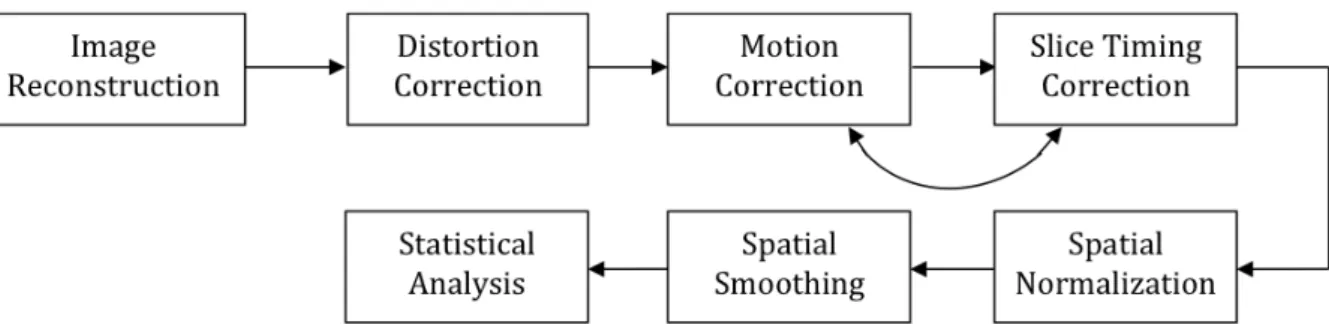
FIGURE 2.1–SCHEMATICS OF THE PREPROCESSING PIPELINE ... 14
FIGURE 2.2–BOLD TIME COURSE OF AN EXAMPLE SUBJECT ... 16
FIGURE 2.3– SHCEMATICS OF THE PRINCIPLES OF SEED-BASED FUNCTIONAL CONNECTIVITY ANALYSIS ... 19
FIGURE 3.1–HEAT MAP RESULTING FROM THE TACTILE STIMULATION OF THE LEFT HAND ... 28
FIGURE 3.2–HEAT MAP RESULTING FROM THE TACTILE STIMULATION OF THE RIGHT HAND ... 28
FIGURE 3.3–HEAT MAP RESULTING FROM THE LANGUAGE COMPREHENSION PARADIGM ... 29
FIGURE 3.4-HEAT MAP RESULTING FROM THE TENNIS PARADIGM ... 34!
FIGURE 3.5-HEAT MAP RESULTING FROM THE SPINNING SPIRALS PARADIGM ... 35!
FIGURE 3.6-HEAT MAP RESULTING FROM THE CHECKERBOARD PARADIGM ... 36!
FIGURE 4.1-MIGRAINES ... 40!
FIGURE 4.2-MINOR STROKE ... 41!
FIGURE 4.3-TIA ... 42!
FIGURE 4.4-LESION MASK AND OPPOSITE HEMISPHERE SEED MASK OVERLAID ON TOP OF THE NORMALISED STRUCTURAL T1 ... 47
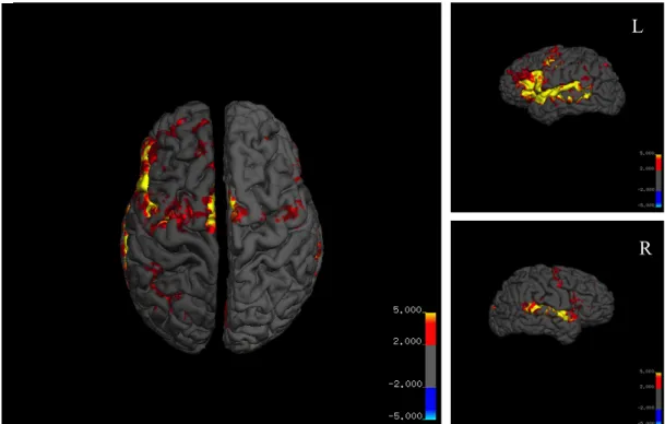
FIGURE 4.5-INCREASED CONNECTIVITY FOR MIGRAINE PATIENTS WHEN COMPARED WITH HEALTHY VOLUNTEERS USING WGBC ... 50!
FIGURE 4.6-WEIGHTED GLOBAL CONNECTIVITY MAP OF ONE EXAMPLE SUBJECT ... 51!
FIGURE 4.7-DIFFUSION WEIGHTED IMAGES (DWI) PRESENTING WITH LESIONS FOR TWO REPRESENTATIVE PATIENTS ... 52!
FIGURE 4.8-LESION MASK DRAWN ON TOP OF THE DIFFUSION WEIGHTED IMAGING SCANS ... 52!
FIGURE 5.1-GLASS BRAIN AND DESIGN MATRIX RESULTING FROM THE STATISTICAL TESTING OF THE CONNECTIVITY MAPS RESULTING FROM THE USE OF A RIGHT CEREBELLUM ROI ... 64
FIGURE 5.2-SIGNIFICANT DECREASES IN FUNCTIONAL CONNECTIVITY AFTER EXERCISE BETWEEN THE HIPPOCAMPUS AND THE CEREBELLAR VERMIS ... 66
FIGURE 5.3-DECREASED FUNCTIONAL CONNECTIVITY BETWEEN THE PCC AND THE LEFT PARIETAL LOBE ... 64
FIGURE 6.1-LOW COGNITIVE PARADIGM PRESENTED TO THE PARTICIPANTS WITH PICTURES OF A) BUILDINGS AND B) LANDSCAPES.ONCE THE BUTTON WAS PRESSED A MESSAGE SAYING “BUTTON PRESSED” WOULD BE DISPLAYED ... 79!
FIGURE 6.2-28 COMPONENTS IDENTIFIED AS RSN ... 83!
FIGURE 6.3-SIMILARITY MATRIX RESULTING FROM THE COMPUTATION OF PEARSON'S CORRELATION OF ALL THE VOXELS WITHIN THE TEMPLATE ... 90!
FIGURE 6.4-PLOT OF THE PEARSON CORRELATION COEFFICIENT OBTAINED FOR THE UNIQUE CORRELATIONS .... 96!
FIGURE 6.5-PLOT OF THE SPEARMAN CORRELATION COEFFICIENT OBTAINED FOR THE UNIQUE CORRELATIONS .. 96!
FIGURE 6.6–PLOT OF THE AVERAGE ICC VALUES FOR EACH NETWORK ... 98
FIGURE 6.7–WMH ... 100
xvi
FIGURE 6.9–PROEMINIENT CISTERNA MAGNA ... 101
!
!
!
!
!
!
!
List of Tables
!
TABLE 3.1–PARADIGM DESCRIPTION FROM MISS SUSA MERZ DOCUMENTATION ... 29
TABLE 3.2–BRIEF DESCRIPTION OF PARTICIPANT EXCLUSION AND REASONS ... 31
TABLE 4.1–SUMMARY OF THE MOST COMMON DIAGNOSIS AND THEIR PROGNOSTIC ... 41
TABLE 4.2–MINOR STROKE PATIENT’S INFORMATION REGARDING LESION’S SIZE AND LOCATION ... 47
TABLE 4.3–INFORMATION REGARDING CLUSTERS LOCATION – WGBC METHOD ... 54
TABLE 5.1–REGIONS OF INTEREST USED FOR SEED BASED CONNECTIVITY ANALYSIS ... 66
TABLE 5.2–INFORMATION REGARDING CLUSTER LOCATION – RIGHT CEREBELLUM SEED ... 70
TABLE 5.3–INFORMATION REGARDING CLUSTER LOCATION – LEFT HIPPOCAMPUS SEED ... 70
TABLE 6.1–SCANNING ORDER FOR BOTH BASELINE AND FOLLOW-UP SCANS ... 83
TABLE 6.2–THRESHOLDS FOR THE T-MAPS OF EACH COMPONENT FROM THE ALLEN ET AL. PAPER ... 87
TABLE 6.3–SEED REGIONS USED FOR THE SEED-BASED CONNECTIVITY ANALYSIS ... 90
TABLE 6.4–UNIQUE CORRELATION VALUES FOR EACH COMPONENT AND AVERAGED PER SUBJECT ... 98
TABLE 6.5–ICC VALUES AVERAGED WITHIN EACH COMPONENT ... 100
TABLE 6.6–CLUSTERS OF SIGNIFICANT DIFFERENCES BETWEEN THE CORRELATIO MAPS FROM BASELINE AND
FOLLOW-UP ... 102
!
!
!
!
!
!
xviii
List of Abbreviations
!
3D Three Dimensional
Aud Primary auditory cortex
BOLD Blood Oxygen-level Dependent
BA Brodmann Area
CSF Cerebrospinal Fluid
DMN Default-mode Network
DoC Disorders of Consciousness DWI Diffusion Weighted Imaging
EPI Echo Planar Imaging
FC Functional Connectivity
fcMRI Functional Connectivity Magnetic Resonance Imaging
FEF Frontal eye fields
fMRI Functional Magnetic Resonance Imaging
FOV Field of View
FWE Family wise error
FWHM Full width at half maximum
GE Gradient Echo
GM Grey matter
HF Hippocampal formation
ICA Independent Component Analysis ICC Intraclass Correlation Coefficient
IPS Intraparietal sulcus
LatPar lateral parietal cortex
M1 Primary Motor Cortex
MCS Minimally Conscious State
Mot Primary motor cortex
mPFC medial Prefrontal cortex
MRI Magnetic Resonance Imaging
MT Medial temporal area
NBS Network Based Statistic
PCC Posterior cingulate cortex
RF Radiofrequency
ROI Region of Interest
rs-fcMRI Resting state Functional connectivity Magnetic Resonance Imaging RSN Resting state networks
S1 Primary Somatosensory Cortex
S2 Secondary Somatosensory Cortex
SE Spin Echo
SnPM Statistical non-Parametric Mapping SPM Statistical Parametric Mapping
TE Echo Time
TIA Transient Ischaemic Attacks
TR Repetition Time
V1 Primary Visual Cortex
Vis Primary visual cortex
VS Vegetative State
wGBC Weighted Global Brain Connectivity
xx
!
!
!
!
!
!
!
!
!
!
!
!
!
Chapter 1
Introduction
1.1 Rationale
Research has extensively been developed during the last decade using functional connectivity MRI. This method has proved to efficiently distinguish between healthy and diseased populations and has also provided us with a greater insight into brain mechanisms. Particularly during resting periods, this methodology has raised the interest of researchers and practitioners since it does not require the performance of a particular task. We therefore focus our attention into the different analytical methodologies to perform functional connectivity analysis using different populations and study designs.
1.2 Project Aims
Resting state fMRI has found large application in the study of pathological states where the intrinsic activity was found to be related to the severity of the disease. Regardless of the still early stages of development, the clinical applications of this technique demonstrate great promise. The aim of this dissertation is to investigate functional connectivity in pathological cases as well as connectivity differences arising from exercise.
Nevertheless, the concept of resting state is complex and probably elusive (Morcom, Fletcher 2007a) and up until today there is no established methodology for resting state fMRI studies.
CHAPTER 1. INTRODUCTION
2
It is therefore expected that the reliability of functional connectivity during resting state is affected due to inter-subject and inter-session variability.
Several papers have been published studying the reliability and consistency of resting state fMRI measurements (Wang et al. 2011, Fiecas et al. 2013, Yan et al. 2009, Guo et al. 2012, Shehzad et al. 2009, Patriat et al. 2013) and networks such as the Default-mode network (DMN) (Greicius et al. 2003, Fox et al. 2005, Fransson 2005), when using conditions such as eyes open, eyes closed and passive visual preprocessing tasks. To our knowledge, no study has been published investigating these statistical parameters in the presence of a low cognitive demand task. With the RESTATE study we aim to quantify possible differences and their statistical significance between the two conditions, subjects and scans.
1.3 Methodology
Every study described during the course of this dissertation resulted from the interest to study functional connectivity under different conditions and diseases. All of them went through a process of study design, ethical approval, recruitment and data acquisition.
The author took part in the RESTATE study design, execution and analysis. The following general methodology was applied to all separate studies described in this dissertation.
1. The study started with a literature review of what has been previously done in order to design a suitable study protocol. The literature review provides important clues on how to efficiently outline the purpose of the study regarding what has been previously investigated and what sort of contributions can be achieved to provide better knowledge towards the work of researchers and the general public.
2. A proposal had to be written to the ethical committee in charge of the area in order to obtain ethical approval for the study. In the particular case of the RESTATE study it was designed as an amendment to the ethics application previously submitted for the TIA cfMRI study.
RESTING STATE FMRI EXPERIMENTAL AND ANALYTICAL METHODOLOGY: A FUNCTIONAL CONNECTIVITY ANALYSIS
3. After ethical approval a proposal had to be submitted to the Research and Development (R&D) department and approved before the study could officially start.
4. The recruitment started after approval of the R&D. Patient/volunteers were carefully screened to ensure that they were suitable for the specific research.
5. Data management is of increased importance with studies involving patients. For the studies involving NHS stroke patients all information had to be stored on the main site file at the Aberdeen Biomedical Imaging Centre as well as in a site file located at the Stroke-Unit of the Aberdeen Royal Infirmary (NHS Grampian).
6. At last, the structural scans were screened by a radiologist to identify any possible accidental findings and if these existed they were reported to the participant as well as to their General Practitioner (GP). The data was then processed to obtain the functional connectivity results.
CHAPTER 1. INTRODUCTION
4
1.4 Thesis Outline
This dissertation features a series of connectivity studies developed at the Aberdeen Biomedical Imaging Centre during a period of 9 months. The methodology used to perform functional connectivity MRI analysis is common to all of the separate studies even though the their aims and outcomes vary. Therefore, this dissertation is outlined in a way so that each chapter represents a specific study. The particular purpose, aims, methods and results of each study are discussed in each of these chapters. The studies are organised chronologically as the work was developed.
! Chapter 1. Introduction – This chapter is a brief overview of the structure used throughout the dissertation as well as of the work developed.
! Chapter 2. Background – In order to contextualize the work developed this chapter includes an introduction to MRI, resting state techniques and functional connectivity methodologies. The various scanning procedures used in the studies are also described in this chapter.
! Chapter 3. DoC study - The first study focuses on Disorders of Consciousness (DoC) and how the use of new imaging techniques such as fMRI can help in the assessment of these patients. The contribution of the author to this study was in ascertaining the validity of the paradigms designed to assess consciousness in DoC patients. In order to do so, the author used an in-house built method to quantify the activation induced by the paradigms in a group of healthy volunteers. This analysis was performed to test the paradigm and use the results as a point for improvement.
! Chapter 4. TIA cfMRI study -The second project is focused on the study of stroke patients presenting at the Aberdeen Royal Infirmary and who are given an uncertain diagnosis. This study aims to ascertain the advantages of including a functional MRI sequence into the already established neurovascular MRI protocol.
RESTING STATE FMRI EXPERIMENTAL AND ANALYTICAL METHODOLOGY: A FUNCTIONAL CONNECTIVITY ANALYSIS
! Chapter 5. PECON study- The third study was designed to be an integrating part of the TIA cfMRI study by assessing the effects of an acute session of exercise in functional connectivity using a group of healthy volunteers.
! Chapter 6. RESTATE study - The fourth and last study chapter describes the RESTATE study, which focuses on the study of resting state fMRI acquisition techniques and the investigation of their reliability and reproducibility. The study was designed and put into practice during the course of this internship and this chapter provides a description of the whole process from ethical approval, volunteer recruitment, scanning procedure and equipment, cognitive assessment, the paradigm used, data analysis and results.
! Chapter 7. Closing Remarks – This last chapter describes the achievements accomplished during the course of the internship and their contribution to future research.
Chapter 2
Background
2.1 Magnetic Resonance Imaging
Magnetic Resonance (MR) is an imaging method that uses protons and their magnetic properties to generate an image. This modality takes advantage of the great abundance of the hydrogen nucleus in tissue in the form of water (each water molecule containing two hydrogen nuclei/protons) and particularly in the human body constituting up to 70% to 90% of most tissues. Moreover, the presence of a single positively charged proton in each hydrogen nucleus gives it a relatively large magnetic moment. When exposed to a strong static magnetic field (B0), the majority of the magnetic moments (or spins) of the hydrogen
nuclei will align parallel to the static field. The use of a short oscillating magnetic field at the Larmor frequency (i.e. RF pulse) results in the precession of the net magnetization towards the xy plane. The xy-magnetization will straight after start to diphase, along with the regrowth of the magnetisation in the direction of the external magnetic field (z-direction). Magnetic field gradients are used to cause the nuclei at different locations to precess at different speeds providing us with spatial information. Severe alterations in the tissue water content as well as in their magnetic properties can be found in cases of disease or injuries. MR can not only be used to obtain structural anatomical images and investigate pathologies, but it can also be applied to study organ function, the chemical composition of tissues and to provide an insight into brain activity.
CHAPTER 2.BACKGROUND
8
2.2 Imaging Techniques
2.2.1
Noise in functional magnetic resonance imaging sequences
The noise characteristics of data obtained using functional magnetic resonance imaging (fMRI) can pose a great challenge for the analysis process. The signal under focus represents less than 2% or 3% of the total BOLD (Blood oxygenation level dependent) response, reflecting a very small effect size. The majority of the signal is dominated by physiological noise (Kruger, Glover 2001) and scanner drift (Bianciardi et al. 2009). The noise present in the data has several sources that can be broken down into true noise and unaccounted-for-signal. True noise results from thermal motion of electrons residing inside the bore of the magnet or within the equipment that is used to collect the raw data. A second source of true noise is related with brain physiology. The observed noise in the fMRI data is also the result of various other contributions that can be described as unaccounted-for signal. Head motion, scanner drift, and uncontrolled cognitive activity on the part of the subject are some of the sources for this type of noise. Scanner drift is the result of slow but constant changes in the strength of the magnetic field inside the bore over the course of the scanning session and is modelled during data analysis. The subject under study should ideally lie very still in the scanner since even small movements of the head position can cause movement artefacts. Although these are accounted for and corrected during the preprocessing steps, large head movements can be problematic to correct. Physiological fluctuations resulting from heartbeat and respiration can be corrected by monitoring and recording these and use them as nuisance variables (regressors in the GLM model) during data analysis. Regarding the spontaneous low-frequency BOLD fluctuations, unrelated to the paradigm, and due to unconstrained cognitive activity, these are impossible to correct due to its unpredictable nature regarding time and location and are currently a relevant topic under extensive study.
2.2.2
Echo-Planar Imaging – EPI
Echo planar imaging (EPI) is one of the most used imaging sequences finding application in diffusion, perfusion and functional magnetic resonance imaging (fMRI). The methodology behind this sequence involves the acquisition of all k-space lines in one repetition time and using a single radio-frequency excitation. It provides us with images with decreased motion
RESTING STATE FMRI EXPERIMENTAL AND ANALYTICAL METHODOLOGY: A FUNCTIONAL CONNECTIVITY ANALYSIS
artefacts with a reduced imaging time, providing us with the ability to image rapid physiological processes of the human body.
2.2.3
T
1–weighted images
T1-weighted images, usually known as ‘anatomical scans’, have very good contrast enabling
a great distinction between the different tissue boundaries. In this particular sequence fluids are usually displayed as dark, water-based tissues have a mid-grey colour and fat-based tissues will be displayed very brightly. The studies described during the course of this dissertation use an ultrafast spoiled gradient echo (GE) sequence: T1-3D TFE (Turbo Field Echo), with a small flip-angle (8˚) and a very short repetition time (TR = 8.2 ms). This sequence uses an optimised k-space filling procedure to reduce the acquisition time. However, the drawback of a small flip angle and very short TR is poor T1-weighting. Therefore this GE pulse sequence uses an initial 180 degrees inversion pulse to prepare the magnetization and provide contrast enhancement before starting acquiring data.
2.2.4
T
2–weighted images
T2-weighted images acquired using a spin echo sequence will require a long TR and echo
time (TE), making them more time-consumable than T1-weighted images. When using this
type of sequence the fluids will appear very bright and both water- and fat-based tissues will be displayed on the middle of the grey scale. This is one of the preferred sequences used for pathological scans since it enables the easy distinction of collections of abnormal fluid. Long T2s will provide more signal and thus images obtained using this combination will be brighter
than using short T2s. T2*-weighted images are closely related to T2-weighted ones, having
basically the same contrast. The main difference is due to susceptibility effects responsible for creating field inhomogeneities that speed the transverse relaxation decay. SE sequences can correct for this effect but GE cannot. So GE sequences will result in images with a combined effect of T2 and magnetic field inhomogeneity, being this relaxation known as T2*.
For the TIA cfMRI study the acquired images were T2*-weighted using a GE sequence
CHAPTER 2.BACKGROUND
10
2.2.5
Neurovascular protocol
The consultant neuroradiologist on the site, Dr. Arnab Rana, originally developed the neurovascular protocol used in the TIA cfMRI study.
The local minor-stroke MRI protocol includes: 1) T1 structural image;
2) T2* weighted sequence;
3) Fluid attenuation inversion recovery (FLAIR) pulse sequence; 4) Diffusion-weighted imaging (DWI) sequence.
For the purpose of the TIA cfMRI study an Echo-Planar Imaging functional MRI sequence was also included in the scanning protocol. Each of these sequences has a specific diagnostic value, even though they complement each others.
The T1 structural image is used to identify bleeding, tumours and other structural
abnormalities such as cortical laminar necrosis. The T2* gradient echo structural image will
exhibit an enhanced contrast in the presence of microbleeds therefore is used to confirm intracerebral haematomas. If a heaematoma is diagnosed then the use of blood thinning drugs is not recommended to prevent a potential stroke. The FLAIR sequence is included in this protocol once the inversion recovery nulls the CSF signal resulting in an easier identification of infarcts at the cortical surface compared to the standard T2SE. This sequence also shows
infarcts that are older than the hyper-acute lesion. Finally, DWI will identify infarcted regions as diffusion restriction.
Apart from the structural and the functional sequences, all of the other sequences are acquired in an axial plane. The reason behind this choice is related to an easier diagnose using this particular plane when compared with the coronal or sagittal.
RESTING STATE FMRI EXPERIMENTAL AND ANALYTICAL METHODOLOGY: A FUNCTIONAL CONNECTIVITY ANALYSIS
The neurovascular protocol parameters are as follows:
A gradient-echo echo-planar sequence (EPI) was used to obtain the functional images (used in the posterior functional connectivity analysis) with an acquisition time of 10 minutes (30ms echo time; 2s repetition time; 78˚ flip angle; 96!96 matrix size; 240!240 mm2 field of view; 32 slices; 3.5 mm slice thickness; 1 mm inter-slice gap; SENSE parallel imaging method with two-fold acceleration; 300 dynamic scans; 4 dummy scans). A high-resolution T1-weighted structural scan was also obtained in 5 minutes and 58 seconds, using fast three-dimensional gradient-echo imaging (8.2s repetition time; 3.8ms echo time; 8˚ flip angle; 240!240!125 matrix size; 240!240!160 mm3 field of view; 1.0!1.0!1.0 mm3 voxel size). A
T2*-weighted image was acquired with 706 ms repetition time; 16.11ms echo time; 18˚ flip
angle; 232!229!131 matrix size; 230!131!182 mm3 field of view; 4.50!4.5!4.5 mm3 voxel size; 24 slices; slice thickness 4.5 mm. The parameters used to obtain the FLAIR images are 11000ms repetition time; 125 ms echo time; 120˚ refocusing pulse; 0.65!0.87 mm2 voxel size; 29 slices; 4 mm slice thickness; Finally, a diffusion-weighted imaging (DWI) scan was acquired in 1 min and 30 s (b value of 1000; 152!152 matrix size; 230x230 mm2 field of view; 24 slices; 4.5 mm slice thickness; 1 mm inter-slice gap; SENSE parallel imaging method with two-fold acceleration).
CHAPTER 2.BACKGROUND
12
2.3 Resting state fMRI
During periods of resting wakefulness the human brain presents spontaneous, low frequency, fluctuations of the blood oxygenation level dependent (BOLD) signal. In 1995 Biswal and colleagues(Biswal et al. 1995) published the results of a study that focused on explaining the meaning of these oscillations. The authors first identified a region of interest in the left somatosensory cortex by using a traditional fMRI experimental design during which the subjects were performing bilateral finger tapping. The same subjects were posteriorly scanned during a period of rest, without performing any sort of cognitive, motor or language task. The results reflected high correlation between the seed region in the left somatosensory cortex and the homologous areas in the contralateral hemisphere.
One of the major motivations for studying spontaneous activity in the brain focuses in understanding brain energy metabolism systems. The brain represents only 2% of total body mass but is responsible for the consumption of 20% of body’s energy (Raichle, Mintun 2006). Task related increases in neuronal metabolism are usually small (<5%) when compared with its large resting energy consumption(Raichle, Mintun 2006).
The first studies identifying the presence of spatial patterns with coherent signal fluctuations in the human brain were performed in the late 90’s and early 2000 using both fMRI (Biswal et al. 1995, Lowe, Mock & Sorenson 1998) and positron emission tomography (PET) (Friston et al. 1993, Shulman et al. 1997, Raichle et al. 2001a). These patterns have been named “intrinsic connectivity networks” (Seeley et al. 2007) or “resting state networks” (RSNs) (Greicius et al. 2003). These RSN are located in grey matter regions and several studies have suggested the importance of some of these networks supporting core perceptual and cognitive processes therefore strengthening the hypothesis that these reflect functional systems with intrinsic energy demands. The neuron population enclosed within each networks is thought to be firing together with a common functional purpose. The patterns displayed by RSN are reliable and reproducible across a range of analysis techniques, conscious states and both in an individual subject and group level (Greicius, Menon 2004, Damoiseaux et al. 2006, Shehzad et al. 2009).
These BOLD signal fluctuations are intrinsically generated by the brain and do not happen as a result of an input from the outside environment (e.g. being asked to perform a task) or an output (e.g. performing the task). Therefore, fMRI studies of spontaneous activity attempt to
RESTING STATE FMRI EXPERIMENTAL AND ANALYTICAL METHODOLOGY: A FUNCTIONAL CONNECTIVITY ANALYSIS
minimize changes in sensory input and ask the participants to refrain from making any specific cognitive task. This scanning protocol has been commonly named ‘resting state fMRI’ since subjects are usually instructed to simply lie in the scanner and refrain from falling asleep. However, there is still some debate on whether the low frequency BOLD fluctuations observed during ‘resting state’ represent true intrinsic neuronal activity or are just the result of ‘mind wandering’ and conscious mentation (Morcom, Fletcher 2007b). Up until today, most of the studies performed seem to suggest that although spontaneous individual behaviour is likely to contribute to resting state BOLD fluctuations, this is unlikely to be the main source of the signal. Supporting this hypothesis are the studies reporting similar spatial location of BOLD correlations across different behavioural states, including different resting conditions, task performance, sleep and even anaesthesia. Furthermore brain activity evoked by the performance of a task seems to be distinct from and only superimposed on the underlying spontaneous activity. A 2006 study performed by Nir and colleagues (Nir et al. 2006) reported that spontaneous cognition, such as mental imagery, results in patterns of neuronal activity in visual regions that are distinct from the patterns observed in spontaneous activity. These studies support the idea that unconstrained behaviour experienced by the participants inside the scanner will result in BOLD modulations that are in addition to, and not the source of, spontaneous coherent BOLD fluctuations. Therefore one can divide the spontaneous BOLD oscillations into two different components: resulting from unconstrained behaviour and the intrinsic activity underlying the first and persisting across different states (Fox, Raichle 2007).
Two very important data analysis issues should be considered when studying resting state data: how to account for non-neuronal noise and how to identify spatial patterns of spontaneous activity. As mentioned in the previous section, non-neuronal components (e.g. cardiac and respiratory activity) can be measured during data acquisition and removed from data through linear regression. Independent component analysis is a technique that can be used to isolate noise sources from the BOLD data itself, along with the regression of signals that are common to all the voxels (e.g. the global signal) or signals from regions that are likely to have a high degree of physiological noise compared to the amount of neuronal activity (e.g. ventricles or white matter).
In the early days of resting state analysis the attention was focused on how this state could potentially represent a baseline for comparisons with activation studies. The
CHAPTER 2.BACKGROUND
14
acknowledgement of a true baseline would provide a new tool for block-design fMRI experiments as well as the correction of the influence that this baseline might have on the signal under study. However the lack of agreement on whether ‘resting state’ represents a true neuronal baseline or just a physiological response has moved the current functional connectivity studies to focus on the dynamic properties of these low frequency oscillations which can provide new clues to the mechanisms underlying brain function.
2.3.1
Resting state networks
The low frequency BOLD fluctuations observed during the ‘resting state’ (i.e., in the absence of an external stimulation task) and described in the previous section were not only found to be reproducible but also to show temporal correlations between different areas of the brain. To these distant regions that are thus hypothesised to be working on the same process, we give the name of resting state networks. The most studied and reproducible network is the default-mode network (DMN) that is thought to be involved in memory consolidation and keeping a certain level of awareness even when resting.
The first evidence for the default-mode hypothesis came from a PET study carried out by Raichle et al. in 2001(Raichle et al. 2001a). In this study the volunteers were asked to rest quietly with their eyes closed. It was found that consistent regions of the brain were active at rest but decreased their activity when cognitive tasks were performed. The authors then suggested the existence of an organised, baseline default mode network (DMN) of brain function. Not long after, Greicius et al., 2003(Greicius et al. 2003)firstly identified the DMN using functional MRI. A study performed by the same team also revealed that even thought this network is affected by the performance of cognitive tasks (presenting a decreased magnitude) its activity persists throughout both experimental and rest periods if the experiment is not sufficiently challenging(Greicius et al. 2004).
Numerous studies(Greicius et al. 2003, Shulman et al. 1997, Mazoyer et al. 2001)hypothesise the existence of two large opposing networks in the brain: the “task-negative” including the DMN and the “task-positive” composed by networks involving attention and task-based systems such as the somatosensory or visual.
RESTING STATE FMRI EXPERIMENTAL AND ANALYTICAL METHODOLOGY: A FUNCTIONAL CONNECTIVITY ANALYSIS
Several other resting state networks have been identified up until today and investigation suggests the existence of at least 6 consistent networks: Visual, Auditory, Dorsal and ventral attention, default-mode network, somatosensory network and frontoparietal network. Although there is no agreement on the literature regarding neither the names nor their divisions the existence of significantly agreeable resting state networks is already an established fact.Once again, these patterns of activated regions are consistent across subjects, states of cognitive development, degrees of consciousness, to some degree even across species, and under pharmacological manipulation.
CHAPTER 2.BACKGROUND
16
2.4 fMRI analysis
After data acquisition and before model estimation the data has to be preprocessed. During this stage the images are realigned with each other, the functional scans are co-registered to the structural image and there is usually also a normalization step that ensures that all the brains are in the same image space. Figure 2.1 summarises the most common preprocessing pipeline procedure.
We designate as ‘native space’ the original coordinate system as the images were acquired from the MRI scanner. The brain of different individuals will not necessarily line up in the native space due to different brain sizes, and even the same individual will have his or her brain in a different position during different scans. Therefore there is a need for a standardised/stereotactic space to register the images with. The studies performed and described on the forthcoming chapters will use the MNI (Montreal Neurological Institute) stereotactic coordinate space by default.
Motion correction (realignment and reslice) is another step that has to be performed during fMRI data analysis. The studies described in this dissertation used a rigid-body spatial transformation model (with six-parameters), which assumes that the effects of motions do not change the shape of the brain, just its position and orientation.
Slice timing correction is a process that is not implemented in all studies. Nonetheless, slice timing correction is important and it is used because different slices are acquired in sequence therefore the BOLD signal is sampled at different time points in different parts of the brain. One would ideally like to have the signal for the whole brain at the same time point. This correction method works at the voxel level examining the time course and shifting it by a
RESTING STATE FMRI EXPERIMENTAL AND ANALYTICAL METHODOLOGY: A FUNCTIONAL CONNECTIVITY ANALYSIS
small amount and using interpolation with the points actually sampled in order to obtain the time course we would have if we sampled all the voxels at the same time.
Normalization of data with the presence of lesions can prove to be difficult due to existence of areas with missing signal. This can result in substantial lack of information to perform a correct spatial normalization. The standard way to deal with this problem is to use cost function masking, in which a portion of the image (in this case the area with the lesion) is excluded from the cost function computation during registration. For the particular case of the patients involved in the fcMRI study of a stroke population (Chapter 3) the participants only presented with small lesions such as minor ischaemic attacks. As it has been described before, the normalization of standard stroke lesions would use a lesion masking procedure, however, the lesions in the dataset of chapter 3 are of a relatively small size and have proven not to interfere with the normalisation of the data.
As mentioned before artifacts should always be corrected during data preprocessing. When carrying out connectivity analysis one has to be extremely careful with artifacts such as head motion since these can cause spurious connections between regions of the brain. The typical nuisance trends considered to be removed from the resting state fMRI data include the six motion parameters, the average signal from white matter regions, ventricular signal, global mean signal, and physiologic signals such as the heart rate and respiration (if available) (Cohen et al. 2008, Fox et al. 2005). Opinions differ on the use of global signal regression and whether this step should or not be implemented. The main reason supporting the use of global signal regression is related to information on non-neuronal signal artifacts (such as components related to motion and respiration) that are encompassed in this variable. Nevertheless recent studies discourage the use of this variable as a regressor. A more detailed overview about this topic can found in the Chapter 5 of this dissertation.
When using spatial smoothing we assure a more homogeneous signal since the data in one voxel is averaged with its neighbours. It is important to avoid using too much smoothing otherwise most of the information can also be lost. It is a fine compromise and the usual values for smoothing range between a Gaussian kernel of 4 and 8 mm.
The last step of fMRI analysis is the statistical testing of the results obtained with the aim of testing/validating a hypothesis. This procedure usually involves the use of t-tests and will provide a valid method to verify if for e.g. two conditions elicit a different brain response or
CHAPTER 2.BACKGROUND
18
2.5 Functional connectivity MRI (fcMRI)
The analysis of brain connectivity can be divided into three types: Anatomical connectivity (AC), Functional Connectivity (FC) and Effective Connectivity (EC). Anatomical connectivity, as the name implies, is the study of the anatomical connections between different brain regions. On the other side, functional connectivity assumes the possibility of an anatomical pathway but is mainly focused on the study of temporal correlations in BOLD fluctuations between different brain areas. Finally, effective connectivity is the specific study of causal influence between at least 2 regions, and the direct and/or indirect influence that these regions exert upon each other (Varsou, Macleod & Schwarzbauer 2013). Apart from their fundamental differences these types of connectivity are usually found to interact with each other. Hence the measured connectivity may be the result of a combination between anatomical, effective and functional networks. The studies in this dissertation are based on functional connectivity and therefore any future references on brain connectivity will relate to this specific type of connectivity.
As mentioned before, functional connectivity targets the study of temporal correlations between different brain areas in the fMRI data. This is a non-invasive technique used to study the large-scale neural networks in the brain (Bullmore, Sporns 2009, Salvador et al. 2005, Zhang, Raichle 2010).
Low-frequency spontaneous oscillations in the BOLD signal are correlated over time between regions within the same working brain systems. Figure 2.2 depicts the BOLD time course of an example subject measure in (top) both left (green) and right (red) primary motor cortex where can clearly be seen a pattern of correlated fluctuations; and (bottom) the absence of correlation between motor (green) and visual (blue) regions.
RESTING STATE FMRI EXPERIMENTAL AND ANALYTICAL METHODOLOGY: A FUNCTIONAL CONNECTIVITY ANALYSIS
Functional connectivity MRI applied during ‘resting state’ has proved to be a good segregation method between health and diseased population. Resting state fMRI has also shown that neuronal network patterns change with increasing age(Meunier et al. 2009) and it has just started to be used in the study of the neonatal and fetal development (Schöpf et al. 2012). On the other hand, the resting state networks on their own have also provided clues regarding brain function. The DMN has shown to be affected by ageing (Damoiseaux et al. 2008) and disrupted in several neuropsychiatric disorders such as mild cognitive impairment (Sorg et al. 2007), Alzheimer’s disease (Greicius et al. 2004, He et al. 2009), schizophrenia (Calhoun, Eichele & Pearlson 2009, Zhou et al. 2007), depression (Greicius et al. 2007), and autism (Kennedy, Redcay & Courchesne 2006, Müller et al. 2011). Regarding autism spectrum disorders the study of resting state activity has shown that multiple networks are affected by this condition presenting each presenting some sort of dysfunction. Depressive disorder studies show some evidence for abnormal hyperconnectivity, moreover the study published by Perrin et al. (Perrin et al. 2012) analysed depression patients before and after electroconvulsive therapy (ECT) treatment and reported a decreased connectivity in the left dorsolateral prefrontal cortex after treatment. The patients included in the study had reported improvement in symptom intensity after the ECT treatment, which was found to be correlated
Figure 2.2 - BOLD time course of an example subject: top – time course of two regions located in the
left (green) and right (red) primary motor cortex; bottom – time course of two regions located in the left (green) primary motor cortex and the left (blue) primary visual cortex.
CHAPTER 2.BACKGROUND
20
with the decreased connectivity pattern identified. Patients suffering from Alzheimer’s exhibit abnormal neuronal patterns as well as Parkinson’s (Wu et al. 2009) (connectivity disrupted in motor areas), epilepsy and multiple sclerosis.
Some of the most widely used methodologies to investigate functional connectivity in fMRI data include seed-based analysis, independent component analysis (ICA) and graph theory methods. Graph measures were not used in any of the studies described in this dissertation so they will not be described any further. The two forthcoming sections (Section 2.5.1 and Section 2.5.2) will give a brief overview of the theory behind seed-based and weighted global connectivity measures as well as of their strengths and weaknesses. Section 2.5.3 will focus on independent component analysis.
It is important to note that subtle changes in the analytic approach of resting state data, for example using slightly different spatial seeds in seed-based correlation, or altering the model order dimensionality estimation in ICA, can have a significant impact on the spatial characteristics of the RSNs identified.
2.5.1
Seed-correlation analysis
Seed-based correlation mapping is a methodology based in extracting the BOLD signal time course from a “seed” region of interest (ROI). The time courses from all the other voxels in the brain are also extracted and a correlation measure is computed between these and the seed’s time course (Figure 2.3). The signal from the seed region is either averaged before computation of the correlation measure or after by averaging the correlation values of the voxels inside the ROI. The Pearson product-moment correlation method is the most widely used measure of functional connectivity. Nevertheless, when using this method one has to carefully remove the non-neuronal contributions (such as head movement or any other confounding variables) insofar as is possible to ensure that the calculated correlations truly reflect neuronal activity. In addition, low pass filtering of the data should be performed under the assumption of temporal sample independence, to address the high noise content of fMRI data. This step attempts to minimise artificial correlation emanating from noise processes such as synchronised cardiac and respiratory signals (Birn et al. 2006). The physiological noise resulting from respiratory and cardiac function is concentrated at relatively high frequencies (>0.1Hz) (Cordes et al. 2001a, Lowe, Mock & Sorenson 1998, Thomas,
RESTING STATE FMRI EXPERIMENTAL AND ANALYTICAL METHODOLOGY: A FUNCTIONAL CONNECTIVITY ANALYSIS
Harshman & Menon 2002) while scanner drift is localised to frequencies bellow 0.01Hz. Signal resulting from spontaneous neuronal activity is mostly confined to frequencies between 0.01Hz – 0.1Hz (Biswal et al. 1995, Cordes et al. 2001a, Demirci et al. 2009, Salvador et al. 2005). Therefore the majority of spontaneous BOLD studies perform low-pass filtering of the data at a cut-off of 0.08 or 0.1Hz.
The primary advantage of seed correlation analysis (SCA) over other methods is that this approach provides straightforward, sensitive and easy to interpret results, showing the network of regions most strongly functionally connected with the seed voxel or region of interest. SCA is an attractive approach for many researchers due to its inherent simplicity, sensitivity and ease of interpretation. In 2009 Shehzad et al. (Shehzad et al. 2009) published a study with an assessment of the test-retest reliability of the most widely used connectivity measures and SCA has proved to provide moderate to high reliability in identifying RSN connectivity relationships.
Nonetheless, this method requires a priori assumptions regarding seed location, which in turn can be considered to bias the connectivity findings towards specific, smaller or overlapping sub-systems, rather than the larger, distinct networks. Moreover, the fundamental problem resides in the fact that there are as many possible networks to be derived, as there are possible seeds. Some seeds are regarded as being very reliable (e.g. posterior cingulate cortex) in determining specific networks (e.g. DMN). However small changes in the seed coordinates can prove to completely distort the resulting connectivity map.
Figure 2.3 - Schematic of the principles of