1415
LIFE TABLE ANALYSIS OF THE PERFORMANCE OF APHID SITOBION AVENAE
(HOMOPTERA: APHIDIDAE) NYMPHS EXPOSED TO A STATIC MAGNETIC FIELD
JUAN HE, HUAN-HUAN GAO, ZHU CAO, WALTER MONIKA and HUI-YAN ZHAO*
Institute of Plant Protection, College of Plant Protection, Northwest Agriculture and Forest University, State Key Laboratory of Crop Stress Biology in Arid Areas, Yangling, Shaanxi 712100, China
Abstract - Using the age-stage two-sex life table, this work was undertaken in order to determine the efect of static mag-netic ields (SMFs) at two lux densities (0.176T and 0.065T) applied at increasing times of duration (0.25, 0.5, 1 and 2 h) on the development, fecundity and reproduction of the aphid, Sitobion avenae. Exposed nymphs had a statistically signiicant shortened irst instar period and adult longevity and prolonged fourth instar periods compared to controls. here were signiicant diferences in the population parameters for two exposure combinations, 0.176T for 0.5 h and 0.065T for 1 h. he intrinsic rate of increase (r), net reproductive rate (R0) and mean generation time (T) were 0.1165, 3.5 and 11.7 days, respectively, 0.176 T for 0.5 h and -0.0198, 0.7 and 11.8 days, respectively, 0.065T for 1 h. We therefore recommend using the age-stage, two-sex life table to study the efect of the static magnetic ield on development and growth of the aphid,
Sitobion avenae.
Key words: Life table, static magnetic ield (SMF), Sitobion avenae
INTRODUCTION
he magnetic ield (MF) has been studied for bio-medical use for some time. Technological develop-ment has led to an overwhelming increase in the presence of electromagnetic ields (EMF), with many harmful efects increasingly observed in living sys-tems.
Many authors have studied the efect of a mag-netic ield on living systems in laboratory condi-tions. here have been reports about the inluence of a magnetic ield on the behavior of unicellular organisms (Frankel, 1984), enzyme reactions (Nos-sol et al., 1993), replication and transcription mecha-nisms (Goodman et al., 1987), mutations (Giorgi et al., 1992) and higher systems (Gould, 1980). Fun-damental processes (growth, development,
orienta-tion) were also studied in living systems, including mortality (Ramirez et al., 1983), wing development (Stamenković-Radak et al., 2001), behavior and me-tabolism (Pan et al., 2004) etc.
MATERIALS AND METHODS
Plant source and insects
he wheat used for feeding the aphids (Triticum aestivum Linn) was planted in a plastic pot (9×9×10 cm) illed with nutrient substrate (a mixture of soil, sand and soil-peat compound substrates) in a green-house at 15-18°C with, and exposed to a 16 h pho-toperiod, using artiicial light of 800 lux. he host plant at stages 12 to 13 was used to rear the aphids (Zadoks et al., 1974). Ten wheat seeds were planted in each plastic pot (9×9×10 cm).
he organisms used in this study were obtained from a colony of S. avenae collected in the Insect Ecology and Integrated Pest Management Laborato-ry in Northwest A&F University, Yangling, Shaanxi, China, in April 2010, and maintained in the labora-tory. One wingless adult aphid was reared on wheat plants for 4-5 consecutive generations at 21 ± 0.5°C temperature, 75 ± 5% RH (relative humidity), and a photoperiod of 16:8h (L: D) in a climate-controlled chamber. he adults began parthenogenetic repro-duction and the population at that time was a mono-clonal colony (Du et al., 2007).
Physical treatment
A moderate static magnetic ield was used. he ield gradient and exposure time were determined by a previous experiment with 0.176 T and 0.065 T for 0.25 h, 0.5 h, 1 h and 2 h (He et al., 2012). Glass Petri dishes with treated aphids were placed on the center of the magnet’s surface. One 24-nymph cluster was taken from the monoclonal colony: 8 nymphs were exposed to control, 8 to 0.065 T and 8 to 0.17 T for 0.25 h. Another set of 24 nymphs from the mono-clonal colony was similarly exposed but for 0.5 h, a third set of 24 nymphs was exposed for 1 h, and a fourth set of 24 nymphs similarly taken and exposed for 2 h. Each of the 8 treated nymphs were individu-ally reared in one pot of the plant using a clip cage (0.6 cm in diameter and 0.3 cm in height). All the tested insects were placed at 21 ± 0.5°C temperature, 75 ± 5% RH, and a photoperiod of 16:8h (L: D) in a
climate-controlled chamber. Parallel control experi-ments were performed with the samples not being exposed. he experiment was repeated three times with a new monoclonal colony under the same con-ditions.
Life history
It is known that four instars occurred in S. avenae ontogenesis. he newborn exposed nymphs were considered instar zero and the irst time of S. ave-nae molting was recorded to irst instar, followed by the second, third and fourth instar. Survivability of S. avenae and a time marker of development stage’s onset were evaluated during exposure to an SMF for 24 h. he nymphs exposed to SMFs were observed daily. When the experimental nymphs were in adult age and parthenogenetic, all the newborn nymphs were removed and recorded daily from birth to death. Development times, total pre-oviposition pe-riod (TPOP), age-speciic survival rate (lx), and
age-speciic fecundity (mx) were recorded daily until the
death of all individuals. Development times for each nymphal instar and lifetime fecundity were calcu-lated. All of the time-speciic life table parameters of Sitobion avenae were also used to calculate the fecun-dity, development duration (T), net reproductive rate (R0), innate capacity of increase (r) and inite rate of increase (e) according to the data.
Statistical analysis
he variable developmental rate among individuals was taken into consideration, the age-stage and two-sex life table were used to analyze the life history data (Chi and Liu, 1985; Chi, 1988) by the computer pro-gram TWOSEXMSChart (Chi, 2008). he intrinsic rate of increase (r) was estimated by using the Euler-Lotka formula:
1
0 ) 1 (=
∑
∞ = + − x x x x rm
l
e
with age indexed from 0age-stage distribution and is calculated as
r
R
T
=
(ln
0)
/
.he data on nymphal development, life fecundi-ty and adult longevifecundi-ty were compared by three-way analysis of variance (ANOVA) with SMF intensities, exposure times and replication as factors. Lifetime parameters were compared by two-way analysis of variance with SMF intensities and exposure times as factors. hese data among treatments were com-pared by the Student-Newman-Keuls (SNK) method ater one-way ANOVA, with either SMF intensities or exposure times as a factor. All the statistical analy-ses were processed with SPSS 11.5 statistical package (Lu, 2002).
RESULTS
Multi-way ANOvAs of SMF intensities and exposure time efects on S. avenae
he development periods and life fecundity were not afected by replication for any of the parameters. here were also no signiicant interactions between replication and SMF intensities or exposure times. hus, these data were used in comparison among the SMF intensities and exposure times.
Development periods and fecundity
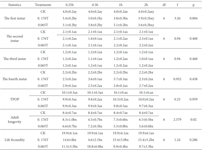
he efects of a magnetic ield on the developmental periods and fecundity are shown in Table 1. As can be seen, there were no diferences between the con-trol and MF groups in the second instar, third instar and total pre-oviposition period (TPOP). here was a signiicant acceleration of Sitobion avenae develop-ment at the irst instar (p = 0.004) while it exhibited shortened adult longevity (p = 0.02) compared with the control. In both the 0.176T for 0.5 h, and 0.065T for 1 h combinations, it was accelerated by 1.0 days and 0.9 days respectively, and shortened by 3.9 days and 3.1 days, respectively.
he prolonged periods of fourth instar and the negative life fecundities were also observed, com-paring with the control group. However, statistical
analysis of the data revealed the absence of signii-cant diferences (the fourth instars p = 0.458; life fe-cundity p = 0.286).
Population parameters
Table 2 shows that the variation of population pa-rameters (the intrinsic rate of increase r, net repro-ductive rate R0, mean generation time T and inite
rate of increase e) were inluenced by the SMFs. All these parameters were statistically diferent between the SMFs and control; but there were no signiicant diferences between exposure times and between SMF intensities. In these cases, the intrinsic rates of increase were 0.1165±0.0424 in 0.176T SMF for 0.5 h and 0.0168±0.0485 in 0.065T SMF for 1 h, all of which were signiicantly lower than the control (0.2257±0.0112). he efect of the SMF on net repro-ductive rate also resulted in a signiicant decrease in 0.176T for 0.5 h (3.5±1.5) and 0.065T for 1 h (0.7±0.3) compared with the control group (19.8±3.1).
Interaction of SMF intensities and exposure times
he statistical analysis exhibited a signiicant difer-ence of interaction between intensities and exposure times (Fig. 1). For the combination of 0.176T for 0.5 h and 0.065T for 1 h, the developmental periods of irst instar and adult longevity were signiicant lower than those of the other combinations; meanwhile the population parameters of the intrinsic rate of increase (r) and net reproductive rate (R0) of these
two combinations were also signiicantly lower than those of the other combinations. he above data are in accordance with the efects established in the de-velopment periods and population parameters.
DISCUSSION
development time, reproduction and adult longevity of S. avenae.
Giorgi et al.(1992) deduced that the MF exposure of D. melanogaster led to an increase in body size and the number of wing hypodermal cells. hey conclud-ed that the MF, as a physical mutagenic agent, could afect some processes during the embryonic and lar-val development of insects. In our experiment, the irst instar of Sitobion avenae was signiicantly short-ened under the SMF exposures, indicating that the
aphid’s embryo was likely inluenced by the static magnetic ields. Because the reproduction type of S. avenae is typically parthenogenesis and viviparous, the embryos had developed completely within the mothers’ ovarioles before the ofspring’s gave birth (Zhao et al., 1992).
Sex ratio and occurrence of sex-linked reces-sive lethal mutations have been reported in D. mela-nogaster by Gotz (1977) in the presence of MF during insect development. Ramirez et al. (1983) recorded
Table 1. Efect of 0.176 T and 0.065 T SMF exposure on the developmental periods of instar stages, total pre-oviposition period and life fecundity (mean±SE) of S. avenae.
Statistics Treatments 0.25h 0.5h 1h 2h df f p
he irst instar
CK 4.0±0.2ay 4.0±0.2ay 4.0±0.2ax 4.0±0.2axy
6 3.26 0.004
0. 176T 3.4±0.2by 3.0±0.1by 3.8±0.3bx 3.9±0.1bxy
0.065T 3.1±0.2by 3.8±0.2by 3.1±0.2bx 3.6±0.2bxy
he second instar
CK 2.1±0.1ax 2.1±0.1ax 2.1±0.1ax 2.1±0.1ax
6 0.94 0.468
0. 176T 2.1±0.2ax 1.8±0.1ax 2.1±0.2ax 2.3±0.1ax
0.065T 2.1±0.1ax 2.1±0.1ax 2.2±0.2ax 2.2±0.2ax
he third instar
CK 1.2±0.1ax 1.2±0.1ax 1.2±0.1ax 1.2±0.1ax
6 0.94 0.468
0. 176T 1.2±0.2ax 1.1±0.1ax 1.2±0.2ax 1.0±0.1ax
0.065T 1.2±0.1ax 1.2±0.1ax 1.2±0.2ax 1.2±0.2ax
he fourth instar
CK 2.2±0.2bx 2.2±0.2bx 2.2±0.2bx 2.2±0.2bx
6 0.952 0.458
0. 176T 2.5±0.2ax 3.6±0.1ax 3.7±0.3ax 2.5±0.2ax
0.065T 2.9±0.2ax 2.5±0.2ax 2.8±0.2ax 2.7±0.2ax
TPOP
CK 10.1±0.3ax 10.1±0.3ax 10.1±0.ax 10.1±0.ax
6 0.25 0.959
0. 176T 9.9±0.3ax 9.8±0.2ax 10.3±0.2ax 10.0±0.2ax
0.065T 9.9±0.3ax 9.9±0.3ax 9.8±0.5ax 9.7±0.3ax
Adult longevity
CK 8.4±0.7ax 8.4±0.7ax 8.4±0.7ax 8.4±0.7ax
6 2.579 0.02
0. 176T 8.3±1.0bx 4.5±0.7bx 7.3±0.6bx 6.5±0.5bx
0.065T 6.6±0.7bx 7.2±0.5bx 5.3±0.8bx 5.6±0.6bx
Life fecundity
CK 19.9±6.1ax 19.9±6.1ax 19.9±6.1ax 19.9±6.1ax
6 1.24 0.286
0. 176T 14.6±4bx 4.6±2.1bx 15.4±5.0bx 15.4±5.2bx
0.065T 11.5±3.5bx 18.8±6.0bx 0.9±0.4bx 9.7±3.3bx
CK – control group; TPOP – Total pre-oviposition period of female counted from birth.
that the total viability of exposed D. melanogaster was lower than the control group. he develop-mental dynamics in the preadult of mealworm was signiicant in mortality and deformities (Tenebrio molitor) when exposed to a constant MF of 0.325 T. In our work, clear diferences were observed in the last instar of the insects and adult longevity. Our re-sults are in agreement with the data of the previously cited authors of Ma and Chu (1993), whose studies reliably conirmed the inluence of ELF-EMF on the processes of D. melanogaster embryo development.
MF also contributed to stimulation of the neu-roendocrine system. his means the inluence of MF on an insect’s neuroendocrine system was produced directly through the nervous system (Klimovskaya and Maslova, 1981) or indirectly through hormone
regulatory pathways (Zagorskaya, 1981; Reiter and Richardson, 1992). Meanwhile, it can be assumed that a physical reaction can promote diferent dy-namics in the insect of an inhibitory or stimula-tive character. he inhibitory level would be an ir-reversible disturbance of the internal equilibrium with the possibilities of biostructural damages, and this change could lead to permanent disturbances throughout the life span (Martin, 1988), even with a lethal outcome (Khalil and Qassem, 1991). How-ever, the simulative level is reversible; the biosys-tem re-equilibrates without consequence ater the MF is removed. hus, an immediate neuroendo-crine efect accelerated the irst instar, returning to a normal physiological condition in the later instar period. Moreover, this change would demand some redistribution of resource and would be quickly
Table 2. Efect of 0.176T and 0.065T SMF exposure on the intrinsic rate of increase (r), net reproductive rate (R0), mean generation time (T) and inite rate of increase (e) (mean±SE) of S. avenae.
Population
pa-rameters Treatments 0.25h 0.5h 1h 2h
r
CK 0.2257±0.0112ax 0.2257±0.0112ax 0.2257±0.0112ay 0.2257±0.0112ax
0.176T 0.2032±0.0161bx 0.1165±0.0424bx 0.196±0.0150by 0.2013±0.0565bx 0.065T 0.1765±0.0199cx 0.2137±0.0650bx 0.0168±0.0485by 0.1808±0.0076bx
R0
CK 19.8±3.1ax 19.8±3.1ax 19.8±3.1ax 19.8±3.1ax
0.176T 14.6±3.0bx 3.5±1.5bx 12.9±2.3bx 12.9±2.0bx
0.065T 9.7±2.4bx 15.8±2.5bx 0.7±0.3bx 8.5±1.9bx
T
CK 13.2±0.2ax 13.2±0.2ax 13.2±0.2ax 13.2±0.2ax
0.176T 13.3±0.4bx 11.7±0.2bx 13.1±0.3bx 12.7±0.2bx
0.065T 13.0±0.4bx 12.9±0.2bx 11.8±0.8bx 12.1±0.3bx
e
CK 1.2531±0.0150ax 1.2531±0.0150ax 1.2531±0.0150ay 1.2531±0.0150ax
0.176T 1.2252±0.0198bx 1.1225±0.0467bx 1.2164±0.0180by 1.223±0.0138bx 0.065T 1.1928±0.0237cx 1.2382±0.0161cx 0.9791±0.0463cy 1.1957±0.0242cx
r R0 T e
Df 6 6 6 6
F 9.171 4.529 2.906 9.5
P 0.000 0.000 0.009 0.000
a,b means within a column sharing the same letter are not signiicantly different in magnetic ield and x,y means within a line sharing the same letter are not signiicantly different in treatment time (a=0.05, Student-Newman-Keuls Test).
surpassed by the simulative SMF exposure. Such inluences may inally lead to negative efects on the longevity of fourth instar nymphs and the female aphids.
In addition, the intrinsic rate of natural increase r is a key demographic parameter that has been used to summarize the qualities of an animal in relation to its capacity to increase (Andrewartha and Birch, 1954). In our study, the statistically signiicant difer-ence of r and R0, of S. avenae between treated groups
and control group could indicate that an external MF changed the aphid’s immature developmental time, progeny production and reproduction.
here is also a dose efect of a magnetic ield on the biological system. It includes time thresholds that investigate in which ield efects are observed only ater a certain time of exposure (Wilson, 1981); transient responses that study in which ield expo-sure induces a biological efect for only a short time ater a change in the exposure (Byus, 1986); and ield threshold efects that appear only when the ield strength exceeds a certain threshold value (Libof , 1984). he static magnetic ields (0.05-0.2T) had an inhibitory impact on the spinal cord neurons in vitro of rat and were also dose-efect related to the inten-sity. We obtained the remarkable efects of two SMF combinations (0.176T 0.5 h and 0.065 T 1 h) that
Fig. 1. he interaction of SMF exposure time and intensity on the developmental periods and population parameters of S. avenae
(p<0.01).
were in agreement with the time and ield thresholds of MF.
Finally, many authors have already demonstrated that MF biological efects were related to the modula-tion of ion low, DNA synthesis and RNA transcrip-tion, interaction of normal cells with the hormones, neurotransmitters, and growth factors (Libof et al., 1984; Stuchly and Esselle, 1992; Frey, 1993; Yim and Jeong, 2006). hus, more detailed examinations of the various molecular mechanisms and SMF-inten-sity are required to explain the mechanisms of static magnetic ield on aphids.
REFERENCES
Andrewartha, H. G. and L. C. Birch (1954). he distribution and abundance of animals. University of Chicago Press, Chi-cago.
Arthur, D. R. (2003). Mechanism of action of moderate- intensity static magnetic ields on biological systems. Cell Biochem. Biophys. 39, 162-173.
Byus, C.v., Pieper, S. e. and W. R. Adey (1986). he efects of low-energy 60-Hz Environmental Electromagnetic ield upon the growth-related enzyme ornithine decarboxylase.
Carcinogenesis 8.
Chi, H. (1988). Life-table analysis incorporating both sex and variable development rate among individuals. environ. entomol. 17, 26-31.
Chi, H. and H. Liu (1985). Two new methods for study of insect population ecology. Bull. Inst. Zool. Acad. Sinica. 24, 225-240.
Chi, H. (2008). TWOSEX-MSChart: computer program for age-stage, two-sex life table analysis. <http://140. 120. 197. 183/Ecology>.
Du, e.X., Zhao, H. Y. and J. W. Guo (2007). UV-induced ecologi-cal response and COI-mutation of Myzus persicae. Journal of Northwest A&F University (Nat. Sci. ed.). 35, 123-126.
Frankel, R. B. (1984). Magnetic guidance of organisms. Rev. Bio-phys Bioeng. 13, 85-103.
Frey, A. H. (1993). Electromagnetic ield interactions with bio-logical systems. Fed Am Soc exp Biol. 7, 272-281.
Giorgi, G., Guerra, D., Pezzoli, C., Cavicchi. S. and F. Bersani
(1992). Genetic efects of static magnetic ields. Body size increase and lethal mutations induced in populations of
Drosophila melanogaster ater chronic exposure. Gene.Sel. evol. 24, 393-413.
Goodman, D. (1982). Optimal life histories, optimal notation and the value of reproductive value. Am. Nat. 119, 803-823.
Goodman, R., Abbott. J. and A. S. Henderson (1987). Transcrip-tional patterns in the X chromosome of Sciara coprophila
following exposure to magnetic ields. Bioelectromagnet-ics. 8, l-7.
Gotz, K. G. and G. Gotz (1977). Normal entwicklung der liege Drosophila in niederfrequenten magnetfeldern. Z. Natur-forsch. 32, 125-132.
Gould, J. L. (1980) he case for magnetic sensitivity in birds and bees (such as it is). Am. Sci. 68, 256-267.
He, J., Gao, H. H., Zhao, H. Y., Monika, W., Hu, Z. Q. and X. S. Hu
(2012). Efect of static magnetic ield on the viability and fecundity of aphid Sitobion avenae (Homoptera: Aphidi-dae) under laboratory conditions. Archives of Biological Science. 64, 693-702.
Kale, P. G. and J. W. Baum (1980). Genetic efects of strong mag-netic ields in Drosophila melanogaster: II Lack of interac-tion between homogenous ields and ission neutron-plus-gamma radiation. envir. Mutagen. 2, 179-l 86.
Khalil, A. M. and W. Qassem (1991). Cytogenetic efects of puls-ing electromagnetic ield on human lymphocytes in vitro:
chromosome aberrations, sister-chromatid exchanges and cell kinetics. Mutat. Res. 247, 141-146.
Klimovskaja, L. D. and A. F. Maslova (1981). Constant magnetic ields and reticular efects on the adrenergic and cholin-ergic system. Kosmitch. Biol. Aviakosm. Med. 15, 74-76.
Libof, A. R., Williams, T. J., Strong, D. M. and R. J. Wistar (1984). Time-varying magnetic ields: Efect on DNA synthesis.
Science. 223, 818-820.
Lu, W. D. (2002). Data analysis with SPSS for windows. Publish-ing house of electronics industry, BeijPublish-ing, China.
Martin, A. H. (1988). Magnetic ields and time dependent efects on development. Bioelectromagnetics. 9, 393-396.
Ma, T. H. and K. C. Chu (1993). Efect of extremely low frequen-cy (ELF) electromagnetic ield on developing embryos of the fruit ly (Drosophila melanogaster L.). Mutat. Res. 303, 35-39.
Nossol, B., Buse, G. and J. Silny (1993). Inluence of weak static and 50 Hz magnetic ields on the redox activity of cyto-chrome-C oxidase. Bioelectromagnetics. 14, 361-372.
Pan, H. J. and X. H. Liu (2004). Apparent biological efect of strong magnetic ield on mosquito egg hatching. Bioelec-tromagnetics. 25, 84-91.
Prolić, Z.M. and v. Nenadović (1995). he inluence of a perma-nent magnetic ield on the process of adult emergence in
Ramirez, e., Monteagudo, J. L., Garcia. M. and J. M. Delgado
(1983). Bioelectromagnetics. 4, 315-326.
Rauš, S., Todorović, D. and Z. Prolić (2009). Viability of old-house borer (Hylotrupes bajulus) larvae exposed to a con-stant magnetic ield of 98 mT under laboratory conditions.
Arch. Biol. Sci., Belgrade. 61, 129-134.
Reiter., R. J. and B. A. Richardson (1992). Magnetic ield efects on pineal indoleamine metabolism and possible biological consequences. FASeB. J. 6, 2283-2287.
Stamenković-Radak, M., Kitanović, I., Prolić, Z., Tomisić, I., Stojković, B. and M. Andjelković (2001). Efect of a perma-nent magnetic ield on wing size parameters in Drosophila melanogaster.Bioelectromagnetics. 22, 365-369.
Stuchly, M. A. and K. P. esselle (1992). Factors afecting neural stimulation with magnetic ields. Bioelectromagnetics. 13, 191-204.
Wilson, B. W., Anderson, L. e., Hilton, D. L. and R. D. Phillips
(1981). Chronic exposure to 60-HZ electric ields: efects on pineal function in the rat. Bioelectromagnetics. 2, 371-380.
Yim, S. H. and J. H. Jeong (2006). Environmental magnetic ields and its biological efect. Kor J Gerontol. 16, 6–10.
Zadoks, J. C., Chang, T. T. and C. F. Konzak (1974). A decimal code for the growth stages of cereals. Weed Research. 14, 415-421.
Zagorskaja, e. A. (1981). Efect of the constant magnetic ield on the endocrine system. Kosmitch. Biol. Aviakosm. Med 15, 14-17.