online | memorias.ioc.fiocruz.br
Despite decades of investigations, tuberculosis and leprosy caused by Mycobacterium tuberculosis and My-cobacterium leprae, respectively, still represent major threats to human health. If M. leprae is a major con-cern in developing countries with an incidence of about 230.000 new cases per year (WHO 2010a), M. tubercu-losis remains the world’s leading cause of death due to a single infectious agent with 1.5 million deaths annually (WHO 2010b). Although these two bacterial species dis-play different tropisms for human tissues, they both trig-ger infections characterized by a systemic dissemination step which requires specific interactions of the pathogen with host cell (Pessolani et al. 2003). The mechanisms responsible for the systemic mycobacterial dissemination remain poorly understood and their deciphering consti-tutes a major challenge since it may lead to the develop-ment of new prophylactic and/or therapeutic strategies. To better understand the molecular interplay between mycobacteria and host cells, we have initiated investiga-tions dealing with the identification and
characteriza-tion of mycobacterial adhesins that interact with human host cells. These studies led us to identify a 22-kDa sur-face-exposed heparin-binding haemagglutinin adhesin (HBHA) which is involved in the interaction of M. tuber-culosis and M. leprae with epithelial cells, but not with professional phagocytes (Menozzi et al. 2006, Lima et al. 2009). In addition, HBHA which binds heparan sulphate-containing receptors through its C-terminal lysine-rich domain (Delogu & Brennan 1999, Pethe et al. 2000) has been shown to be required for extrapulmonary dissemi-nation (Pethe et al. 2001, Mueller-Ortiz et al. 2002). Re-cently, HBHA was shown to trigger receptor-mediated transcytosis, suggesting that mycobacteria may dissem-inate via a direct passage through an epithelial barrier (Menozzi et al. 2006). Besides HBHA, the histone-like protein (Hlp) has also been implicated in the attachment of pathogenic mycobacteria to host cells. Mycobacte-rial Hlp, a positively-charged, surface-exposed molecule with roughly twice the size of other bacterial Hlps, is a highly conserved protein shared by all mycobacterial species (Lefrançois et al. 2011). This protein was initially described as a laminin-binding protein (LBP) involved in M. leprae-Schwann cell (SC) interaction (Shimoji et al. 1999, Marques et al. 2000). More recently LBP/Hlp has been shown to also play a major role in mediating the ad-hesion of mycobacteriato epithelial respiratory cells by interacting with proteoglycan-containing receptors such as heparan sulphate and hyaluronic acid (HA) (Aoki et al. 2004, Lefrançois et al. 2011).
Financial support: FIOCRUZ/PAPES III, INSERM, Institut Pasteur de Lille
+ Corresponding author: [email protected] Received 31 March 2012
Accepted 17 July 2012
Mycobacterial
laminin-binding histone-like protein
mediates collagen-dependent cytoadherence
André Alves Dias1, Dominique Raze2, 3, 4, 5, Cristiana Soares de Lima1,
Maria Angela de Melo Marques6, Hervé Drobecq7, Anne-Sophie Debrie2, 3, 4, 5,
Michelle Lopes Ribeiro-Guimarães1, Franck Biet8, Maria Cristina Vidal Pessolani1/+
1Laboratório de Microbiologia Celular, Instituto Oswaldo Cruz-Fiocruz, Rio de Janeiro, RJ, Brasil 2Inserm, U 1019, Lille, France 3CNRS, UMR 8204 7CNRS, UMR 8525, Lille, France 4Institut Pasteur de Lille, Centre for Infection and Immunity of Lille (CIIL), Lille, France
5Université Lille Nord de France, Lille, France 6Department of Microbiology, Immunology and Patology, Colorado State University,
Fort Collins, USA 8INRA, UR1282, Infectiologie et Santé Publique, Centre de Tours, Nouzilly, France
When grown in the presence of exogenous collagen I, Mycobacterium bovis BCG was shown to form clumps. Scanning electron microscopy examination of these clumps revealed the presence of collagen fibres cross-linking the bacilli. Since collagen is a major constituent of the eukaryotic extracellular matrices, we assayed BCG cy-toadherence in the presence of exogenous collagen I. Collagen increased the interaction of the bacilli with A549 type II pneumocytes or U937 macrophages, suggesting that BCG is able to recruit collagen to facilitate its attach-ment to host cells. Using an affinity chromatography approach, we have isolated a BCG collagen-binding protein corresponding to the previously described mycobacterial laminin-binding histone-like protein (LBP/Hlp), a highly conserved protein associated with the mycobacterial cell wall. Moreover, Mycobacterium leprae LBP/Hlp, a well-characterized adhesin, was also able to bind collagen I. Finally, using recombinant fragments of M. leprae LBP/ Hlp, we mapped the collagen-binding activity within the C-terminal domain of the adhesin. Since this protein was already shown to be involved in the recognition of laminin and heparan sulphate-containing proteoglycans, the pres-ent observations reinforce the adhesive activities of LBP/Hlp, which can be therefore considered as a multifaceted mycobacterial adhesin, playing an important role in both leprosy and tuberculosis pathogenesis.
Because collagenous proteins are the major constitu-ents of the extracellular matrices of epithelial cells and may serve as receptors for bacterial adherence (Kreis & Vale 1993), we have conducted experiments to know whether mycobacteria may directly interact with colla-gen. In the present study, we show that Mycobacterium bovis BCG grown in the presence of exogenous collagen I forms clusters. In addition, cytoadherence assays have revealed that the interaction of M. bovis BCG with pneu-mocytes and macrophages is increased in the presence of exogenous collagen or following a pretreatment of the mycobacteria with collagen. These observations indicated that M.bovis BCG is able to recruit collagen, suggesting the presence of a surface-exposed collagen-binding re-ceptor. This receptor has been purified by affinity chro-matography and demonstrated to be the LBP/Hlp protein. Finally, we mapped the collagen-binding activity of LBP/ Hlp within the C-terminal domain of the protein.
MATERIALS AND METHODS
Mycobacterial strains and growth conditions - M. bovis BCG (strain 1173P2, World Health Organization, Stockholm, Sweden) was grown at 37ºC in tissue cul-ture grade Roux flasks (Nunc) using Sauton medium supplemented or not with type 1 calf skin collagen (Col-lagen S, Roche, Mannheim, Germany) at the concentra-tion of 25 µg/mL. Mycobacterium smegmatis wild-type (wt) mc2155 and the mutant for the hlp gene (Δhlp) were
kindly provided by Thomas Dick of Singapore Univer-sity, Singapore. The mutant was generated as previously described by Lee et al. (1998). Both of the strains were grown in Middlebrook 7H9 broth (Difco, Detroit, MI, USA) supplemented with 10% albumin, dextrose and NaCl (ADC) and 0.05% Tween-80 and 0.5% glycerol un-der agitation. Mycobacteria were acid-fast stained by the Kinyoun method and visualised under light microscope.
Mycobacterial adherence assays - Exponentially-growing M. bovis BCG (OD600 of 0.4) were collected by low-speed centrifugation (6.500 g for 10 min at 4ºC) and carefully resuspended in 50 mM Tris-HCl (pH 7.3) con-taining 150 mM NaCl [Tris buffered saline (TBS)]. The mycobacterial cytoadherence was then assayed as de-scribed previously (Menozzi et al. 1996) using A549 cells (human type II pneumocytes; ATCC, CCL 185) or U937 cells (human macrophages; ATCC, CCL 1593) grown in 24-well tissue culture trays. The adherence assays were performed at a multiplicity of infection of 10 in the ab-sence or the preab-sence of increasing concentrations of type 1 collagen. Following 2 h of incubation at 37ºC, the cells were washed three times with TBS, lysed by adding 1 mL distilled water containing 0.05% (wt/vol) sodium deoxycholate and serial dilutions were plated onto 7H11 medium for colony forming units (CFUs) counting. Cy-toadherence is expressed as the percentage of CFUs pres-ent in the inoculum that remain associated with target cells after the washing step. To investigate the effect of trypsin or collagen pretreatment on the M. bovis BCG cytoadherence, the bacteria were incubated for 30 min. at 37ºC in TBS supplemented with porcine pancreas trypsin (Sigma) or collagen I, respectively. After three washes
with TBS, the bacilli were passed five times through a 28-gauge needle to unravel possible clumps and finally submitted to the adherence assay as described above.
Affinity chromatography on collagen-Sepharose - Collagen I was covalently linked to CNBr-activated Sepharose 4B (Amersham) according to the manufac-turer’s recommendations. Briefly, 5 mL of gel swollen in distilled water was washed with 20 mL 1 mM HCl followed by 200 mL phosphate buffered saline (PBS) (10 mM phosphate buffer pH 7.2, 0.15 M NaCl). The gel was then resuspended in 25 mL PBS containing col-lagen I at the concentration of 1 mg/mL and incubated overnight at 4ºC under gentle agitation. The remaining active groups were blocked by washing the gel with 200 mL 100 mM Tris-HCl pH 8.0. The collagen cou-pling efficiency ranged between 95-99%. Fractions con-taining soluble extracts of M. bovis BCG were prepared as described previously (Menozzi et al. 1996) and chro-matographed at a flow rate of 1.5 mL/min over 5 mL of collagen-Sepharose matrix packed in a glass column (1-cm diameter) and equilibrated with PBS. At the end of the sample loading step, the gel was washed with 100 mL PBS and the bound material was eluted by a 0-1 M NaCl linear gradient in 100 mL PBS. Fractions of 1 mL were collected and their protein concentrations were deter-mined by using the bicinchoninic acid method (Pierce) and bovine serum albumin (BSA) as a standard.
Protein identification - The protein band correspond-ing to the M. bovis BCG collagen-binding protein was excised from the Coomassie-stained polyacrylamide gel, washed twice with 50% acetonitrile prepared in 20 mM ammonium hydrogenocarbonate and finally digested overnight within the gel fragment using 50 ng trypsin (Promega, Madison, WI, USA). The resulting peptides were eluted, desalted with a ZIPTIP C18 column (Mil-lipore, Billerica, MA) and spotted on a Maldi plate with
0.5 µL of freshly dissolved α-cyano-4-hydroxycinnaminic
acid at 5 mg/mL in 50% acetonitrile and 0.1% trifluoroa-cetic acid. After drying, the spots were washed with 3 µL of 20 mM diammonium citrate pH 4.5 and mass spec-trometry analyses were performed by using a matrix-as-sisted laser desorption ionization/time-of-flight Voyager-DE-STR (Applied Biosystems, Palo Alto, CA). Peptides were analyzed by using the following setting parameters: positive and reflector modes, acceleration voltage of 20 kV, grid voltage of 61%, 90 ns of delayed extraction time and low mass gate 500 Da. The spectra were cali-brated externally by using the [M+H+] monoisotopic ions
of peptides resulting from trypsin-digested lyzozyme. Database search based on the peptide masses observed was performed against the NCBInr database using the MS-FIT algorithm (Protein Prospector; 128.40.158.151/ mshome3.4.htm) or the ProFound algorithm (prowl.rock-efeller.edu/profound_bin/WebProFound.exe).
trans-ferred onto nitrocellulose membranes (Protran BA85, Schleicher & Schuell) as described. Immobilized pro-teins were probed with the 5G9 anti-rLBP/Hlp monoclo-nal antibody (1:1000) (Marques et al. 2000). The mem-branes were then washed three times with TBS/T and incubated with a goat anti-mouse alkaline phosphate-conjugated antibody. The substrates nitro blue tetrazo-lium and 5-bromo-4-chloro-3-indolyl phosphate were used for colour development.
Scanning electron microscopy (SEM) - M. bovis BCG grown in Sauton medium to mid log phase in the absence or the presence of 25 µg/mL collagen I were resuspended in PBS (OD600 of ~1.0). The bacteria were then fixed for 5 h at room temperature (RT) in a 1.25% (v/v) glutaral-dehyde solution prepared in 100 mM sodium cacodylate buffer (pH 7.0). A similar fixation step was applied to a collagen I solution at 50 µg/mL prepared in PBS. Next, the samples were filtered through 25 mm diameter/0.2 µm porosity Anodisc (Whatman) and the filters were rinsed five times for 10 min in 25 mL cacodylate buffer. Post-fixation was performed for 3 h in 1% OsO4 solution prepared in cacodylate buffer, followed by five washes in ultrapure water. The samples underwent progressive dehydration by successive soaking in 50%, 70%, 95% and 100% ethanol. Soaking in isopentyl acetate was performed before critical point drying in CO2, using an EMSDCOPE CPD 750 apparatus. The filters were then attached to large SEM stubs and coated with gold/pal-ladium by cathodic spreading in a Polaron E5100 coater. Sample observation and microphotographs were done in a JEOL JSM35CF scanning electron microscope, oper-ating at a voltage of 10 kV.
Preparation of recombinant full-length LBP/Hlp and truncated forms - The recombinant M. bovis BCG LBP/Hlp protein was obtained using the same proce-dure previously described for cloning and expression of the Mycobacterium avium subsp. paratuberculosis LBP/Hlp-encoding gene (Lefrançois et al. 2011). The M. bovis LBP/Hlp-encoding gene (GenBank accession GQ259334) was amplified by polymerase chain reac-tion from chromosomal DNA of M. bovis BCG strain 1173P2 using the Pfu DNA polymerase (Promega) and two synthetic oligonucleotides (Sigma) with the fol-lowing sequences: 5’TATACATATGCACCACCACCA CCACCACATGAACAAAGCAGAGCTCATTGACG -3’ (BCG-LBP/Hlp S) and 5’-TATAGCGGCCGCCT- ATTTGCGACCCCGCCGAGCGG-3’ (BCG-LBP/Hlp AS), containing an NdeI and NotI sites, respectively. The oligonucleotide BCG-LBP/Hlp S sequence was designed to produce a hybrid protein carrying a His-tag at the N-terminal position used for affinity purification of the re-combinant protein. Rere-combinant full-length M. leprae LBP/Hlp (rLBP/Hlp) and truncated forms of the protein corresponding to the N-terminal (residues 1-110; rLBP/ Hlp-N) and the C-terminal (residues 111-200; rLBP/ Hlp-C) were obtained as previously described (Marques et al. 2000, Lima et al. 2005).
Binding assays in microplates of recombinant BCG LBP/Hlp to collagen I - To investigate the capacity of M. bovis BCG rLBP/Hlp to bind to collagen, 1.0 μg/mL of
collagen I (Sigma, St. Louis, MO) in 50 μL 0.1 M car -bonate-bicarbonate buffer (pH 9.6) was used to coat the wells of a polystyrene microplate (Corning, New York, NY). The microtitre plate was incubated overnight at 4ºC. The wells were then washed with PBS and blocked for
2 h with 200 μL PBS-3% BSA at RT. After washing with PBS/0.05% Tween 20 (PBS/T), 50 μL of increasing con -centrations of M. bovis BCG rLBP/Hlp were added to the wells and incubated at 37ºC for 2 h. The wells were then rinsed with PBS/T and incubated with the anti-LBP/Hlp monoclonal antibody 5G9 (1:500) for 1 h at 37ºC. After washing with PBS/T, rabbit anti-mouse IgG peroxidase conjugate (Sigma; 1:1000) was added and incubated for an additional 50 min at 37ºC. Peroxidase activity was re-vealed with hydrogen peroxide and o-phenylenediamine (OPD). The reaction was stopped with HCl and read at 490 nm with a TitertekPlus microplate reader (ICN Bio-medicals Inc, Costa Mesa, CA). Control wells coated with BSA were included in all binding assays.
Mapping the collagen-binding site of M. leprae LBP/ Hlp - To monitor the binding of different types of solu-ble collagens to M. leprae rLBP/Hlp or truncated forms, 5 µg/mL of each protein in 0.1 M carbonate buffer pH 9.6 (50 µL) were used to coat the wells of polystyrene mi-croplates (Corning, New York, NY). Plates were incubated overnight at 4ºC. The wells were then washed with PBS and blocked for 2 h at RT with 200 µL PBS containing 2% BSA. Upon washing with PBS/T, 50 µL of increasing concentrations of biotinylated collagens I, III, IV or VI (Sigma; 0-80 µg/mL) were added to the wells and incuba-tion was performed at RT for 2 h. The wells were rinsed with PBS/T and incubated with streptavidin-peroxidase (Pierce, Rockford, IL) at 0.5 µg/mL. Peroxidase activ-ity was revealed with hydrogen peroxide and OPD. The reaction was stopped with HCl and read at 490 nm in a TitertekPlus microplate reader. In control wells, collagen was omitted and specific collagen-binding activity was determined by subtracting the absorbency resulting from non-specific binding detected in the control wells.
RESULTS
cultures performed in the presence of collagen I (Fig. 1B, Supplementary data). An amorphous and fibrillar material in contact with the bacteria was also observed within these aggregates. Because this material could represent collagen fibres cross-linked by glutaraldehyde during the sample preparation, collagen I was fixed and observed in the same conditions. Collagen appeared as long and entangled filaments exhibiting various thick-nesses (Supplementary data). Since this aspect was quite similar to that of the extracellular material detected within the mycobacterial clumps, it suggests that M. bo-vis BCG may interact directly with collagen fibres.
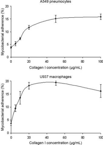
Exogenous collagen I increases the cytoadherence of M. bovis BCG - Because collagen is a predominant con-stituent of the extracellular matrices of epithelial cells (Kreis & Vale 1993), we assayed the cytoadherence of M. bovis BCG in the presence of increasing collagen I concentrations ranging from 0-100 µg/mL. These assays were performed using human type II A549 pneumocytes, but also U937 human macrophages since mycobacteria display a tropism for monocyte-derived phagocytic cells (Stokes et al. 1993, Ernst 1998). As shown in Fig. 2, a dose-dependent effect was observed for both cell lines. Compared with the control, a ca. three-fold increase in adherence was observed with the A549 pneumocytes in the presence of 40 µg/mL collagen I. The use of high-er collagen concentrations did not furthhigh-er increase the mycobacterial adherence, indicating a saturable mecha-nism. The effect of collagen I on the interaction of M. bovis BCG with U937 macrophages was also shown
to be saturable, but it appeared more pronounced since a four-fold increase in adherence was observed in the presence of 20 µg/mL collagen I.
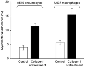
Pretreatment of M. bovis BCG with collagen I in-creases its cytoadherence - In order to investigate the molecular mechanism leading to the increased cytoad-herence of M. bovis BCG in the presence of exogenous collagen I, the bacilli were incubated in TBS containing 50 µg/mL collagen I prior to the cytoadherence assay. At the end of 30 min incubation at 37ºC, the mycobacteria were washed twice with TBS and then assayed for their capability to adhere to A549 pneumocytes and U937 macrophages. As shown in Fig. 3, such a collagen I pre-treatment induced a ca. three-fold increase in the myco-bacterial adherence onto both A549 pneumocytes and U937 macrophages. This observation suggests that M. bovis BCG may recruit exogenous collagen which may in turn favour the attachment of the bacilli to the eu-karyotic cell surface. Such a recruitment was confirmed by the SDS-PAGE analysis of M. bovis BCG pretreated with collagen I followed by three washing steps with TBS. Indeed, the Coomassie blue staining of the gel re-vealed that collagen was present in the whole-cell lysate derived from mycobacteria pretreated with collagen (data not shown). This finding indicates that collagen I binds compounds probably associated with the M.bovis BCG surface. Therefore, these compounds could act as adhesins through their collagen-binding activity.
A proteinaceous M. bovis BCG compound is required for the collagen-dependent adherence - The recruitment of collagen by M. bovis BCG led us to characterize the mycobacterial surface compounds involved in this
activ-Fig. 1: Mycobacterium bovis BCG grown in the absence or the pres-ence of collagen I. Bacilli grown in the abspres-ence (A) or the prespres-ence (B) of 25 µg/mL collagen I were fixed and observed by scanning elec-tron microscopy. The arrow indicates collagen fibres that surround mycobacteria.
Fig. 2: effect of exogenous collagen I on the Mycobacterium bovis
ity. We first assayed the cytoadherence of mycobacteria pretreated for 30 min with porcine pancreas trypsin at the concentrations of 0.1 or 1 µg/mL. These conditions were chosen because neither modified the viability of M. bovis BCG, as observed by CFUs determination following the trypsin pretreatment (data not shown). Pretreatment of the bacilli with trypsin at 1 µg/mL did not significantly modi-fy the mycobacterial interaction with U937 macrophages, suggesting that this interaction does not mainly involve proteinaceous adhesins (Supplementary data). In contrast, such a trypsin pretreatment induced a ca. 60% reduction of the M. bovis BCG attachment to the A549 pneumocytes. This finding is in agreement with the fact that HBHA, which is the major epithelial adhesin produced by both M. bovis BCG and M. tuberculosis, exhibits a surface-ex-posed heparin-binding adhesive domain highly sensitive to proteolytic degradation (Pethe et al. 2002, Dupres et al. 2005). When bacilli pretreated with 0.1 µg/mL trypsin were used in the cytoadherence assay run in the presence of 50 µg/mL exogenous collagen I, the increase in
myco-bacterial adherence to both A549 pneumocytes and U937 macrophages was reduced by ca. 55% compared to un-treated M. bovis BCG assayed in the presence of collagen (Supplementary data). This inhibitory effect of trypsin was confirmed by using M. bovis BCG pretreated with 1 µg/mL trypsin, as shown by the complete abolition of the collagen-induced stimulation of adherence onto A549 pneumocytes and U937 macrophages. This observation suggests that M. bovis BCG surface proteins are involved in the collagen-dependent cytoadherence.
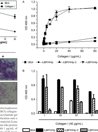
LBP/Hlp binds immobilized collagen I - In order to isolate the M. bovis BCG compounds that bind colla-gen I, exponentially-growing mycobacteria were lyzed by sonication and centrifuged. Then, the centrifugation supernatant corresponding to the soluble material was collected and directly chromatographed onto a colla-gen I-Sepharose matrix equilibrated in PBS. Fractions corresponding to the eluted material were analyzed by SDS-PAGE and immunoblotting (Fig. 4A). Coomassie blue staining of the polyacrylamide gel revealed the presence of a protein exhibiting an apparent molecu-lar weight of ca. 32 kDa. The gel band containing this protein was excised and submitted to trypsin digestion. Mass spectrometry analysis of the resulting peptides re-vealed peptide masses corresponding to the previously described mycobacterial LBP/Hlp. To confirm the colla-gen-binding capacity of BCG LBP/Hlp, the recombinant protein was successfully obtained (Supplementary data) and shown in microplate binding-assays to interact with collagen I (Fig. 4B). Finally, we cultivated M. smegmatis
wt and Δlbp/hlp strains in 7H9 medium in the absence or presence of collagen I at the concentration of 25 µg/ mL. When cultures reached an OD600nm of 1.8, bacteria were fixed, acid-fast stained and visualised under light microscope. Fig. 4C shows that, as expected, the mutant strain was unable to form large clumps, in contrast to the wt strain, indicating a decrease capacity to interact with collagen fibres when compared with the wt strain. These results confirmed the direct interaction between the mycobacterial LBP/Hlp and collagen.
Recombinant M. leprae LBP/Hlp binds collagen - M. leprae LBP/Hlp shares 84% sequence identity with the BCG homologue and it has been characterized as an important adhesin mediating bacterial interaction with host cells (Shimoji et al. 1999, Marques et al. 2000, Lima et al. 2005, Portugal et al. 2008). Next, we then tested the capacity of recombinant M. leprae LBP/Hlp to bind collagen in a solid phase interaction assay. For this purpose, microplate wells were coated with M. le- prae rLBP/Hlp and following a blocking step with BSA, increasing concentrations of biotinylated collagen I were added to the wells. The complexes were finally developed using a streptavidin-peroxidase conjugate. As shown in Fig. 5A, BSA displayed no significant collagen-binding activity for collagen I. In contrast, M. leprae rLBP/Hlp was demonstrated to bind collagen I in a dose-dependent and saturable manner.
Recombinant LBP/Hlp binds different collagen types via its C-terminal domain - The NaCl elution of M. bovis BCG LBP/Hlp from a chromatographic matrix bearing Fig. 3: effect of collagen pretreatment on the Mycobacterium bovis
covalently immobilized collagen I suggested that ionic in-teractions were more likely involved in the adhesin inter-action with collagen. The C-terminal half of LBP/Hlp is highly positively charged and exhibits homology with the eukaryotic class H1 histones (Lee et al. 1998, Prabhakar et al. 1998). In a recent investigation, we have used trun-cated recombinant M. leprae LBP/Hlp molecules corre-sponding to the N-terminal (residues 1-110, rLBP/Hlp-N) or the C-terminal (residues 111-200, rLBP/Hlp-C) do-mains and showed that the C-terminal half of the protein constitutes the major laminin and heparin-binding site of the adhesin (Lima et al. 2005, Portugal et al. 2008). We then tested if the C-terminal domain of LBP/Hlp was the collagen-interacting site of the protein. For this purpose,
we assayed the collagen-binding activity of the M. leprae LBP/Hlp truncated forms which were previously shown to coat microplate wells with identical efficiencies (Lima et al. 2005). Fig. 5A shows overlapping of the collagen I-binding curves obtained with rLBP/Hlp and rLBP/Hlp-C, while no collagen I-binding activity was observed using rLBP/Hlp-N. This finding indicates that the interaction of LBP/Hlp with collagen I is mediated by the C-terminal half of the adhesin. In contrast, rLBP/Hlp-N displayed very limited binding capacity to collagen I. Finally, we next tested the capacity of full-length rLBP/Hlp and trun-cated forms to bind to other collagen types. As shown in Fig. 5B, the rLBP/Hlp and rLBP/Hlp-C were also able to significantly bind collagens III, IV and VI. These results
Fig. 5: Mycobacterium leprae laminin-binding histone-like protein (LBP/Hlp) binds to different collagen types by its C-terminal domain. Microplate wells coated with 5 µg/mL of full-length M. leprae recom-binant-LBP/Hlp, truncated N-terminal (rLBP/Hlp-N) or C-terminal (rLBP/Hlp-C) domains were incubated with increasing concentra-tions of biotinylated collagen I (A) or 40 µg/mL of collagens I, III, IV or VI (B). Binding is expressed in absorbency units at 490 nm. Data represent the mean +/- standard deviation of a typical experiment done in duplicate. Five experiments were performed with similar re-sults. BSA: bovine serum albumin.
Fig. 4A: sodium dodecyl sulphate polyacrylamide gel electrophoresis and immunoblotting analyses of Mycobacterium bovis BCG collagen-binding protein. The Coomassie-blue staining of a polyacrylamide gel containing a clarified lysate of M. bovis BCG prior application onto a collagen-Sepharose matrix (Lane 1) and the salt-eluted material (Lane 2); B: recombinant M. bovis BCG laminin-binding histone-like protein (LBP/Hlp) binds collagen. Microplate wells coated with 1 µg/mL of collagen I were incubated with increasing concentrations of M. bovis
BCG recombinant-LBP/Hlp. The wells were then incubated with the anti-LBP/Hlp monoclonal antibody 5G9 and, finally, with a rabbit an-ti-mouse IgG peroxidase conjugate; C: the Mycobacterium smegmatis
wild-type (wt), but not the Δhlp strain, was able to form large clumps
in the presence of collagen I. M. smegmatis wt and the mutant for the
hlp gene (Δhlp) were grown in complete Middlebrook 7H9 broth
sup-plemented or not with type 1 calf skin collagen at the concentration
of 25 μg/mL. When cultures reached an OD600nm of 1.8, mycobacteria
suggest that, similarly to its interaction with heparin and laminin, LBP/Hlp binds collagen mainly through inter-actions involving the lysine-rich repeats present in the C-terminal domain of the adhesin.
DISCUSSION
Because specific interactions with host cells repre-sent a crucial and early step in any infectious process, pathogenic microorganisms have evolved adhesins ca-pable to bind membrane receptors or compounds present in the eukaryotic extracellular matrices (Westerlund & Korhonen 1993). Pathogenic mycobacteria synthesize a wide panel of adhesins involved in the bacterial interac-tion with epithelial cells and/or monocyte-derived pro-fessional phagocytes (Hoppe et al. 1997, Sidobre et al. 2000, Diaz-Silvestre et al. 2005, Kinhikar et al. 2006, Ragas et al. 2007, Hickey et al. 2010). Several proteina-ceous mycobacterial adhesins have been demonstrated to recognize mammalian extracellular matrix compo-nents. Such an adhesive activity has been demonstrated for the proteins belonging to the antigen 85 complex and the fibronectin attachment protein, which both bind fi-bronectin (Abou-Zeid et al. 1991, Peake et al. 1993, Zhao et al. 1999), the HBHA which interacts with heparan sulphate-containing proteoglycans (Delogu & Brennan 1999, Pethe et al. 2002) and the LBP/Hlp (also called MDP1) that binds laminin, heparan sulphate chains and HA (Shimoji et al. 1999, Aoki et al. 2004, Lima et al. 2005, Lefrançois et al. 2011).
Evidence strongly suggests that LBP/Hlp displays a bi-functional role in the biology of mycobacteria, acting both in nuclei acid metabolism (Prabhakar et al. 1998) and as an adhesin. In this regard, LBP/Hlp was initially characterized as a LBP, mediating the binding and inva-sion of SC by M. leprae (Shimoji et al. 1999, Marques et al. 2000). Recombinant LBP/Hlp was able to avidly bind laminin in solid phase assays and the exogenous addition of the protein significantly enhances bacterial binding to SC (Marques et al. 2000). These data, together with the recent finding indicating that M. leprae actively expresses LBP/Hlp during the human infection (Lima et al. 2005), have reinforced the involvement of LBP/Hlp in the suc-cessful invasion and colonization of the peripheral nerve system by M. leprae. More recently, LBP/Hlp was also shown to play an important role in mycobacteria-lung epi-thelial cell interaction. Both M. tuberculosis and M. bovis BCG were shown to bind and invade A549 human lung epithelial cells through the interaction of LBP/Hlp with HA presence on the cell surface (Aoki et al. 2004).
Collagenous proteins, which are predominant con-stituents of the mammalian extracellular matrix, can also function as receptors for the adhesion of micro-bial pathogens. The collagen-binding micromicro-bial surface components recognizing adhesive matrix molecules (MSCRAMMs) that are synthesized by both Gram-pos-itive and Gram-negative bacteria constitute a large fami-ly of proteins involved in the bacterial attachment to host tissues (Patti et al. 1994). Therefore, it is not surprising that collagen-binding MSCRAMMs may represent im-portant virulence factors such as the CNA, YadA, Cpa and Dr adhesins, which are produced by
Staphylococ-cus aureus, Yersinia enterocolitica, Streptococcus pyo-genes and Escherichia coli, respectively and which play a role in the establishment and progression and/or per-sistence of the infection (Gripenberg-Lerche et al. 1994, Selvarangan et al. 2004, Xu et al. 2004, Kreikemeyer et al. 2005). Besides adhesins that bind collagen, more recently bacterial collagen-like surface proteins have been characterized and shown to play a role in microbial pathogenesis by mediating attachment to host cell col-lagen receptors (Caswell et al. 2008). So far, no studies have reported the interaction of mycobacteria with col-lagen. Here, we show for the first time that mycobacte-ria may interact directly with collagen proteins via the surface-exposed LBP/Hlp adhesin.
Evidence that mycobacteria can interact with collagen was initially obtained from the observation that collagen I was able to induce aggregation of bacterial cells. Images generated by SEM also showed collagen fibres interact-ing with the surface of bacterial cell. This interaction was confirmed by the observation that exogenous collagen was able to increase mycobacterial adherence to human type II A549 pneumocytes and U937 human macrophages in a saturable and dose-dependent manner. An affinity chro-matography performed on collagen I-Sepharose allowed the purification of a collagen-binding component from BCG cell lysate that was identified by mass spectrometry as the LBP/Hlp protein. Furthermore, when produced in the recombinant form, BCG LBP/Hlp protein was able to bind collagen in microplate solid phase assays confirming that LBP/Hlp is also a collagen-binding protein. LBP/Hlp is a highly conserved protein detected on the surface of all mycobacterial species so far investigated (Lefrançois et al. 2011). To show that LBP/Hlp is the major collagen-binding protein present on mycobacterial surface, we ana-lyzed the interaction of collagen fibres with M. smegma-tis Δhlp, the only hlp null mutant so far available in the context of mycobacterial species. This mutant has been characterized and used in several previous studies (Lee et al. 1998, Marques et al. 2000, Biet et al. 2007). In contrast to the wt strain, M. smegmatis Δhlp was unable to inter-act and form large clumps when grown in the presence of collagen I, suggesting that LBP/Hlp represents a major component on bacterial surface mediating microbial in-teraction with collagen.
able to bind very similarly to collagen type I, while inter-action with collagen III, IV and VI was partially medi-ated by the C-terminal domain. These data reinforce the idea that LBP/Hlp C-terminal domain is responsible for most if not all of the adhesive property of this protein.
The definition of the molecules used by M. tuberculo-sis and M. leprae to gain access and colonize different tis-sues of the human body may provide clues for the devel-opment of new therapeutic and/or prophylactic strategies to better control tuberculosis and leprosy. The capacity of LBP/Hlp to bind different collagen types here demon-strated reinforces its potential role as a virulence factor of pathogenic mycobacteria. The binding properties of LBP/ Hlp to multiple extracellular matrix components suggest that this protein may play an important role in distinct moments during the natural course of human infection. As previously suggested, early during infection, LBP/ Hlp might play, in conjunction with HBHA, a key role on bacterial attachment to respiratory epithelial cells (Aoki et al. 2004). We can also speculate on the involvement of LBP/Hlp in bacterial dissemination inside the human body that occurs both in leprosy and tuberculosis (Pes-solani et al. 2003). During this process, the bacilli might need to interact with distinct basal membranes, which are essentially composed of laminin and collagen IV (Kreis & Vale 1993). Finally, LBP/Hlp may also participate of the colonization of vulnerable tissues such as the pe-ripheral nerve system by M. leprae through interaction mainly with laminin (Shimoji et al. 1999, Marques et al. 2000) and the bones and joints by M. tuberculosis, which are rich in collagen I (Kreis & Vale 1993).
ACKNOWLEDGEMENTS
To Dr John S Spencer, from the Colorado State Universi-ty, for providing the 5G9 anti-rLBP/Hlp monoclonal antibody, to Elizabeth Pradel and Yves Dufrêne, for critical reading of the manuscript, to Jean-Pierre Tissier, for the technical as-sistance, and to the memory of Franco Dante Menozzi, who died in July 2005.
REFERENCES
Abou-Zeid C, Garbe T, Lathigra R, Wiker HG, Harboe M, Rook GA, Young DB 1991. Genetic and immunological analysis of Myco-bacterium tuberculosis fibronectin-binding proteins. Infect Im-mun 59: 2712-2718.
Aoki K, Matsumoto S, Hirayama Y, Wada T, Ozeki Y, Niki M, Do-menech P, Umemori K, Yamamoto S, Mineda A, Matsumoto M, Kobayashi K 2004. Extracellular mycobacterial DNA-binding protein 1 participates in mycobacterium-lung epithelial cell inter-action through hyaluronic acid. J Biol Chem 279: 39798-39806.
Biet F, Marques MAM, Grayon M, Xavier da Silveira EK, Brennan PJ, Drobecq H, Raze D, Vidal Pessolani MC, Locht C, Menozzi FD 2007. Mycobacterium smegmatis produces an HBHA homo-logue which is not involved in epithelial adherence. Microbes Infect9: 175-182.
Caswell CC, Barczyk M, Keene DR, Lukomska E, Gullberg DE, Lukom-ski S 2008. Identification of the first prokaryotic collagen sequence motif that mediates binding to human collagen receptors, integrins alpha2beta1 and alpha11beta1. J Biol Chem 283: 36168-36175.
Delogu G, Brennan MJ 1999. Functional domains present in the my-cobacterial hemagglutinin, HBHA. J Bacteriol 181: 7464-7469.
Diaz-Silvestre H, Espinosa-Cueto P, Sanchez-Gonzalez A, Esparza-Ceron MA, Pereira-Suarez AL, Bernal-Fernandez G, Espitia C, Mancilla R 2005. The 19-kDa antigen of Mycobacterium tuber-culosis is a major adhesin that binds the mannose receptor of THP-1 monocytic cells and promotes phagocytosis of mycobac-teria. Microb Pathog 39: 97-107.
Dupres V, Menozzi FD, Locht C, Clare BH, Abbott NL, Cuenot S, Bompard C, Raze D, Dufrêne YF 2005. Nanoscale mapping and functional analysis of individual adhesins on living bacteria. Nat Methods 2: 515-520.
Ernst JD 1998. Macrophage receptors for Mycobacterium tuberculo-sis. Infect Immun 66: 1277-1281.
Gripenberg-Lerche C, Skurnik M, Zhang L, Söderström KO, Toivanen P 1994. Role of YadA in arthritogenicity of Yersinia enteroco-litica serotype O:8: experimental studies with rats. Infect Immun 62: 5568-5575.
Hickey TB, Ziltener HJ, Speert DP, Stokes RW 2010. Mycobacterium tuberculosis employs Cpn60.2 as an adhesin that binds CD43 on the macrophage surface. Cell Microbiol 12: 1634-1647.
Hoppe HC, de Wet BJ, Cywes C, Daffé M, Ehlers MR 1997. Iden-tification of phosphatidylinositol mannoside as a mycobacterial adhesin mediating both direct and opsonic binding to nonphago-cytic mammalian cells. Infect Immun 65: 3896-3905.
Kinhikar AG, Vargas D, Li H, Mahaffey SB, Hinds L, Belisle JT, Laal S 2006. Mycobacterium tuberculosis malate synthase is a laminin-binding adhesin. Mol Microbiol 60: 999-1013.
Kreikemeyer B, Nakata M, Oehmcke S, Gschwendtner C, Normann J, Podbielski A 2005. Streptococcus pyogenes collagen type I-binding Cpa surface protein. Expression profile, I-binding charac-teristics, biological functions and potential clinical impact. J Biol Chem 280: 33228-33239.
Kreis T, Vale R 1993. Guidebook to the extracellular matrix and ad-hesion proteins, Oxford University Press, Oxford, 176 pp.
Lee BH, Murugasu-Oei B, Dick T 1998. Upregulation of a histone-like protein in dormant Mycobacterium smegmatis. Mol Gen Genet 260: 475-479.
Lefrançois LH, Pujol C, Bodier CC, Teixeira-Gomez AP, Drobecq H, Rosso ML, Raze D, Dias AA, Hugot JP, Chacon O, Barletta RG, Locht C, Pessolani MCV, Biet F 2011. Characterization of the
Mycobacterium avium subsp. paratuberculosis laminin-binding/ histone-like protein (Lbp/Hlp) which reacts with sera from pa-tients with Crohn’s disease. Microbes Infect 13: 585-594.
Lima CS, Marques MA, Debrie AS, Almeida EC, Silva CA, Brennan PJ, Sarno EN, Menozzi FD, Pessolani MC 2009. Heparin-binding hemagglutinin (HBHA) of Mycobacterium leprae is expressed during infection and enhances bacterial adherence to epithelial cells. FEMS Microbiol Lett 292: 162-169.
Lima CS, Zulianello L, Marques MA, Kim H, Portugal MI, Antunes SL, Menozzi FD, Ottenhoff TH, Brennan PJ, Pessolani MC 2005. Mapping the laminin-binding and adhesive domain of the cell surface-associated Hlp/LBP protein from Mycobacterium leprae.
Microbes Infect 7: 1097-1099.
Marques MAM, Mahapatra S, Nandan D, Dick T, Sarno EN, Brennan PJ, Pessolani MCV 2000. Bacterial and host-derived cationic pro-teins bind alpha2-laminins and enhance Mycobacterium leprae at-tachment to human Schwann cells. Microbes Infect 2: 1407-1417.
Menozzi FD, Rouse JH, Alavi M, Laude-Sharp M, Muller J, Bischoff R, Brennan MJ, Locht C 1996. Identification of a heparin-binding hemagglutinin present in mycobacteria. J Exp Med 184: 993-1001.
Mueller-Ortiz SL, Sepulveda E, Olsen MR, Jagannath C, Wanger AR, Norris SJ 2002. Decreased infectivity despite unaltered C3 binding by a DeltahbhA mutant of Mycobacterium tuberculosis.
Infect Immun 70: 6751-3760.
Patti JM, Allen BL, McGavin MJ, Höök M 1994. MSCRAMM-me-diated adherence of microorganisms to host tissues. Annu Rev Microbiol 48: 585-617.
Peake P, Gooley A, Britton WJ 1993. Mechanism of interaction of the 85B secreted protein of Mycobacterium bovis with fibronectin.
Infect Immun 61: 4828-4834.
Pessolani MCV, Marques MA, Reddy VM, Locht C, Menozzi FD 2003. Systemic dissemination in tuberculosis and leprosy: do mycobacterial adhesins play a role? Microbes Infect 5: 677-684.
Pethe K, Alonso S, Biet F, Delogu G, Brennan MJ, Locht C, Menozzi FD 2001. The heparin-binding haemagglutinin of M. tuberculosis is re-quired for extrapulmonary dissemination. Nature 412: 190-194.
Pethe K, Aumercier M, Fort E, Gatot C, Locht C, Menozzi FD 2000. Characterization of the heparin-binding site of the mycobacte-rial heparin-binding hemagglutinin adhesin. J Biol Chem 275: 14273-14280.
Pethe K, Bifani P, Drobecq H, Sergheraert C, Debrie AS, Locht C, Menozzi FD 2002. Mycobacterial heparin-binding hemaggluti-nin and lamihemaggluti-nin-binding protein share antigenic methyllysines that confer resistance to proteolysis. Proc Natl Acad Sci USA 99: 10759-10764.
Portugal MI, Todeschini AR, de Lima CS, Silva CA, Mohana-Borges R, Ottenhoff TH, Mendonça-Previato L, Previato JO, Pessolani MC 2008. Characterization of two heparin sulphate-binding sites in the mycobacterial adhesin Hlp. BMC Microbiol 8: 75.
Prabhakar S, Annapurna PS, Jain NK, Dey AB, Tyagi JS, Prasad HK 1998. Identification of an immunogenic histone-like protein (HLP-Mt) of Mycobacterium tuberculosis. Tuber Lung Dis 79: 43-53.
Ragas A, Roussel L, Puzo G, Rivière M 2007. The Mycobacterium tuberculosis cell-surface glycoprotein apa as a potential adhesin to colonize target cells via the innate immune system pulmonary C-type lectin surfactant protein A. J Biol Chem 282: 5133-5142.
Selvarangan R, Goluszko P, Singhal J, Carnoy C, Moseley S, Hudson B, Nowicki S, Nowicki B 2004. Interaction of Dr adhesin with collagen type IV is a critical step in Escherichia coli renal persis-tence. Infect Immun 72: 4827-4835.
Shimoji Y, Ng V, Matsumura K, Fischetti VA, Rambukkana A 1999. A 21-kDa surface protein of Mycobacterium leprae binds periph-eral nerve laminin-2 and mediates Schwann cell invasion. Proc Natl Acad Sci USA 96: 9857-9862.
Sidobre S, Nigou J, Puzo G, Rivière M 2000. Lipoglycans are putative ligands for the human pulmonary surfactant protein A attachment to mycobacteria. Critical role of the lipids for lectin-carbohydrate recognition. J Biol Chem 275: 2415-2422.
Stokes RW, Haidl ID, Jefferies WA, Speert DP 1993. Mycobacteria-macrophage interactions. Macrophage phenotype determines the nonopsonic binding of Mycobacterium tuberculosis to murine macrophages. J Immunol 151: 7067-7076.
Westerlund B, Korhonen TK 1993. Bacterial proteins binding to the mammalian extracellular matrix. Mol Microbiol 9: 687-694.
WHO - World Health Organization 2010a. Global leprosy situation 2010. Wkly Epidemio Rec 85: 337-334.
WHO - World Health Organization 2010b. Global tuberculosis control 2010. Wkly Epidemio Rec 85: 69-80.
Xu Y, Rivas JM, Brown EL, Liang X, Höök M 2004. Virulence po-tential of the staphylococcal adhesin CNA in experimental ar-thritis is determined by its affinity for collagen. J Infect Dis 189: 2323-2333.
Purity and integrity of the BCG recombinant-laminin-binding histone-like protein(LBP/Hlp). Purified BCG rLBP/Hlp was subjected to 15% sodium dodecyl sulphate polyacrylamide gel electrophoresis. A: gel stained with silver; B: immunoblotting developed with anti-LBP/Hlp (5G9) monoclonal antibody. The molecular weight markers are indicated on the left.