(1) Universidade Estadual de Maringá (UEM), Departamento de Análises Clínicas e Biomedicina, Avenida Colombo 5790, 87020-900 Maringá, PR, Brazil.
E-mails: [email protected]; [email protected]; [email protected]; [email protected]; [email protected]; [email protected]; paulacampanerut@ gmail.com; [email protected]; [email protected]; [email protected]
(2) Universidade Federal do Paraná, Departamento de Biociências. Rua Pioneiro 2153, Jardim Dallas 85950-000 Palotina, PR, Brazil. E-mail: [email protected]
(3) Universidade Federal do Paraná (UFPR), Departamento de Bioquímica e Biologia Molecular, Centro Politécnico, Jardim das Américas, caixa postal 19046, 81531-990 Curitiba, PR, Brazil. E-mail: [email protected]
(4) Universidade Estadual de Maringá (UEM), Departamento de Farmácia, Avenida Colombo 5790, 87020-900 Maringá, PR, Brazil. E-mail: [email protected]
Correspondence to: Dra. Terezinha Inez Estivalet Svidzinski. Departamento de Análises Clínicas e Biomedicina, Universidade Estadual de Maringá, Av. Colombo 5790, Sala 203, Bloco T20, ORIGINAL ARTICLE
Candida albicans
PROTEIN PROFILE CHANGES IN RESPONSE TO THE BUTANOLIC EXTRACT OF
Sapindus saponaria
L.
Adriana FIORINI(1),Fabio Rogério ROSADO(2), Eliane Martins da Silva BETTEGA(1), Kátia Cristina Sibin MELO(1), Caroline KUKOLJ(3), Patrícia de Souza BONFIM-MENDONÇA(1), Cristiane Suemi SHINOBU-MESQUITA(1), Luciana Dias GHIRALDI(1), Paula Aline Zanetti CAMPANERUT(1),
Isis Regina Grenier CAPOCI(1), Janine Silva Ribeiro GODOY(1), Izabel Cristina Piloto FERREIRA(4) & Terezinha Inez Estivalet SVIDZINSKI(1)
SUMMARY
Candida albicans is an opportunistic human pathogen that is capable of causing superficial and systemic infections in
immunocompromised patients. Extracts of Sapindus saponaria have been used as antimicrobial agents against various organisms.
In the present study, we used a combination of two-dimensional polyacrylamide gel electrophoresis (2D-PAGE) and matrix-assisted laser desorption ionization-time of flight mass spectrometry (MALDI-TOF MS) to identify the changes in protein abundance of C. albicans after exposure to the minimal inhibitory concentration (MIC) and sub-minimal inhibitory concentration (sub-MIC) of the
butanolic extract (BUTE) of S. saponaria and also to fluconazole. A total of six different proteins with greater than 1.5 fold induction
or repression relative to the untreated control cells were identified among the three treatments. In general, proteins/enzymes involved with the glycolysis (GPM1, ENO1, FBA1), amino acid metabolism (ILV5, PDC11) and protein synthesis (ASC1) pathways were detected. In conclusion, our findings reveal antifungal-induced changes in protein abundance of C. albicans. By using the previously identified components of the BUTE of S. saponaria (e.g., saponins and sesquiterpene oligoglycosides), it will be possible to compare
the behavior of compounds with unknown mechanisms of action, and this knowledge will help to focus the subsequent biochemical work aimed at defining the effects of these compounds.
KEYWORDS: Candida albicans; Sapindus saponaria; Two-dimensional gel electrophoresis; Saponins; Mass spectrometry.
INTRODUCTION
Candida albicans is an opportunistic yeast that causes different forms
of candidiasis in human hosts. Normally, C. albicans acts as a commensal organism in the gastrointestinal and genitourinary tracts; it can be isolated from approximately 70% of the population. Nevertheless, it can lead to disease, typically in immunocompromised and neutropenic patients1.
Several antifungal agents have been used to treat Candida infections,
but the effectiveness of many of these fungicides is still being evaluated. The most effective drugs for treating Candida infections are the azoles,
polyenes and echinocandins. Natural compounds are a source of many active compounds that show multiple therapeutic effects2, and because
the existing antifungals can have some toxicity, natural compounds have attracted attention.
Species from the Sapindaceae family are known for their traditional medicinal use as diuretics, stimulants, expectorants, natural surfactants, sedatives, and vermifuges and for their use in the treatment of stomachaches and dermatitis in many parts of the world3. Sapindus
saponaria L. (Sapindaceae), popularly known as “sabão-de-soldado” and
“saboeiro”, is a medium-sized deciduous tree that occurs in the tropics of the Americas and India, where the fruit is used as a soap and as a medicine against ulcers, scabies, joint pain, inflammation4,5,6,7 and skin
lesions caused by fungi8. Sapindus saponaria is a potential candidate for
the treatment of candidiasis in vitro and in vivo9,10,11.
In a recent study, members of our research group isolated and identified the main constituents of the n-BuOH extract (BUTE) of the pericarps of S. saponaria: two acetylated triterpene saponins, S1 and
S2, and also an acyclic oligoglycoside (OGASA-01). The same group also demonstrated an excellent inhibitory in vitro activity of BUTE
against Candida albicans and non-C. albicans isolated from patients
with vulvovaginal candidiasis (VVC)11, indicating that this plant may be
used as an antifungal agent for this pathology. In general, saponins have shown antifungal activity against C. glabrata, C. albicans, Trichosporon beigelii, Penicillium avellaneum, Pyricularia oryzae, Cryptococcus neoformans, Coccidioides immitis, and Saccharomyces cerevisiae, as
mentagrophytes12,13,14. Damke et al.9 showed that S. saponaria BUTE
has antifungal activity in vivo using Wistar rats infected with
azole-susceptible C. albicans (SCA), azole-resistant C. albicans (RCA), and
azole-resistant C. glabrata (RCG). According to Francis et al.10, the main
mechanism for the antifungal activity of the saponins is their interaction with steroids of the fungal membrane.
Proteomics has been used to identify many proteins of Candida
albicans under different conditions, such as adaptive responses to ambient pH, salt, cadmium and peroxide stress, the formation of biofilms and adaptive responses to antifungal agents15,16,17,18. The identification of these
proteins is important because they can become targets for the development of novel therapeutic agents.
In this study, we used a combination of two-dimensional polyacrylamide gel electrophoresis (2D-PAGE) and the matrix-assisted laser desorption ionization-time of flight mass spectrometry (MALDI-TOF MS) to identify the changes in protein abundance of C. albicans
after exposure to the butanolic extract (BUTE) of Sapindus saponaria
and to the commercial antifungal fluconazole. Despite speculation about the possible mechanisms of action of the saponins, there were no prior studies that demonstrated protein changes after Candida albicans
exposure to S. saponaria BUTE.
MATERIAL AND METHODS
Antifungal agents: For the proteomic experiments, the butanolic extract (BUTE) of Sapindus saponaria was prepared at the Laboratory
of Pharmacology of UEM as described below, and a stock solution was diluted in Milli-Q water at 18 mg/mL. The stock solution of fluconazole was prepared immediately before use in 100% DMSO at 500 mg/mL and stored at -20 °C.
Plant and components: Dry fruit pericarps of S. saponaria were collected on the campus of the State University of Maringá, Paraná,
Brazil (UEM). The plant was identified by staff members of the UEM Department of Botany, and an exsiccate was deposited in the Herbarium of this institution (HUM 11710). The dried pericarps of the fruits (450.0 g) of S. saponaria were ground and extracted with EtOH: H2O (9:1) at room temperature by a process of dynamic maceration with constant mechanical stirring. The extraction was carried out in an amber flask at ambient temperature for 6 h per day on six consecutive days. The extract was concentrated under low pressure in a rotary evaporator at a temperature of 40 °C. After the solvent was eliminated, the extract was frozen in liquid nitrogen and lyophilized in a Martin Christ Alpha 1-2 freeze dryer. The lyophilized extract was stored in a closed plastic flask and kept frozen.
Test of antifungal susceptibility: Drug susceptibility assays were
carried out through the microdilution method with some adaptations for natural products19; fluconazole (FLU) (Pfizer Inc., New York, NY, USA)
was dissolved in broth, and the lyophilized n-BuOH extract (BUTE) was dissolved in sterile distilled water to obtain a 10 mg/mL solution of each agent. For proteomic assays, the antifungal susceptibility test used an initial BUTE concentration of 18 mg/mL. The tests were carried out in triplicate in sterile plastic microplates (TPP Zellkultur TestPlate 96F, Switzerland).
Stock solutions of FLU were prepared at 10 times the final concentration and diluted in RPMI-1640 with L-glutamine,
bicarbonate-free, supplemented with 2% dextrose and buffered to pH 7.0 with 0.165 M of 3-(N-morpholino) propanesulfonic acid (MOPS). Each well of the microplate received an increasing concentration of FLU ranging from 0.125 to 64 µg/mL.
Regarding the BUTE test, 100-µL aliquots of RPMI were added to columns 2 to 11. Then, 100- µL aliquots of BUTE were added to columns 1 and 2 of the microplates. From column 2 onward, 1:2 serial dilutions were performed to achieve maximal dilutions of 1:1,024.
For each isolate, negative (only RPMI), positive (RPMI plus inoculum, without antifungal addition), and diluent (alcohol and inoculum) controls were included on the plates. All the assays were carried out in triplicate. The treated microplates were incubated at 35 °C for 48 h. The results of the fluconazole tests were determined in a microplate reader (Asys Hitech GmbH, Eugendorf/Austria), and the BUTE tests were evaluated by visual comparison using mirror reflex.
The MIC of FLU was defined as the first column with a significant growth reduction (~50%) when compared to the growth of the positive control. Concerning the BUTE, the results of the MIC were determined as the lowest concentration that was capable of inhibiting 100% of the yeast growth compared to the corresponding positive control. The MIC50 and MIC90 for FLU and BUTE were defined as the MIC capable of inhibiting 50% and 90% of the isolate, respectively.
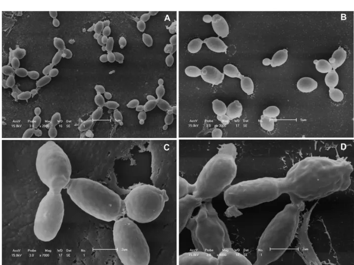
Scanning electron microscopy for the study of S. saponaria BUTE effect onC. albicanscells: Scanning electron microscopy was
performed to verify the effect of S. saponaria BUTE on cell morphology
of C. albicans. Aliquots of 1 mL of C. albicans ATCC 90028 cells were collected after 2 and 6 h of contact with 281.24 µg/mL of the extract, and the cells were prepared for scanning electron microscopy (SEM). This S. saponaria extract concentration corresponds to the MIC of S. saponaria BUTE observed for the inoculum size used for this assay. At
the 6-h time point, cells were harvested and prepared for the proteomic assay. For the SEM, cells from 1 mL of culture were recovered by brief centrifugation and fixed in 2.5% glutaraldehyde for 2 h. The wells were washed twice with 0.1 M cacodylate and gradually dehydrated in ethanol. After coating with gold, the samples were examined with a Shimadzu SS-550 Superscan electron microscope.
Organism and culture conditions for the proteome assay:C. albicans ATCC 90028 was cultured overnight in 20 mL of YPD broth
(1% yeast extract, 2% peptone, 1% dextrose) in a temperature-controlled incubator at 30 °C. The cultures were diluted in 500 mL of fresh YPD medium to an optical density (OD) of 0.1 at 600 nm and subsequently grown at 30 °C until an OD of 0.2 was reached. After growth to an OD of 0.2, the cells were treated with 750 µg/mL fluconazole diluted
in 0.5% DMSO, or 140.62 µg/mL or 281.24 µg/mL of the BUTE of S.
saponaria, corresponding to the minimal sub-inhibitory (sub-MIC) and inhibitory concentrations (MIC), respectively. Control experiments were performed using 0.5% DMSO (the solvent for the fluconazole) added to the culture, and all cultures were kept at 30 °C in a shaker incubator at 250 rpm (Nova Ética 430/RDBP) in a temperature-controlled incubator for 6 h. After the 6-h incubation, the ODs were measured.
at 4500 rpm (Hettich® Rotina 420). The pellet of cells was washed twice
with ice-cold PBS buffer and once with ultrapure water. The cells were re-suspended in 500 µL of 7 M urea, 2 M thiourea, 4% CHAPS and 50 mM DTT buffer containing 1x Protease Inhibitor Mix (GE Healthcare Bio-Science AB, Piscataway, MA, USA), and the suspension was sonicated on ice (30 bursts of 10 seconds ON, 30 seconds OFF with 40% amplitude). The lysates were clarified by centrifugation at 14,000 rpm (Eppendorf® 5430 R) for 20 min at 4 °C, and the protein concentrations
were determined using Bradford assays21 and BSA as a standard.
Isoelectric focusing and 2DE analysis: Proteins were prepared for isoelectric focusing using the 2D Clean-Up kit (GE Healthcare Bio-Science AB, Piscataway, MA, USA) and re-suspended in DeStreak Rehydration Solution (GE Healthcare). The protein concentration was verified by the Bradford assay and adjusted to 250 µg in 250 µL of DeStreak Rehydration Solution with 0.5% IPG pH 3-10 buffer. The samples were applied to isoelectric focusing (IEF) strips (3-10 pH range, 13 cm; GE Healthcare), which were overlaid with 0.6 mL DryStrip Cover Fluid (GE Healthcare) and rehydrated overnight at room temperature. Isoelectric focusing was performed using an IPGphor II IEF System (GE Healthcare Bio-Science AB, Uppsala, Sweden) at 20 °C, 500 V for 1 h, 1,000 V for 1 h, 8,000 V for 2.5 h, and 8,000 V for 30 min.
Thereafter, the strips were equilibrated for 15 min with gentle shaking in a solution of 6 M urea, 2% SDS, 30% glycerol, 75 mM Tris-HCl pH 8.8, 0.002% bromophenol blue, and 1% DTT. The strips were then equilibrated for 15 min in a second buffer containing 2.5% iodoacetamide instead of DTT.
The equilibrated strips were overlaid on homogeneous 12% (1.0 mm thick) SDS-PAGE gels, electrophoresed at 100 V/ 10 mA/ 4 W per gel for 15 min and then 300 V/ 50 mA/ 60 W for approximately 4 h at 15 °C using a Hoefer SE 600 Ruby power supply (GE Healthcare Bio-Science AB, Uppsala, Sweden) with 12 µL of ColorPlus Prestained Protein Ladder (New England, Biolabs, Beverly, MA, USA). The proteins were detected by a modified Neuhoff Blue Silver Coomassie Colloidal Blue staining protocol22 and maintained in 5% acetic acid. The gels were scanned at
an optical resolution of 300 dpi on a high-resolution image scanner (GE Healthcare, USA), and the images were analyzed using the ImageMaster Platinum 2D v6.0 software (GE Healthcare, USA). The differentially expressed proteins were selected for identification.
Protein Identification via In-gel Digestion and Mass Spectrometry:
Protein spots were excised from the 2DE gels and subjected to in-gel digestion with sequencing-grade trypsin (Promega, USA) as described by Shevchenko23 with minor modifications; the digested proteins were
then analyzed using mass spectrometry. After incubation at 37 °C for 16 h, aliquots of each hydrolyzed sample were mixed with a saturated matrix solution of a-cyano-4-hydroxycinnamic acid, spotted onto MALDI target plates and allowed to air dry. Mass spectra (MS) were acquired using a MALDI-TOF/TOF Autoflex II spectrometer (Bruker Daltonics, Germany) with the MASCOT search engine (Matrix Science Ltd., UK) against the “Fungi” subset of the SwissProt database (http://www.expasy. ch/sprot/) or NCBInr (http://www.ncbi.nlm.nih.gov/).
RESULTS
Morphological features ofC. albicans after exposure toS. saponaria
BUTE: SEM was performed to observe general morphological changes in C. albicans cells after exposure to S. saponaria BUTE (Fig. 1).
Comparisons were made between the control C. albicans cells (untreated)
and the C. albicans cells treated with a concentration of 281.24 µg/mL of
BUTE. The untreated-cells were generally smooth-walled bodies after both 2 h and 6 h of culture (Fig. 1A and 1B). The BUTE-treated cells presented surface irregularities, including convolutions (Fig. 1C and 1D).
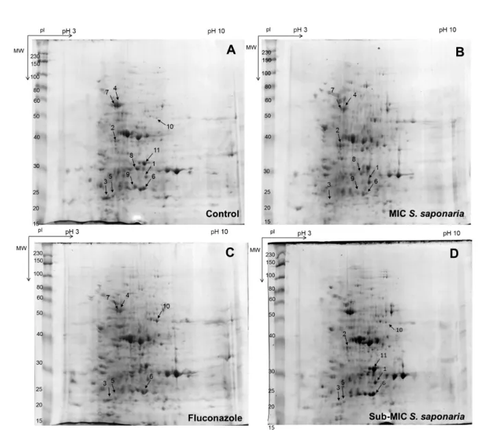
Comparison of the effects of fluconazole and the MIC and sub-MIC of S. saponaria BUTE on the protein expression pattern of C. albicans
Changes in the C. albicans proteome were analyzed by comparing
cells treated with the MIC and sub-MIC of S. saponaria BUTE and cells
treated with fluconazole with untreated control cells. The minimum inhibitory concentration (MIC) of S. saponaria BUTE (281.24 µg/mL) was determined at 0.2 O.D. The high concentration of fluconazole, 750 µg/mL, is due to a large amount of inoculum and takes into account the conditions observed by Bruneau et al.24. Among the three conditions, a
total of six proteins, not including individual protein isoforms, that had greater than 1.5-fold induction or repression relative to their amounts in untreated control cells were identified by in gel enzymatic digestion and MALDI-TOF-TOF (Fig. 2). The molecular proprieties and changes in the relative levels of expression are shown in Table 1, and the putative biological functions of these proteins are shown in Table 2. In general, the proteins are involved in glycolysis and amino acid metabolism.
Proteins that are responsive for the MIC of S. saponaria BUTE:
The six proteins that were responsive for the MIC of BUTE of S.
saponaria based on MALDI-TOF-TOF analysis are displayed in Fig.
2B. Among these, two proteins were induced, and four proteins were repressed. The induced proteins were guanine nucleotide-binding protein subunit beta-like protein (ACS1) and likely mitochondrial ketol-acid reductoisomerase (ILV5). The expression of fructose-bisphosphate aldolase (FBA1), pyruvate decarboxylase (PDC11), enolase 1 (ENO1), and phosphoglycerate mutase (GPM1) was reduced.
Proteins that are responsive for the sub-MIC of S. saponaria BUTE: The six proteins that were responsive to the sub-MIC of S. saponaria BUTE based on MALDI-TOF-TOF analysis are displayed in
Fig. 2C. Among them, four proteins were induced, and 2 proteins were repressed. The induced proteins were guanine nucleotide-binding protein subunit beta-like protein (ACS1), phosphoglycerate mutase (GPM1), peroxisomal catalase (CTA1) and fructose-bisphosphate aldolase (FBA1). The expression of likely mitochondrial ketol-acid (ILV5) and fructose-bisphosphate aldolase (FBA1) was decreased.
Fluconazole-responsive proteins: The four proteins that were responsive to fluconazole according to MALDI-TOF-TOF analysis are displayed in Fig. 2D. Among them, one protein was induced, and three proteins were repressed. The induced protein was peroxisomal catalase (CTA1). The expression of fructose-bisphosphate aldolase (FBA1), pyruvate decarboxylase (PDC11) and phosphoglycerate mutase (GPM1) was decreased.
DISCUSSION
of C. albicans cells exposed to three treatments (MIC and sub-MIC of
BUTE of S. saponaria and fluconazole). Proteins/ enzymes involved
in glycolysis (GPM1, ENO1, FBA1), amino acid metabolism (ILV5, PDC11) and protein synthesis (ASC1) pathways were detected. It is difficult to obtain a clear interpretation of the mechanism of action of
S. saponaria BUTE from this preliminary study of proteome changes.
However, some observations can be made. Several of the biological processes are associated with carbohydrate metabolism, as observed by Martínez-Gomariz et al.25. Comparing the effects of the two S.
saponaria BUTE concentrations on protein expression, we observed
that more proteins had a decreased abundance after treatment with the MIC than with the sub-MIC. These included mainly proteins involved in glycolysis. Some proteins appeared as multiple spots on the gels, such as fructose-bisphosphate aldolase, pyruvate decarboxylase and phosphoglycerate mutase. The variation in the spots specifically occurs in the pI values. This may be caused by post-translational modifications such as phosphorylation. However, we cannot rule out that some of the spots may be due to the experimental procedure; this was observed for spot number 11, related to fructose-bisphosphate aldolase, which was found to be the same protein in spots 3 and 5 but with different pIs and MWs.
The ASC1 protein (guanine nucleotide-binding protein subunit beta-like protein) was found to be upregulated in C. albicans after exposure
to both the MIC and sub-MIC of S. saponaria BUTE, with increased
values of 2.51 and 1.60 fold, respectively. Located at the head of the 40S ribosomal subunit, in the vicinity of the mRNA exit channel, ASC1 serves as a scaffold protein that can recruit other proteins to the ribosome. This protein is also a signal transducer that plays a pivotal role in cellular adhesion and virulence by regulating specific gene expression in C. albicans. It is also repressed in the stationary phase26 and presents a role
in the induction of filamentation and biofilm formation27. This protein had
no differential expression in cells treated with fluconazole. Given that fluconazole is a fungistatic antifungal and that C. albicans cells enter a stationary phase when they stop growing, ASC1 may be repressed due to fluconazole (data not shown).
Enzymes involved in the glycolytic pathway such as FBA1 (fructose-bisphosphate aldolase), PDC11 (pyruvate decarboxylase) and GPM1
(phosphoglycerate mutase) were found to be downregulated in C.
albicans cells treated with both the MIC of S. saponaria BUTE and
fluconazole. ENO1 (enolase) was downregulated only in C. albicans cells
treated with fluconazole. MALDI-TOF analysis of the sub-MIC-treated cell proteins showed three possible isoforms of FBA1; two of these isoforms were downregulated, and the other was upregulated. This protein is involved in a reversible reaction that is required for both glycolysis and gluconeogenesis. It is known that different antifungal agents have varying
effects on the expression of GPM1 in C. albicans. Hoehamer et al.16 have
shown that GPM1 mRNA increases during fluconazole exposure but is downregulated when cells are exposed to ketoconazole. One reasonable cause of the decreased expression of GPM1 in C. albicans cells exposed to
fluconazole may be the growth phase of the harvested cells (the stationary phase). GPM1 mRNA levels increase during the exponential growth phase in yeast and then decrease to relatively low levels in the stationary phase. Additionally, the MIC of S. saponaria BUTE used in this work may have had a deleterious effect on C. albicans strains, preventing their growth
and decreasing GPM1 expression.
Shirtliff et al.28 used proteomic analysis to demonstrate that several
glycolytic enzymes (e.g., glyceraldehyde 3-phosphate dehydrogenase, enolase, phosphoglycerate mutase and pyruvate kinase) were
downregulated when C. albicans was exposed to farnesol, a natural
15-carbon organic compound and acyclic sesquiterpene alcohol that
is used by C. albicans as a quorum-sensing molecule, inhibiting
filamentation. S. saponaria BUTE also contains sesquiterpenes, and one possible effect of this component may be the downregulation of these glycolytic enzymes. However, this interpretation remains speculative because we were unable to test the saponins and sesquiterpenes of S. saponaria BUTE separately.
The protein ILV5 (likely mitochondrial ketol-acid reductoisomerase) had a differential expression in C. albicans after exposure to the MIC
and sub-MIC of S. saponaria BUTE, with 2.09-fold upregulation and
1.60-fold downregulation, respectively, compared to the untreated control cells. Ketol-acid reductoisomerase is described as an antigenic; GlcNAc, amino acid starvation (3-AT) induced; macrophage-repressed protein that is present in the exponential and stationary phases26,29,30.31.
Table 1
List of Sapindus saponaria and Fluconazole responsive proteins identified by MALDI-TOF mass spectrometry (difference at least 1.5-fold)
Spot no. MS/MS Score MS score Protein cover-age % Fold change1 Gel isoform pI Mr Protein ID
MIC S. saponaria
Upregulated Proteins
1 Nd 162 45 2.51 6.07 34533.62 ASC1
2 48 199 48/37 2.09 6.17 44935 ILV5
Downregulated Proteins
3 28 149 5/32 5.10 1 5.69 48/37 FBA1
4 Nd 162 21 1.82 1 5.39 62401.92 PDC11
5 Nd 168 34 2.46 2 5.69 39190.76 FBA1
6 26 82 4/19 4.06 1 5.79 27437.46 GPM1
7 5 312 2/43 3.10 2 5.39 62401.92 PDC11
8 43 159 3/26 1.93 5.54 47202.50 ENO1
9 22 136 4/28 1.60 2 5.79 27437.46 GPM1
Sub-MIC S. saponaria
Upregulated Proteins
1 Nd 162 45 1.60 6.07 34533.62 ASC1
6 26 82 4/19 1.84 5.79 27437.46 GPM1
10 51 115 1/21 2.05 6.03 55029.13 CTA1
11 Nd 252 53 1.87 1 5.69 39190.76 FBA1
Downregulated Proteins
2 48 199 48/37 1.60 6.17 44935 ILV5
3 28 149 5/32 4.13 2 5.69 39190.76 FBA1
5 Nd 168 34 2.72 3 5.69 39190.76 FBA1
Fluconazole Upregulated Proteins
10 51 115 1/21 4.87 6.03 55029.13 CTA1
Downregulated Proteins
3 28 149 5/32 2.15 1 5.69 39190.76 FBA1
5 Nd 168 34 2.58 2 5.69 39190.76 FBA1
4 Nd 162 21 3.35 1 5.39 62401.92 PDC11
7 5 312 2/43 1.88 2 5.39 62401.92 PDC11
6 26 82 4/19 1.60 5.79 27437.46 GPM1
1Fold change: Change in expression between treated cells and control cells. Nd: Not done. The spot numbers are those indicated on Fig. 2.
exposed to the sub-MIC of S. saponaria BUTE and fluconazole, but
the MIC did not cause differential expression of this protein. It has been reported that CTA1 plays a role in resistance to oxidative stress32,
virulence, core stress responses, and other functions that are regulated by fluconazole.
In conclusion, our findings reveal antifungal-induced changes in
protein abundance of C. albicans. By using the previously identified
components of the S. saponaria BUTE, such as saponins and
Table 2
Function of the proteins exhibiting a greater than 1.5-fold induction and repression which were identified by means of in-gel enzymatic digestion and MALDI-TOF-TOF
Protein ID SwissProt or NCBI Accession number Metabolic process Function
ASC1 P83774 Ribosomal protein A component of the 40S ribosomal subunit and a signal transducer
ILV5 gi|68483473 Branched-chain amino acid biosynthetic process Involved in branched chain amino acid biosynthesis.
FBA1 Q9URB4 Glycolysis
Catalyzes the aldol condensation of dihydroxyacetone phos-phate with glyceraldehyde 3-phosphos-phate to form fructose 1,6-bisphosphate in gluconeogenesis and the reverse reaction in glycolysis.
PDC11 P83779 Glycolysis Catalyses the decarboxylation of pyruvic acid to acetaldehyde and carbon dioxide.
ENO1 P30575 Glycolysis
Catalyzes conversion of 2-phosphoglycerate to phosphoenol-pyruvate during glycolysis and the reverse reaction during gluconeogenesis.
GPM1 P82612 Glycolysis Catalyses conversion of 2-phospho-D-glycerate to 3-phospho-D-glycerate.
ACKNOWLEDGEMENTS
We thank the Coordination for the Improvement of Higher Education Personnel/Postdoctoral National Program (CAPES/PNPD) for supporting this study.
CONFLICT OF INTEREST
The authors declare that there are no conflicts of interest.
REFERENCES
1. Kabir MA, Hussain MA, Ahmad Z. Candida albicans: a model organism for studying fungal pathogens. ISRN Microbiol. 2012;2012:538694.
2. Matos FJ. Farmácias vivas : sistema de utilização de plantas medicinais projetado para pequenas comunidades. 4ª ed. rev. ampl. Fortaleza : UFC : SEBRAE/CE; 2002. 3. Cavalcanti SB, Teles HL, Silva DH, Furlan M, Young MC, Bolzani V. New Tetra-acetylated
oligosaccharide diterpene from Cupania vernalis. J Braz Chem Soc. 2001;12:413-6. 4. Abdel-Wahab SM, Selim MA. Lipids and flavonoids of S. saponaria. Fitoterapia.
1985;56:167-8.
5. Meyer Albiero AL, Aboin Sertié JA, Bacchi EM. Antiulcer activity of Sapindus saponaria
L. in the rat. J Ethnopharmacol. 2002;82:41-4.
6. Braga R. Plantas do Nordeste, especialmente do Ceará. 4ª ed. Mossoró: Universitária; 1984.
7. Pio Corrêa M. Dicionário das plantas úteis do Brasil e das exóticas cultivadas. Rio de Janeiro: Ministério da Agricultura : Instituto Brasileiro de Desenvolvimento Florestal; 1984.
8. Murgu M, Santos LF, Souza GD, Daolio C, Schneider B, Ferreira AG, et al. Hydroxilation of a hederagenin derived saponin by a Xylareaceous fungus found in fruits of Sapindus saponaria. J Braz Chem Soc. 2008;19:831-5.
9. Damke E, Tsuzuki JK, Cortez DA, Ferreira IC, Bertoni TA, Batista MR, et al. In vivo
activity of Sapindus saponaria against azole-susceptible and-resistant human vaginal
Candida species. BMC Complement Altern Med. 2011;11:35.
10. Francis G, Kerem Z, Makkar HP, Becker K. The biological action saponins in animal systems: a review. Br J Nutr. 2002;88:587-605.
11. Tsuzuki JK, Svidzinski TI, Shinobu CS, Silva LF, Rodrigues-Filho E, Cortez DA, et al. Antifungal activity of the extracts and saponins from Sapindus saponaria L. An Acad Bras Cienc. 2007;79:577-83.
12. Du Z, Zhu N, Ze-Ren-Wang-Mu N, Shen Y. Two new antifungal saponins from the Tibetan herbal medicine Clematis tangutica. Planta Med. 2003;69:547-51.
13. Lee MW, Kim SU, Hahn DR. Antifungal activity of modified hederagenin from the leaves of Kalopanax pictum var. Biol Pharm Bull. 2001;24:718-9.
14. Tamura Y, Mizutani K, Ikeda T, Ohtani K, Kasai R, Yamasaki K, et al. Antimicrobial activies of saponins of pericarps of Sapindus mukurossi on dermatophytes. Nat Med. 2001;55:11-6.
15. Heilmann CJ, Sorgo AG, Mohammadi S, Sosinska GJ, de Koster CG, Brul S, et al. Surface stress induces a conserved cell wall stress response in the pathogenic fungus Candida albicans. Eukaryot Cell. 2013;12:254-64.
16. Hoehamer CF, Cummings ED, Hilliard GM, Rogers PD. Changes in the proteome of
Candida albicans in response to azole, polyene, and echinocandinantifungal agents. Antimicrob Agents Chemother. 2010;54:1655-64.
17. Sosinska GJ, de Koning LJ, de Groot PW, Manders EM, Dekker HL, Hellingwerf KJ, et al. Mass spectrometric quantification of the adaptations in the wall proteome of
Candida albicans in response to ambient pH. Microbiology. 2011;157(Pt 1):136-46. 18. Thomas DP, Bachmann SP, Lopez-Ribot JL. Proteomics for the analysis of the Candida
19. Clinical and Laboratory Standards Institute. Reference method for broth dilution antifungal susceptibility testing of yeast : approved standard. 3rd ed. Wayne: Clinical and Laboratory Standards Institute; 2008.
20. Shin JH, Kee SJ, Shin MG, Kim SH, Shin DH, Lee SK, et al. Biofilm production by isolates of Candida species recovered from nonneutropenic patients: comparison of bloodstream isolates with isolates from other sources. J Clin Microbiol. 2002;40:1244-8.
21. Bradford MM. A rapid and sensitive method for the quantitation of microgram quantities of protein utilizing the principle of protein–dye binding. Anal Biochem. 1976;72:248-54.
22. Candiano G, Bruschi M, Musante L, Santucci L, Ghiggeri GM, Carnemolla B, et al. Blue silver: a very sensitive colloidal Coomassie G-250 staining for proteome analysis. Electrophoresis. 2004;25:1327-33.
23. Shevchenko A, Wilm M, Vorm O, Mann M. Mass spectrometric sequencing of proteins silver-stained polyacrylamide gels. Anal Chem. 1996;68:850-8.
24. Bruneau JM, Maillet I, Tagat E, Legrand R, Supatto F, Fudali C, et al. Drug induced proteome changes in Candida albicans: comparison of the effect of beta(1,3) glucan synthase inhibitors and two triazoles, fluconazole and itraconazole. Proteomics. 2003;3:325-36.
25. Martínez-Gomariz M, Perumal P, Mekala S, Nombela C, Chaffin WL, Gil C. Proteomic analysis of cytoplasmic and surface proteins from yeast cells, hyphae, and biofilms of Candida albicans. Proteomics. 2009;9:2230-52.
26. Kusch H, Engelmann S, Bode R, Albrecht D, Morschhäuser J, Hecker M. A proteomic view of Candida albicans yeast cell metabolism in exponential and stationary growth phases. Int J Med Microbiol. 2008;298:291-318.
27. Liu X, Nie X, Ding Y, Chen J. Asc1, a WD-repeat protein, is required for hyphal development and virulence in Candida albicans. Acta Biochim Biophys Sin (Shanghai). 2010;42:793-800.
28. Shirtliff ME, Krom BP, Meijering RA, Peters BM, Zhu J, Scheper MA, et al. Farnesol-induced apoptosis in Candida albicans. Antimicrob Agents Chemother. 2009;53:2392-401.
29. Fernández-Arenas E, Cabezón V, Bermejo C, Arroyo J, Nombela C, Diez-Orejas R, et al. Integrated proteomics and genomics strategies bring new insight into Candida albicans response upon macrophage interaction. Mol Cell Proteomics. 2007;6:460-78. 30. Pitarch A, Abian J, Carrascal M, Sánchez M, Nombela C, Gil C. Proteomics-based identification of novel Candida albicans antigens for diagnosis of systemic candidiasis in patients with underlying hematological malignancies. Proteomics. 2004;4:3084-106.
31. Yin Z, Stead D, Selway L, Walker J, Riba-Garcia I, McLnerney T, et al. Proteomic response to amino acid starvation in Candida albicans and Saccharomyces cerevisiae. Proteomics. 2004;4:2425-36.
32. Wysong DR, Christin L, Sugar AM, Robbins PW, Diamond RD. Cloning and sequencing of a Candida albicans catalase gene and effects of disruption of this gene. Infect Immun. 1998; 66:1953-61.