Fátima C. Pereira
Dissertation presented to obtain the Ph.D degree in Biology
Instituto de Tecnologia Química e Biológica António Xavier | Universidade Nova de Lisboa
Oeiras,
Spore differentiation in relation to the infectious
Fátima C. Pereira
Dissertation presented to obtain the Ph.D degree in Biology
Instituto de Tecnologia Química e Biológica António Xavier | Universidade Nova de LisboaOeiras, May, 2014
To Instituto de Tecnologia Química e Biológica António Xavier of Universidade Nova de Lisboa, for receiving me as a PhD student, for providing all the working conditions and the scientific environment for the execution of this work.
To Fundação para a Ciência e Tecnologia (FCT) and European Molecular Biology Organization (EMBO), for the financial support.
To Instituto Gulbenkian de Ciência, for my initial training in the PhD Programme (PDIGC), and to all my colleagues at the time.
To my supervisor Dr. Adriano O. Henriques, for the opportunity to work in his lab and for his continuous help and support throughout this journey. For all I learned from him and for sharing with me his passion for science.
To Dr. Bruno Dupuy, Prof. Isabelle Martin-‐Verstraete and Dr. Claire Janoir for welcoming me in their labs, providing me a rich scientific environment as well as excellent working conditions. A special merci to Dr. Bruno Dupuy for his constant interest and support during the development of this work.
To Mónica Serrano for all the helpful discussions, enthusiasm and sense of humour. Thanks for your assistance and positive attitude.
times when this journey seemed endless. To Teresa, for her friendship and relaxed moments, having lunch at the beach or watching a movie. To Filipa, for showing me how to always laugh of the misfortune. To Rita, for the free hugs, encouragement and contagious joy.
To Laure Saujet, for our excellent scientific interaction that resulted in joint papers.
To Marc Monot, Laure Saujet, Emilie Camiade, Evelyne Couture-‐Tosi, Ana Antunes, Rita Tomé and Sandra Hoys, for the lab support and scientific help during my visits to Institut Pasteur and to Faculté de Pharmacie, Paris.
To Patrícia Amaral, for her valuable help with the image on the cover. To all of my friends who have supported me during the dark times and celebrated with me through the good. A special thank to Guida, for her unconditional friendship and loyalty.
To my parents for their unconditional love, they will always be an inspiration to me. Finally, to my brother, for his constant support and encouragement throughout my life.
the sigma factors are conserved in C. difficile, a reduced temporal segregation between the activities of early and late sigma factors is observed when in comparison with the model organism B. subtilis. We further observed that the activity of σE is partially independent of σF, despite the fact that the forespore product SpoIIR is required for pro-‐σE processing. In addition, σG activity is not dependent on σE, and the activity of σK does not require σG. In agreement, our genome-‐wide analysis of gene expression during spore formation confirmed a weaker connection between the mother cell and the forespore in C. difficile. This global transcriptional analysis allowed the identification of 225 genes controlled by each of these sporulation sigma factors: 25 in the σF regulon, 97 σE-‐controlled genes, 50 σG-‐dependent genes and 56 genes under σK control. Our analysis of a mutant for the transcriptional regulator SpoIIID also demonstrates that many of the σE target genes here identified are either repressed or activated by this transcriptional regulator, including sigK, which is activated. While most of the genes known to be essential for sporulation in B. subtilis are conserved, many other genes, especially in the σE and σK regulons, are novel. Our analysis supports the view that the top level in the control of the sporulation network, defined by the cell type-‐specific sigma factors is conserved in evolution, together with a core of target genes possibly representing the conserved machinery for endosporulation, while others are group specific.
properties that allowed it to adapt to its host and to efficiently colonize it.
caracterização dos mutantes dos factores sigma de esporulação de C. difficile, tendo sido estabelecidos a janela temporal e a célula em que ocorre a sua transcrição e actividade. Em geral, os resultados aqui apresentados indicam que, enquanto que os principais períodos de actividade dos factores sigma são conservados em C. difficile, existe uma redução na segregação temporal entre a actividade dos factores sigma iniciais ou tardios quando em comparação com o organismo modelo B. subtilis. Observou-‐se ainda que a actividade de σE é parcialmente independente de σF, embora o papel da proteína SpoIIR do pré-‐esporo na sinalização da activação proteolítica de pro-‐σE seja mantido. Para além disso, a actividade de σG também não depende de σE, assim como a actividade de σK não requer σG. Em consonância com estes resultados, uma análise global da expressão genética de C. difficile durante a esporulação confirmou uma ligação mais ténue entre os programas genéticos do pré-‐esporo e da célula-‐mãe. Esta análise transcripcional global permitiu ainda a identificação de 225 genes sob o controlo dos factores sigma de esporulação: 25 destes pertencem ao regulão σF, 97 são controlados por σE, 50 são dependentes de σG e 56 estão sob controlo de σK. Uma análise do mutante para o regulador transcripcional auxiliar SpoIIID demonstrou ainda que muitos dos genes pertencentes ao regulão σE são reprimidos ou ativados por este regulador, como é o caso de sigK, activado por SpoIIID. Enquanto que a maioria dos genes essenciais para a esporulação em B. subtilis são conservados, muitos outros, sobretudo nos regulões σE e σK, são novos. A nossa análise sugere que o nível superior de controlo da esporulação, definido pelos 4 factores sigma específicos do pré-‐esporo ou da célula mãe é conservado, juntamente com um grupo de genes alvo que possivelmente define a maquinaria mínima para a esporulação, enquanto que outros são específicos de determinados grupos ou organismos.
superfície do esporo, e é necessária para a eficiente colonização de ratinhos axénicos por C. difficile. Os esporos de um mutante sp17 não são capazes de montar uma estrutura polar que é observada nos esporos selvagens, e que provavelmente desempenha um papel na aderência do esporo ao epitélio do cólon. O trabalho aqui desenvolvido estabelece assim uma ligação directa entre a superfície do esporo e a colonização. Além disso, Sp17 é exclusivo de C. difficile, o que sugere que a superfície do esporo neste organismo tem propriedades estruturais e funcionais distintas que representam, provavelmente adaptações à colonização dos seus hospedeiros.
This Thesis is divided into six Chapters.
Chapter 1 provides an introduction to the enteric pathogen and spore former Clostridium difficile, with emphasis on sporulation, a determinant factor of pathogenesis. Since our knowledge about spore formation and composition is limited in C. difficile, the overview of sporulation provided by this Chapter is based on the model organism Bacillus subtilis. The text is however supplemented with available information about sporulation in C. difficile and other Clostridial spore formers.
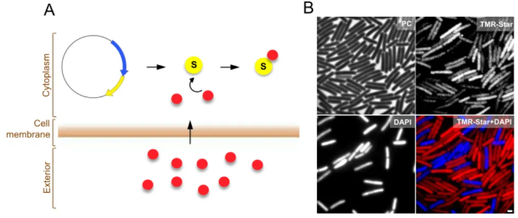
Chapter 2 reports the development of a fluorescent reporter for single cell analysis of gene expression in C. difficile. We provide evidence that this fluorescent reporter, which is based on the SNAP-‐tag technology, can be employed as a quantitative reporter to monitor gene expression in this organism. Our results further demonstrate its applicability at the translational level, to determine the cellular localization of proteins.
organism B. subtilis; iii) the cell-‐cell signaling pathways that operate in B. subtilis are simpler or absent in C. difficile, resulting in a less tight control of the spore morphogenesis; iv) most members of the σE and σK regulons lack homologues in B. subtilis, suggesting a distinct composition for the surface layers of the C. difficile spore.
Chapter 5 reports the identification and characterization of Sp17, an abundant component of the spore coat, identified on Chapter 4 as a previously uncharacterized member of the σK regulon. The Chapter evidences a role for Sp17 in the assembly of the spore surface layers and in host colonization. The chapter establishes a direct link between the spore surface and host colonization in C. difficile.
Lastly, Chapter 6 presents a general discussion of the results and points to future lines of work.
The results presented in Chapters 2, 3 and 4 are based on published material, as indicated in the beginning of each Chapter. Note however that some unpublished material was added when appropriate. Some of the results presented on Chapter 5 are also based on published material, but most of the results presented in this Chapter constitute the basis for a future manuscript.
CHAPTER 1 – Introduction 1
THE ENTERIC PATHOGEN CLOSTRIDIUM DIFFICILE 1 Antibiotics and CDI 4 The changing epidemiology 6 Diagnosis 8 Treatment 9
Conventional Treatment 9 Reconstitution of the colonic microbiota 9
Pathogenesis 10
Toxin production 10 Cell surface and adhesion 12
SPORE FORMATION 13 An overview of the sporulation process 14 Regulation of sporulation 15
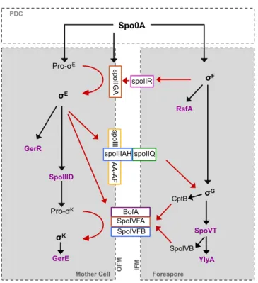
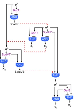
The forespore line of gene expression 16 The mother cell line of gene expression 21 Cell-‐cell communication during sporulation 24
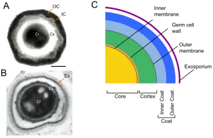
Regulation of sporulation in Clostridia 25 Spore morphology 27
Spore core and cortex 28 Spore coat 29 Exosporium 31
Spore germination and outgrowth 32 AIMS OF THIS WORK 33 REFERENCES 34
CHAPTER 2 – Development of a fluorescent reporter for single cell
analysis of gene expression and protein localization in C. difficile 55
SUMMARY 57 INTRODUCTION 58 MATERIALS AND METHODS 62 RESULTS 66 Specific labeling of C. difficile cells with the TMR-‐Star SNAP substrate 66 Optimization of SNAP labeling times 68 Maximal accumulation of SNAP under the Ptet control 69
ACKNOWLEDGEMENTS 78 REFERENCES 79
CHAPTER 3 – The pathway of spore differentiation in C. difficile 83
SUMMARY 85 INTRODUCTION 86 MATERIALS AND METHODS 89 RESULTS 100
Sporulation in sporulation medium (SM) 100 Stages of sporulation 102 Spore staining by FM4-‐64 103 Spore ultrastructure 106 Disruption of the genes for the sporulation sigma factors 108 Morphological characterization of the sigF, sigE, and sigG mutants 111 Functional analysis of the sigK gene 113 Localizing the expression of the sporulation-‐specific sig genes 116 Localizing the activity of σF and σE 121
Requirements for the activity of σG and σK 124
DISCUSSION 128 Transcription of sigF and sigE, and activity of σF and σE 128
Production and activity of σG 130
Production and activity of σK 132
ACKNOWLEDGEMENTS 135 REFERENCES 136
CHAPTER 4 – Genome-‐wide analysis of cell-‐type specific gene
transcription during spore formation in C. difficile 145
SUMMARY 147 INTRODUCTION 148 MATERIALS AND METHODS 151 RESULTS 158 Overview of the transcriptome data 158 Overview of the four cell type-‐specific σ regulons 159
The σF regulon 160 The σE regulon 161 The σG regulon 163 The σK regulon 165
Characterization of the SpoIIID regulon 173 Control of sigK transcription 176
Communication between the forespore and the mother cell 178
Absence of a strict control of the σE regulon by σF 178 The role of SpoIIR in the regulatory cascade 180 The loose regulation of σG-‐dependent genes by σE 183 The absence of control of the σK regulon by σG 185
DISCUSSION 187 ACKNOWLEDGEMENTS 190 REFERENCES 191
CHAPTER 5 –
A C. difficile spore surface protein with a role in host
colonization 199
SUMMARY 201 INTRODUCTION 202 MATERIALS AND METHODS 205 RESULTS 214
CD1581 is required for efficient colonization in a mouse
axenic model 214
CD1581 is expressed in the mother cell chamber of
developing cells, under the control of regulatory protein σK 215
Sp17 is an abundant component of the C. difficile spore coat 218 Sp17 undergoes multimerization at the spore surface 221 Sp17 is surface exposed 222 An sp17 mutant fails to assemble the spore outer coat 225 Sp17 is required for the assembly of a spore polar appendage 227 Germination is altered in sp17 mutant spores 230 DISCUSSION 232 ACKNOWLEDGEMENTS 235 REFERENCES 236
CHAPTER 6 –
Discussion 243
The sporulation regulatory network of C. difficile 245 The σF, σE, σG and σK regulons of C. difficile 251
A link between the C. difficile spore surface and host colonization 253
Chapter 1
Introduction
THE ENTERIC PATHOGEN
CLOSTRIDIUM DIFFICILE
We are not alone. A vast array of microorganisms lives on or inside
the human body, with our intestine carrying the highest density and
diversity of microbial cells that inhabit us. Our gut harbours more than 1000
of bacteria phylotypes and this microbial diversity is highly variable over
time and across populations. Most (90%) of our gut microbiota belongs to
only two phyla: the Bacteriodetes and the Firmicutes. Actinobacteria,
Proteobacteria and Verrucomicrobia are also represented, but as minor
constituents (Lozupone et al., 2012). The trillions of bacteria that live in the
human gut form a complex ecological community that helps in food
digestion, protects against enteropathogens, and stimulate the immune
system (Backhed et al., 2004;
Stecher and Hardt, 2011; Littman and Pamer,
2011). The host, in turn, provides habitat and a constant influx of nutrients
(Berry et al., 2013).
Besides sustaining a large number and diversity of commensal
microbes, the human gut also provides habitat to a panel of harmful
pathogenic microorganisms. The enteropathogen Clostridium difficile is a
gram positive spore forming anaerobe that inhabits the mammalian gut. This
organism is able to produce two potent cytotoxins and cause infection
(Carter et al., 2010; Carroll and Bartlett, 2011; Rupnik et al., 2009; Dawson et
al., 2009; Shen, 2012).
C. difficile was first identified and described in 1935 by Hall and
O´Toole as part of the intestinal microflora in neonates (Hall and O´Toole,
1935). Later, in 1977, Bartlett et al. isolated C. difficile from the faeces of
hamsters with clindamycin-‐induced colitis, confirming this pathogen as the
cause of antibiotic-‐induced disease in animals
(Bartlett et al., 1977). One year
the causative agent of antibiotic-‐associated diarrhoea and colitis not only in
animals, but also in humans (Larson et al., 1978; Bartlett et al., 1978).
Symptoms of the disease caused by C. difficile infection (CDI) can range from
mild diarrhoea, abdominal pain, fever and leucocytosis, to more severe
symptoms such as pseudomembranous colitis, toxic megacolon, bowel
perforation, sepsis and death (Rupnik et al., 2009; Carroll and Bartlett, 2011;
Dawson et al., 2009).
Antibiotics and CDI
C. difficile is an enteric pathogen that relies on the disturbance of the
normal gut microbiota to expand in the gut and cause infection (Britton and
Young, 2012; Carroll and Bartlett, 2011; Rupnik et al., 2009). Individuals
with a normal, balanced microbiota are usually resistant to infection by C.
difficile. This is because the commensal bacteria that inhabit the gut are able
to directly or indirectly prevent colonization by this pathogen (Figure 1.1)
(Britton and Young, 2012). Direct mechanisms for the inhibition of C. difficile
growth involve the production of antimicrobial compounds, such as Thuricin
CD, a bacteriocin produced by the intestinal bacterium Bacillus thuringiensis
with narrow activity against C. difficile (Rea et al., 2010). Another direct
mechanism by which the gut microbiota prevents C. difficile colonization is
via the transformation of bile acids, which have profound effects on C. difficile
spore germination and vegetative growth (see also the Spore germination
section) (Sorg and Sonenshein, 2008; Sorg and Sonenshein, 2010; Francis et
al., 2013; Theriot et al., 2014).
Commensal bacteria may also contribute to inhibition of C. difficile
growth in the intestine through indirect mechanisms, such as nutrient
exhaustion or the stimulation of the host immune system (Figure 1.1). As an
example of the first, the access to sialic acid, which is liberated upon
depletion of the commensal microbiota, is determinant for the expansion of
colonization of the gut by non-‐toxicogenic C. difficile strains prevents the
colonization by most toxinogenic strains, in a process referred to as “niche
exclusion” (Nagaro et al., 2013). Regarding the second mechanism, the
commensal microbiota produces microbial associated molecular patterns
(MAMPs) that stimulate the production of adaptive or innate effectors by the
host immune system, preventing C. difficile expansion in the gut (Littman and
Pamer, 2011; Britton and Young, 2012).
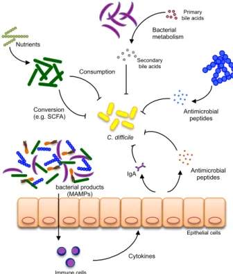
Figure 1.1. Potential mechanisms involved in colonization resistance against C.
difficile in the gut. Direct inhibition of C. difficile by the gut microbiota can occur via
competition for nutrients, conversion of host or diet compounds into secondary metabolites that inhibit C. difficile growth (e.g., bile acids) or production of primary compounds with bacteriostatic activity against C. difficile (e.g., bacteriocins). Indirect control of C. difficile
expansion can occur via the stimulation of the host immune system through the production of microbial associated molecular patterns (MAMPs). Detection of MAMPs by the host induces the activation of an host immune cascade and the production of innate (e.g., antimicrobial peptides) or adaptive (e.g., IgA) immune effectors. Adapted from Britton and Young, 2012.
During antibiotic treatment, C. difficile can colonize the gut because,
antibiotics. Resistance to antibiotics such as erythromycin, chloramphenicol
or tetracycline is largely mediated by transposons that are present in the C.
difficile genome
(Lyras et al., 2004; Hussain et al., 2005;
Sebaihia et al.,
2006). In contrast, resistance to fluoroquinolones is mediated by mutations
in specific genes, such as point mutations in the A or B subunits of DNA
gyrase (gyrA or gyrB)(He et al., 2013)
.
The changing epidemiology
CDI is mainly a nosocomial infection, associated with elderly and
debilitated patients, long hospital stays, prolonged periods of antibiotic
therapy and the uptake of immunosuppressors (Kuipers and Surawicz, 2008;
Rupnik et al., 2009;
Carroll and Bartlett, 2011). It is estimated that 7-‐17% of
hospitalized patients carry toxinogenic strains of C. difficile (Poutanen and
Simor, 2004; Liao et al., 2012). The incidence of CDI has risen dramatically in
the turn of this century, and it is nowadays the most common cause of
antibiotic associated-‐diarrhoea in the industrialised world. This was because
new, highly virulent C. difficile strains have emerged worldwide to cause
outbreaks of increased disease severity and higher recurrence, morbidity
and mortality rates (Kelly and LaMont, 2008;
Brazier, 2008; Cartman et al.,
2010; Bartlett, 2010;
Dawson et al., 2009;
Deneve et al., 2009). Most of these
strains belong to a particular PCR-‐ribotype, the 027 ribotype (Clements et al.,
2010; O'Connor et al., 2009; Freeman et al., 2010). A factor that contributed
to the increased virulence of 027 strains was the acquisition of
fluoroquinolone resistance (He et al., 2013).
In the UK the appearance of these virulent 027 strains led to a drastic
raise in the number of CDI cases, which have reached historical highs in
2006, with 55000 cases being detected in a single year (Figure 1.2A)
(Brazier, 2012;
Kuijper et al., 2008;
Bauer et al., 2011). Fortunately, the
implementation of prevention measures and the activity of infection control
C D I in ci d e n ce
year year
C D I in ci d e n ce
A
B
Deaths
Deaths (per 1000000 pop) Hospitalization (in thousands)
England and Wales USA
Cases reported 0 10000 20000 30000 40000 50000 60000
2001 2002 2003 2004 2005 2006 2007 2008 2009 2010 201 1 2012 0 20 40 60 80 100 120 140
2001 2002 2003 2004 2005 2006 2007 2008 2009 2010
by CDI in the UK after 2007 (Figure 1.2A) (Jones et al., 2013) (Source: Health
Protection Agency, http://www.hpa.org.uk). Similarly, in the USA the C.
difficile related deaths also drastically rose from 5.7 deaths per billion
inhabitants in 2001 to 23.7 in 2006 (Figure 1.2B) (Source: Centre for Disease
Control, National Centre for Health Statistics, http://www.cdc.gov/nchs/).
The medical costs associated with this disease also represent a major
economic burden, with annual health-‐care costs of hospital-‐onset CDI
estimated in 1.1 billion dollars in the USA and in 3 billion euros in Europe
(data from the European Centre for Disease Prevention and Control,
available at: http://www.ecdc.europa.eu/en/healthtopics/).
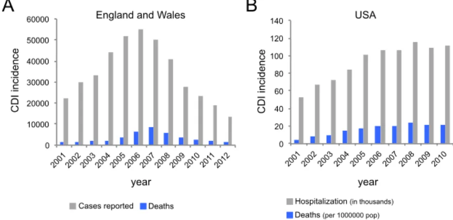
Figure 1.2.CDI incidence. (A) Total number of CDI reported cases (grey) and related
deaths (blue) in England and Wales during the indicated time period. Source: Health Protection Agency, http://www.hpa.org.uk. (B) Hospitalization associated with CDI (in grey; numbers in thousands) and mortality rates (in blue, age-adjusted numbers per 1.000.000 population) in the USA during the indicated time period. Source for mortality data: Centre for Disease Control, National Centre for Health Statistics, http://www.cdc.gov/nchs/; source for hospitalization associated with CDI: Lucado et al., 2009.
In Portugal the first detection of C. difficile 027 strains implicated in a
hospital outbreak dates from January 2012, involving 12 patients, with a
crude mortality rate of 50% (Antunes et al., 2012). The CDI incidence in
Portugal is estimated at 13 cases per 10000 hospital admissions (Bauer et al.,
The appearance of new virulent strains of C. difficile was followed by
an increase in the number of community-‐acquired CDI cases (Freeman et al.,
2010;
Hensgens et al., 2012). Compared with hospitalized patients, the
infected members of the community are younger, with fewer co-‐morbidities,
and in some cases they have not received any antibiotic therapy in the 6
weeks prior to developing CDI, leading to the hypothesis that there is a yet
unknown selection mechanism that favours the emergence of these strains
(Freeman et al., 2010;
Hensgens et al., 2012).
Diagnosis
The first indication of CDI is based on the presence of diarrhoea with
a typical foul-‐smelling odour, conferred by the ability of C. difficile to produce
and tolerate high concentrations of p-‐cresol, a bacteriostatic compound.
However, detection of CDI based solely on the presence of an odour is
obviously not an accurate and definitive diagnosis, and other methods are
available (Carroll and Bartlett, 2011; Burnham and Carroll, 2013). For many
years, the cell culture cytotoxicity neutralization assays (CCCNAs), which
detect the cell toxicity of TcdA and TcdB present in a faecal eluate, were
considered the gold standard. However, the method lacks sensitivity, and
was recently replaced by new and improved diagnosis methods. Among
these are the EIAs (enzyme immunoassays), in which polyclonal or
monoclonal antibodies are used to target the TcdA and TcdB toxins, and the
Glutamate dehydrogenase (GDH) test. GDH is a metabolic enzyme expressed
at high levels in all C. difficile strains, toxinogenic and non-‐toxinogenic (Zheng
et al., 2004). Thus, the GDH test needs to be complemented with EIAs or
other test that allows the detection of toxins (Snell et al., 2004).
Lastly, nucleic acid amplification methods (NAATs), which are based
on the isothermal amplification of conserved regions of the tcdB and tcdA
genes, have recently become available (Goldenberg et al., 2010). In the UK,
first of which should be a NAAT test or a GDH assay, followed by a toxin EIA
test (DH/HCAI/Infectious disease, 2012).
Treatment
Conventional Treatment
Initial therapy for CDI includes stopping all antibiotic therapy, if
possible, followed by the use of metronidazole or vancomycin, depending on
illness severity and co-‐morbidities (Debast et al., 2014). Metronidazole is the
first line agent in treatment for mild to moderate CDI. However, it is
becoming less effective as strains with decreased susceptibility to this
antibiotic are appearing (Baines et al., 2008; Moura et al., 2013). This led to
the adoption of vancomycin as the front-‐line antibiotic, together with
fidaxomicin, a newly FDA approved antibiotic with cure rates equivalent to
vancomycin, but that presents reduced recurrence rates (15% versus 25%)
(Louie et al., 2011). Unlike the other antibiotics, fidaxomicin specifically
inhibits the formation of spores, whose accumulation in the host constitutes
the most common cause of relapse (Babakhani et al., 2012).
Reconstitution of the colonic microbiota
Recurrent CDI is an alarming and important problem. Patients with
multiple episodes of CDI had markedly reduced microbial diversity when
compared to patients that suffered only a single episode of CDI (Chang et al.,
2008). The main problem is that antibiotic treatment for a first infection can
also eliminate many potential protective commensal bacteria, facilitating
reinfection (Kelly and LaMont, 2008). Thus, the gut microbiota needs to be
restored to protect the intestinal epithelium and to prevent residual spores
from causing recurrent disease
(Britton and Young, 2012). For this reason
the development of non-‐antibiotic treatments for CDI has gained much
attention in recent years. Among these, the faecal microbiota transplantation
of the cases (van Nood et al., 2013). However, FMT is not an FDA-‐approved
therapy, it requires time to identify a suitable donor and potentiates the
transmission of undetected pathogens. These, together with the general
patient aversion, represent major impediments to the broad application of
FMT. Thus, a major goal in the field is to find specific bacterial species
present in the gut that confer protection against CDI. A cocktail of only six
cultivable bacterial strains that is able to cure chronic infection in mice was
recently identified (Lawley et al., 2012). This information is of paramount
importance and can now be used to develop standardized treatment
mixtures with the same efficacy as FMT, but increased safety and acceptance.
Pathogenesis
Toxin production
CDI symptoms are primarily caused by the glucosylating toxins TcdA
and TcdB (Carter et al., 2010; Carroll and Bartlett, 2011; Shen, 2012;
Rupnik
et al., 2009). These are classified as large Clostridial toxins (LCTs), due to
their large size (205 and 308 KDa, respectively), cytotoxicity and mechanism
of action
(Just and Gerhard, 2004). Both TcdA and TcdB catalyse the
glycosylation, and hence inactivation, of Rho-‐GTPases (small regulatory
proteins of the eukaryotic actin cytoskeleton), leading to disorganization of
the cytoskeleton, cell rounding and, ultimately, cell death (Voth and Ballard,
2005; Jank and Aktories, 2008; Carter et al., 2010; Shen, 2012). In addition to
its cytotoxic effects, TcdA stimulates cytokine production as well as release
of tumour necrosis factor from activated macrophages (Pothoulakis, 2000).
Genes encoding TcdA and TcdB are located in the pathogenicity locus
(PaLoc) of C. difficile (Figure 1.3A). Besides the tcdA and tcdB genes, the
PaLoc also comprises the tcdE gene, which encodes a holin whose pore
forming activity allows the release of TcdA and TcdB, and two regulatory
genes, tcdC and tcdR. TcdR is a sigma factor and activates tcdA, tcdB and tcdE
TcdR-‐polymerase complex, preventing gene transcription (Figure 1.3A)
(Govind and Dupuy, 2012; Dupuy et al., 2006; Dupuy and Matamouros, 2006)
(Matamouros et al., 2007; Carter et al., 2011). Structural studies of TcdB have
further elucidated the functional domains of these single-‐chain proteins, and
suggest the presence of four structural domains: a C-‐terminal binding
domain containing short combined repetitive oligopeptides (CROPs) for
receptor binding, allowing the subsequent receptor-‐mediated endocytosis; a
middle translocation domain, that upon acidification of the endosome suffers
conformational changes leading to pore formation and translocation of the
N-‐terminal region of the protein into the cytosol; a cysteine protease domain
that is responsible for the auto cleavage of the protein; and finally a catalytic
C-‐terminal domain with glucosyltransferase activity that upon cleavage is
released into the host cell cytoplasm (Reinert et al., 2005; Albesa-‐Jove et al.,
2010; Pruitt and Lacy, 2012). The glycoprotein gp96, located at the
membrane surface of the colon epithelial cells, is a receptor for TcdA (Na et
al., 2008). However, the receptor for TcdB remains unknown.
Some C. difficile strains also produce a third, binary toxin known as
CDT (Clostridium difficile Transferase), coded for by the CDT locus (Figure
1.3B). The CDTb component of CDT binds to the host cells and translocates
CDTa, the catalytic component, that ADP-‐ribosylates actin molecules, leading
to depolymerasion of the actin cytoskeleton, cell rounding, loss of fluid and
cell death (Schwan et al., 2014; Sundriyal et al., 2010). The lipolysis-‐
stimulated lipoprotein receptor (LSR) is the membrane receptor for CDT
uptake by target cells (Papatheodorou et al., 2011). In vitro, CDT induces the
formation of microtubule protrusions that increase the adherence of C.
difficile cells (Schwan et al., 2014). Infection by strains producing CDT is
associated with higher mortality rates than infection by strains that lack the
binary toxin (Bacci et al., 2011). However, strains that produce CDT but do
not produce TcdA or TcdB colonize but do not kill hamsters (Geric et al.,
Activation domain N C B CDTa!
1! 463!
CDTb!
1! 828!
N C Binding/Translocation domain A TcdA! TcdB! 1851!
1! 543! 797!
799!
2366!
542! 1874! 2710!
1!
Translocation domain Catalytic domain
N C
N C
Binding domain
Pore forming region
Cutting domain
tcdB tcdE tcdC
tcdR tcdA
PaLoc (19 kb)
cdtB
cdtR cdtA
CDT locus (4.3 kb)
Catalytic domain
The fact that some pathogenic strains contain truncated versions of
toxin A or B and lack CDT clearly suggests that other factors might account
for the increased virulence observed for these strains.
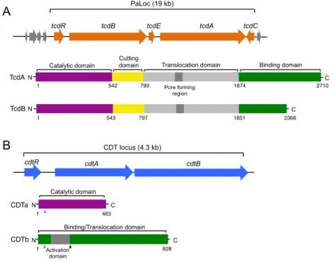
Figure 1.3. C. difficile toxins. (A) Schematic representation of the C. difficile
pathogenicity locus (PaLoc). The structural organization of the two large toxins TcdA and TcdB, encoded by the tcdA and tcdB genes, respectively, is shown in detail. (B) Schematic representation of the C. difficile CDT locus, encoding the binary toxin. Structural domains of the CDTa and CDTb polypeptides are shown in detail. In Cdtb, grey arrowheads point to signal peptide cleavage sites, while the black arrowhead points to the proteolytic activation site. Adapted from Rupnik et al., 2009 and Shen, 2012.
Cell surface and adhesion
Colonization is an important step in C. difficile pathogenesis.
Adhesion factors, such as the S-‐layer proteins, cover the C. difficile cell
surface and facilitate binding to the epithelial cells and components of the
extracellular matrix fibres, allowing colonization (Spigaglia et al., 2013).
Several other proteins located at the cell surface play a role in the adherence
to the intestinal epithelial cells. These include the cell wall proteins Cwp66
fibronectin-‐binding protein (Hennequin et al., 2003), the lipoprotein CD0873
(Kovacs-‐Simon et al., 2014) and the FliC-‐FliD components of the flagella
(Tasteyre et al., 2001). Beyond the role in adhesion, most of these proteins
are immunodominant and are able to induce an immune response in the
host, either inflammatory or regulatory. For these reasons, both the flagellar
antigens FliC and FliD as well as the surface protease Cwp84 have been
proposed as potential vaccine candidates (Pechine et al., 2005).
SPORE FORMATION
Two classes of Firmicutes are able to produce bacterial endospores:
the Bacilli, that includes the extensively studied model organism Bacillus
subtilis, and the Clostridia, to which C. difficile belongs. Spores produced by C.
difficile are crucial for the survival of this anaerobe outside the colonic
environment. Spores constitute the most resilient cell form known, they are
hard to eradicate and can accumulate and persist in the surfaces of health
care institutions for long periods of time, without loosing viability (Nicholson
et al., 2000;
Carroll and Bartlett, 2011;
Maroo and Lamont, 2006; Lawley et
al., 2009). Animals infected with a C. difficile strain that is unable to sporulate
are impaired in C. difficile mice-‐to-‐mice transmission and persistence within
the infected animal (Deakin et al., 2012). These findings highlight the
importance of spores in C. difficile transmission and recurrence. In addition,
higher in vitro sporulation rates have been reported for some 027 strains
responsible for CDI outbreaks, but a straight correlation could not be
established (Merrigan et al., 2010; Akerlund et al., 2008;
Burns et al., 2010a).
Nevertheless, studies carried in animals show that at least one epidemic 027
strain is more efficiently transmitted to uninfected animals than virulent
strains from other ribotypes (Lawley et al., 2012).
Despite the central role played by the spores in C. difficile
comparison with other pathogenesis determinants, such as toxin production.
In an effort to invert this bias, here we focused our attention on the
regulation of sporulation and on the morphology of bacterial spores. The
development of new tools to genetically manipulate C. difficile allowed us to
gain insight on spore formation, structure and composition in this organism
(Underwood et al., 2009; Lawley et al., 2009;
Permpoonpattana et al., 2011;
Permpoonpattana et al., 2013;
Barra-‐Carrasco et al., 2013;
Pizarro-‐Guajardo
et al., 2014;
Burns et al., 2010b; Putnam et al., 2013; Pettit et al., 2014). Still,
most of our knowledge about this process comes from the extensively
studied model organism B. subtilis.
An overview of the sporulation process
In the model organism B. subtilis sporulation is mainly induced by
nutrient exhaustion, and is controlled through the phosphorylation of the
Spo0A response regulator via an expanded “two-‐component” signal
transduction system called the phosphorelay (Burbulys et al., 1991)
(Sonenshein, 2000;
Fujita and Losick, 2005)
.
Once phosphorylated, Spo0Ainduces changes in the transcription of more than 500 genes (Molle et al.,
2003). Phosphorylated Spo0A activates the genes coding for the first
sporulation-‐specific regulators, as well as genes required for the asymmetric
division of the cell
(Rosenbusch et al., 2012;
Errington, 2003; Hilbert and
Piggot, 2004; Higgins and Dworkin, 2012). Asymmetric division originates
two compartments of unequal sizes, the large mother cell compartment and
the small forespore compartment (Figure 1.4A) (Piggot and Coote, 1976;
Stragier and Losick, 1996;
Piggot, 2002;
Errington, 2003). Following
asymmetric division, the mother cell membrane migrates around the
forespore and engulfs it. Once this engulfment process is completed the
forespore becomes a free protoplast inside the mother cell. A thick
concentric layer of peptidoglycan, the cortex, is then synthesized between
layer of dozens of proteins, named spore coat, is deposited around the cortex
(Figure 1.4A). These layers will confer to the newly formed spore the ability
to survive to harsh conditions once it is released from the mother cell (Driks,
1999; Henriques and Moran, 2007;
McKenney et al., 2012). Though
sporulation takes about 8-‐10 hours, vegetative growth is resumed in only a
few minutes, as soon as the spore senses the presence of nutrients in the
surrounding medium, following a process called germination (Figure 1.4A).
Regulation of sporulation
Sporulation involves the expression of a large number of genes. In B.
subtilis, the process is controlled by four RNA polymerase sigma factors that
regulate gene expression during the course of spore morphogenesis (Higgins
and Dworkin, 2012;
Hilbert and Piggot, 2004; Piggot, 2002;
Errington, 2003;
Stragier and Losick, 1996). The sigma factors of sporulation belong to the σ70
family of bacterial sigma factors, which recognize promoters with conserved
elements located near -‐35 and -‐10 with respect to the transcriptional start
site
(Doi and Wang, 1986).
The first sigma factors of sporulation, σF and σE, are activated soon
after asymmetric division and control the early stages of spore assembly. At
later times, these are replaced by σG and σK, respectively, which control the
final stages of spore formation (Figure 1.4A and B). Therefore, inactivation of
both σF and σE results in blockage of the process soon after asymmetric
division, while inactivation of σG and σK results in a blockage of the process
soon after engulfment completion (Piggot and Coote, 1976; Stragier and
Losick, 1996; Piggot, 2002). The activity of these sigma factors is not only
segregated in time, but also in space. While the activities of σF and σG are
restricted to the forespore compartment, the activities of σE and σK are
restricted to the mother cell compartment (Figure 1.4A and B). Activation of
σG and σK requires the preceding σF and σE factors to be active in the
! ! ! ! σF σE σE σF
σK σG
σK σG
Vegetative cycle
A B
Forespore Mother cell Ea rl y Late σF σE
σK σG
a b c d e f Sp o ru la ti o n
2004; Piggot, 2002; Errington, 2003;
Stragier and Losick, 1996). Thus,
asymmetric division sets in motion two parallel programs of gene
expression, the forespore and the mother cell lines of gene expression.
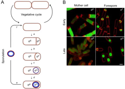
Figure 1.4. Morphological stages and compartmentalized gene expression of B.
subtilis sporulation. (A) The life cycle of B. subtilis. In a nutrient rich medium, the cell
grows and divides by symmetric division (vegetative cycle). However, upon starvation, the cell enters in sporulation. The process begins with an asymmetric cell division (a). The mother cell membrane then migrates around the forespore (b), engulfing it. At the end of this process, the forespore becomes a free protoplast in the mother cell cytoplasm (c). The cortex peptidoglycan (blue) and coat (red) layers are then synthesized and deposited around the developing spore (d). Upon mother cell lysis, a mature spore is released to the surrounding environment, where it remains in a dormant state (e). The spore can then germinate (f). The compartment and main periods of activity of the sporulation σF, σE, σG
and σK sigma factors are indicated. (B) Activity of the sporulation σF, σE, σG and σK factors observed by fluorescence microscopy. Fluorescent microscopy images of sporulating cells carrying fusions of the promoters of the following genes to GFP: σF: yuiC; σE: yhaX; σG:
yhcV; σK: yxeE. Sporulating cells were stained with the membrane dye FM4-64 (red).
Images are the overlap of the green (GFP) and red (FM4-64) channels. Scale bar: 1 µm. The fluorescence images were obtained by Mónica Serrano.
The forespore line of gene expression
The first sporulation-‐specific sigma factor to be activated is σF, in the
forespore compartment. σF is the product of the spoIIAC gene, the third gene
of the spoIIA operon, whose transcription is controlled by phosphorylated
Spo0A and by σH (a sigma factor that governs the transition from the late-‐