Relapse of congenital thrombotic thrombocytopenic
purpura, after spontaneous remission, in a second-trimester
primigravida: case report and review of the literature
Recidiva, após remissão espontânea, de púrpura trombocitopênica trombótica
congênita em primigesta no segundo trimestre de gestação: relato de caso
e revisão da literatura
Donavan de Souza Lúcio
I,
Jacqueline Foelkel Pignatari
II, Marcelo Gil Cliquet
III, Henri Augusto Korkes
IVFaculdade de Ciências Médicas e da Saúde (FCMS), Pontifícia Universidade Católica de São Paulo (PUC-SP), São Paulo (SP), Brazil
ABSTRACT
CONTEXT: Thrombotic microangiopathy syndrome or thrombotic thrombocytopenic purpura-hemolytic uremic syndrome (TTP-HUS) describes distinct diseases sharing common pathological features: microan-giopathic hemolytic anemia and thrombocytopenia, without any other apparent cause.
CASE REPORT: An 18-year-old second-trimester primigravida presented with a history of ifteen days of intense weakness, followed by diarrhea over the past six days. She reported having had low platelets since childhood, but said that she had never had bleeding or menstrual abnormalities. Laboratory investiga-tion showed anemia with schistocytes, thrombocytopenia and hypohaptoglobulinemia. Red blood cell concentrate and platelet transfusions were performed. The hypothesis of TTP or HUS was put forward and ADAMTS13 enzyme activity was investigated. The patient evolved with increasing platelet counts, even without speciic treatment, and she was discharged. One month afterwards, she returned present-ing weakness and swollen face and legs, which had developed one day earlier. The ADAMTS13 activity was less than 5%, without presence of autoantibodies. Regarding the two previous admissions (at 9 and 16 years of age), with similar clinical features, there was spontaneous remission on the irst occasion and, on the second, the diagnosis of TTP was suspected and plasmapheresis was performed, but ADAMTS13 activity was not investigated.
CONCLUSION: To date, this is the only report of congenital TTP with two spontaneous remissions in the literature This report reveals the importance of suspicion of this condition in the presence of microangio-pathic hemolytic anemia and thrombocytopenia without any other apparent cause.
RESUMO
CONTEXTO: A síndrome de microangiopatia trombótica, ou púrpura trombocitopênica trombótica-sín-drome hemolítico urêmica (PTT-SHU), descreve doenças diversas com clínica e achados patológicos co-muns: anemia hemolítica microangiopática e trombocitopenia, na ausência de outra causa aparente.
RELATO DO CASO: Primigesta de 18 anos no segundo trimestre apresenta-se com quadro de 15 dias de fraqueza intensa seguida por diarreia há seis dias. Relata ter plaquetas baixas desde a infância e nega san-gramentos e anormalidades menstruais. Investigação laboratorial identiicou anemia com esquizócitos, plaquetopenia e hipo-haptoglobulinemia. Foi realizada transfusão de plaquetas e concentrado de hemá-cias. A hipótese de PTT ou SHU foi aventada e realizou-se pesquisa da atividade da enzima ADAMTS13. A paciente evoluiu com elevação das plaquetas, mesmo sem tratamento especíico, tendo alta. Retor-nou após um mês da alta com queixa de fraqueza há um dia e inchaço de face e pernas. A atividade da ADAMTS13 foi menor que 5%, sem autoanticorpos. Nas duas internações anteriores (aos 9 e 16 anos), com quadros similares, houve remissão espontânea na primeira internação e, na segunda, o diagnóstico de PTT foi suspeitado e foi realizada plasmaférese, porém sem a pesquisa da atividade da ADAMTS13.
CONCLUSÃO: Até esta data, este é único relato de TTP congênita com duas remissões espontâneas na literatura. Este relato revela a importância da suspeição desta patologia na presença de anemia hemolítica microangiopática e trombocitopenia sem outra causa aparente.
IMD. Family Medicine Resident, Municipal Health Department, Prefeitura Municipal de Florianópolis (PMF-SC), Florianópolis (SC), Brazil. IIMD. Internal Medicine Resident, Department
of Internal Medicine, Faculdade de Ciências Médicas e da Saúde (FCMS), Pontifícia Universidade Católica de São Paulo (PUC-SP), São Paulo (SP), Brazil.
IIIMD, MSc, PhD. Chairman, Department of
Hematology, Faculdade de Ciências Médicas e da Saúde (FCMS), Pontifícia Universidade Católica de São Paulo (PUC-SP), São Paulo (SP), Brazil. IVMD, MSc. Attending Physician, Department of Obstetrics and Gynecology, Faculdade de Ciências Médicas e da Saúde (FCMS), Pontifícia Universidade Católica de São Paulo (PUC-SP), São Paulo (SP), Brazil.
KEY WORDS:
Purpura, thrombotic thrombocytopenic. Anemia, hemolytic.
Pregnancy, high-risk. Stillbirth.
ADAM proteins
PALAVRAS-CHAVE:
Púrpura trombocitopênica trombótica. Anemia hemolítica.
INTRODUCTION
hrombotic microangiopathy syndrome (TMS) or thrombotic thrombocytopenic purpura-hemolytic uremic syndrome (TTP-HUS) describes distinct diseases sharing common pathological features: microangiopathic hemolytic anemia (deined by the presence of schistocytes in blood smears) and thrombocytope-nia (with or without neurological or renal abnormalities) without any other apparent cause.1-3
he variety of presentations and lack of speciic diagnostic criteria for TTP-HUS hinder and delay its recognition and treat-ment by means of plasmapheresis.1 Children with
microangio-pathic hemolytic anemia, acute renal failure and thrombocytope-nia have been classiied as presenting hemolytic uremic syndrome (HUS). his disease is typically preceded by diarrhea and abdomi-nal pain, caused by Shiga toxins that are produced by bacteria such as Escherichia coli 0157:H7. It has low mortality, and 91% of
typi-cal HUS children survive without plasmapheresis, thus suggesting that TTP and HUS are two diferent syndromes.1 However, their
diagnostic criteria are the same, and although renal failure and neurological abnormalities are characteristic of HUS and TTP, respectively, these features may never occur.1
Several conditions, such as infections, surgery and preg-nancy can precipitate TMS.3 Congenital TTP is the most
fre-quent manifestation of TMS during pregnancy.3,4 This is a rare
disorder with only just over 100 case descriptions worldwide.5
It is caused by mutations in the ADAMTS13 enzyme (“a disinte-grin and metalloprotease with a thrombospondin type 1 motif, member 13”). This is a protease that cleaves von Willebrand factor, which is a multimer synthesized by endothelium that, if degradation does not occur, accumulates and thus leads to spon-taneous formation of microthrombi in the microcirculation.6,7
Congenital TTP is characterized by low ADAMTS13 activity in the absence of anti-ADAMTS13 autoantibodies, which are present in acquired TTP.8
he initial evaluation should investigate possible secondary causes such as pregnancy, autoimmune disease and infection by the human immunodeiciency virus. It needs to rule out other causes of microangiopathy such as neoplasia and disseminated intravascular coagulation.5 Presence of a family history may suggest congenital
cases.5 Its main diferential diagnoses during pregnancy include
disseminated intravascular coagulation and the HELLP syndrome (hemolysis, elevated liver-enzyme levels and low platelets).9
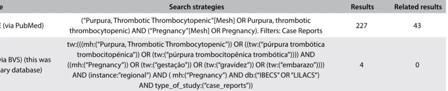
We searched the MEDLINE (via PubMed), LILACS (via BVS) and UpToDate databases for the terms: “Congenital Thrombotic Thrombocytopenic Purpura”, “Hereditary hrombotic hrombocytopenic Purpura”, “Familial hrombotic hrombocytopenic Purpura”, “Upshaw–Schulman syndrome”, “hrombotic hrombocytopenic Purpura”, “Microangiopathic Hemolytic Anemia”, “Pregnancy” and “ADAMTS13” (Table 1). We found 43 cases of congenital thrombotic thrombocytopenic purpura in pregnant women reported in the literature.
Case report
An 18-year-old primigravida, with a fetus of 16 weeks of ges-tational age, presented to the obstetrics emergency department with a condition of intense weakness, followed by diarrhea, that she had had for 15 days. She said that she did not have any fever, headache, bleeding, seizures, neurological abnormalities, urinary abnormalities, rash or shortness of breath, and that she was not using any medications.
She reported that she had had a condition of “low platelets” since childhood (but without any bleeding and with regular men-strual cycle and low), and that she had been admitted to this hos-pital (without remembering any details), where she was followed up by the hematology and nephrology departments, with discharge from the hospital three years before the present case.
In the medical iles relating to this patient, we found two pre-vious admissions to the emergency department of this hospital. At the irst admission, when she was nine years old, she presented with acute hemolysis, uremia and ascites, which progressed to urosep-sis and seizures. She was then kept in hospital for treatment with steroids, antibiotic therapy and peritoneal dialysis. Subsequently, she was followed up as an outpatient by the hematology depart-ment and she evolved with spontaneous and sustained normaliza-tion of platelets and hemoglobin, upon which she was discharged with orientations.
At the second admission, she was 16 years old and presented with abdominal pain, vomiting and hematuria. In the investigation,
Database Search strategies Results Related results
MEDLINE (via PubMed) (“Purpura, Thrombotic Thrombocytopenic”[Mesh] OR Purpura, thrombotic
thrombocytopenic) AND (“Pregnancy”[Mesh] OR Pregnancy). Filters: Case Reports 227 43
LILACS (via BVS) (this was a secondary database)
tw:(((mh:(“Purpura, Thrombotic Thrombocytopenic”)) OR ((tw:(“púrpura trombótica trombocitopénica”)) OR (tw:(“púrpura trombocitopênica trombótica”)))) AND ((mh:(“Pregnancy”)) OR (tw:(“gestação”)) OR (tw:(“gravidez”)) OR (tw:(“embarazo”))))
AND (instance:”regional”) AND ( mh:(“Pregnancy”) AND db:(“IBECS” OR “LILACS”) AND type_of_study:(“case_reports”))
4 0
Coombs-negative hemolytic anemia with thrombocytopenia and elevated blood urea nitrogen were found. An abdominal ultra-sound showed mild alteration of renal texture. She was kept in hospital to receive a methylprednisolone pulse because of the suspicion of rapidly progressive glomerulonephritis. Presence of schistocytes in blood smears suggested a diagnosis of TTP or HUS. Plasmapheresis was performed and steroids were administered, without further investigations. Because her condition evolved with clinical and laboratory improvement, she was discharged with a prescription for prednisone 40 mg/day and was given generic fol-low-up orientations.
On physical examination in the present case, she had an axil-lary temperature of 37.9 °C (100.22 °F), oxygen saturation of 96% in room air, blood pressure of 110 x 80 mmHg, slight jaundice and pallor. Fetal heartbeats were present and normal.
Laboratory tests showed hemoglobin of 4.2 g/dl, 13,000 plate-lets/mm³, hematuria, lactate dehydrogenase (LDH) of 2,014 U/l, erythrocyte sedimentation rate (ESR) of 135 mm/h, and C-reactive protein (CRP) of 62.4 mg/l. Prothrombin and activated thrombo-plastin time were normal. hus, we ordered the tests summarized in Table 2. his investigation showed presence of anemia with reticulocytosis, schistocytes in blood smears, hypohaptoglobu-linemia, let-shit leukocytosis, secondary iron overload due to hemolysis and elevation of D-dimer and antiphospholipid anti-bodies (Table 2).
Red blood cell and platelet transfusions were performed. he hypothesis of TTP or HUS was suggested and we requested an analysis on ADAMTS13 activity. Increasing platelet levels and normalization of hemoglobin were observed, even without spe-ciic treatment. he patient was instructed to continue treatments as an outpatient of the nephrology department. Evaluation of
prothrombin and activated thromboplastin time was requested and both of these were normal.
One month ater having been discharged, she returned to this hospital complaining of weakness, with swollen face and legs that had developed one day earlier. She was in a good general condition, with presence of normal fetal heartbeats. At this new admission, she had the same laboratory abnormalities, but this time her creatinine was 1.6 mg/dl. A hematological assessment was requested: the results from the ADAMTS13 activity analy-sis had just been received. he analyanaly-sis showed that the activity level was less than 5%, without any presence of anti-ADAMTS13 autoantibodies. Ater this conirmation of the presence of con-genital TTP, plasmapheresis was scheduled and plasma infusion was implemented at a dose of 10 ml/kg every 8 hours while wait-ing for the plasmapheresis.
However, plasmapheresis was delayed because of the high risk of bleeding. During this period, she developed paresthesia in the face and limbs and became fatigued. he fetal heartbeats became inaudible and fetal death was conirmed by means of ultrasound. Delivery was induced using misoprostol, which led to expulsion of the conceptus ater 15 hours. Histopathological examination on the fetus showed that it weighed 318 grams and had a gestational age compatible with 16-17 weeks of pregnancy, with a second-trimester hyalinized placenta.
The feasibility of plasmapheresis was confirmed four days after confirmation of the diagnosis and indication of the pro-cedure. Plasma exchange was performed using 16 fresh fro-zen plasma units, with a good response, and the patient was returned to a plasma infusion regimen two days later. Because she responded to maintenance therapy, we then discharged her for outpatient treatment.
*ESR = erythrocyte sedimentation rate; PT/INR = prothrombin time/international normalized ratio; aPTT = activated partial thromboplastin time; LDH = lactate dehydrogenase; CRP = C-reactive protein.
Complete blood count Other tests
Erythrocytes 1.5 x 10⁹ cells/mm³ (4.2-5.2) ESR* 134 mm/h (< 15)
Hemoglobin 5 g/dl (12-15) PT/INR* 12’’/1.03 (9.5-13.5/0.8-1.2)
Hematocrit 12.6% (37-47) aPTT* 26’’ (28-42)
White blood cells 11.6 x 10³ cells/mm³ (5-10) Bilirubin 0.7 mg/dl (< 1.20)
Metamyelocytes 0% (0) Creatinine 0.9 mg/l (0.6-1.1)
Band cells 6% (0) Urea 28 mg/dl (15-40)
Neutrophils 53% (52-72) LDH* 1,072 Ul (200-480)
Lymphocytes 31% (20-30) CRP* 61.7 mg/l (< 6)
Eosinophils 2% (2-4) D-dimer 773 FEU/ml (< 500)
Monocytes 8% (4-8) Fibrinogen 442 mg/dl (160-465)
Platelets 9 x 10³ mm³ (130-450) Complement C3c 122 mg/dl (83-193)
DISCUSSION
hrough descriptions of TTP cases, reviewed in 1966, the clin-ical pentad formed by anemia, thrombocytopenia, fever and neurological and kidney disorders became the diagnostic crite-ria for TTP.10 Nowadays, with the availability of plasmapheresis,
only thrombocytopenia and microangiopathic hemolytic ane-mia are necessary for suspicion of TTP and for implementation of early treatment.11 Ater the irst case report of TTP associated
with pregnancy, subsequent descriptions found the same asso-ciation, irst manifested through gastrointestinal symptoms and then through hypertension closer to term, with serious neuro-logical and renal abnormalities. hese cases evolved to death through disseminated intravascular coagulation, in the absence of plasmapheresis.12 he major features of acquired TTP,
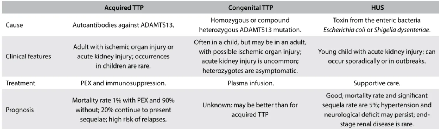
con-genital TTP and hemolytic uremic syndrome are summarized in Table 3.
he incidence of TTP is 1 in 25,000 to 1 in 198,000 pregnan-cies. he outcome is oten unfavorable when it is presented in the second trimester and, without prophylaxis, recurrence in future pregnancies is close to 100%.4 he main factors contributing to
occurrence or recurrence of TTP during pregnancy are a hyperco-agulable state and progressive deiciency of ADAMTS13 over the course of gestation.12 Fibrinogen, factor VIII and von Willebrand
factor levels have been found to increase up to threefold during pregnancy.12,13 he physiological increase in von Willebrand
fac-tor concentration seems to be directly related to the decrease in ADAMTS13 activity. hus, women with congenital ADAMTS13 deiciency may become severely disabled during pregnancy.12
Relapses, deined as acute events of TTP occurring 30 days ater remission, are seen in 20 to 50% of cases. hey are more common in patients with low ADAMTS13 activity or with autoantibodies ater remission.14
Diferentiation between congenital and acquired TTP is needed, through measurement of ADAMTS13 activity and autoantibodies,
since use of immunosuppressive therapy is critical in cases of acquired TTP but is unnecessary in cases of congenital TTP.8
he largest prospective study on pregnancy-associated TTP eval-uated 47 women.8 Fetal loss occurred in 42% of the congenital
TTP cases before the diagnoses, but without further losses in sub-sequent pregnancies if proper management was instituted. Most cases of TTP occurred in the third trimester, and only 15% of the cases occurred before the 20th week. In all pregnancies in which a
diagnosis of TTP was made, labor was induced at the 38th week,
because of the increased risk of complications that is observed in pregnancies that continue to term and beyond. hat study con-cluded that pregnancy is a precipitating factor for TTP; that there is a risk of relapse in further pregnancies; and that, in pregnancies, late-onset congenital TTP occurs more oten than acquired TTP. Treatment with plasma therapy and antithrombotic agents in fur-ther pregnancies has been found to result in improvement of fetal growth and placental histology, without fetal loss.8
A French study assessed 42 women who had had a irst episode of TTP during pregnancy or postpartum.4 he rate of live births
was only 31% and was associated with the time of onset of this condition: 96% of those in whom it occurred during the irst and second trimester presented abortion or fetal death, compared with 17% among those who had the disease during the third trimester. Only two patients attained remission without plasma therapy, and both of them had congenital TTP.4
In the case reported here, the patient had four episodes of TPP: two before pregnancy (at the ages of 9 and 16 years), and two during pregnancy, both in the second trimester of gestation. During the irst and third episodes of TTP, spontaneous remission (i.e. without any plasma therapy) was observed and, as far as we know, this is the only case reported in the literature. he diagnoses was suspected at the time of the second hospitalization and, if the ADAMTS13 activity level had been investigated, this would have had a great impact on the patient’s life and pregnancy outcome,
TTP = thrombotic thrombocytopenic purpura; HUS = hemolytic uremic syndrome; ADAMTS13 = “a disintegrin and metalloprotease with a thrombospondin type 1 motif, member 13”; PEX = plasma exchange.
Acquired TTP Congenital TTP HUS
Cause Autoantibodies against ADAMTS13. Homozygous or compound heterozygous ADAMTS13 mutation.
Toxin from the enteric bacteria Escherichia coli or Shigella dysenteriae.
Clinical features
Adult with ischemic organ injury or acute kidney injury; occurrences
in children are rare.
Often in a child, but may be in an adult, with possible ischemic organ injury;
acute kidney injury is uncommon; heterozygotes are asymptomatic.
Young child with acute kidney injury; can occur sporadically or in outbreaks.
Treatment PEX and immunosuppression. Plasma infusion. Supportive care.
Prognosis
Mortality rate 1% with PEX and 90% without; 20% continue to present
sequelae; high risk of relapses.
Unknown; may be better than for acquired TTP
Good; mortality rate and signiicant sequela rate are 5%; hypertension and
since the diagnosis would have been known and she would prob-ably have had diferent management.
It can be seen that suspicion of this disease is impaired through persistence of the “classical pentad” concept and the diiculty in characterizing signs and symptoms as part of a single syndrome. he diferential diagnoses of microangiopathic hemolytic anemia and thrombocytopenia during pregnancy are summarized in Table 4.
he hemoglobinuria seen in our patient’s previous manifesta-tions and the onset of paresthesia during the current case relect the renal and neurological impairments of the disease that occur in severe cases. he presence of “let shit” leukocytosis, elevated inlammatory markers, leukocyturia and hematuria are oten confused with infectious condition,15 which should always be a
diferential diagnosis and should be ruled out through cultures. It is known that systemic infections may mimic TTP.15 From
anal-ysis on 415 consecutive patients in the Oklahoma TTP Registry who were initially diagnosed with TTP, two distinct groups were identi-ied: 25 patients with measured ADAMTS13 activity whose symp-toms were later attributed to systemic infections; and 62 patients with ADAMTS13 activity < 10% whose symptoms remained assigned to TTP. In the “systemic infections” group, more cases with fever, coma and the classical TTP pentad were identiied, along with more lethal cases. In the TTP group, 31% did not show any neurological abnor-malities, but the presence of relapses and focal deicits was higher and the average platelet and hematocrit levels were lower than in the irst group. his study revealed that the classical clinical pentad is rare among cases of TTP and is much more common in cases of sepsis. Nevertheless, it still has great inluence on many physicians, since there is usually a belief that if more features of the pentad are present, the diagnosis of TTP becomes more likely.15
Since no anti-ADAMTS13 autoantibodies are present in cases of congenital TTP, treatment of acute cases by means of plasma exchange or plasma infusion alone is appropriate.3,5,14 Ater
remis-sion of acute symptoms has been achieved, the treatment should be individualized according each patient’s phenotype. Some patients may require monthly plasma infusions to restore ADAMTS13 and prevent symptomatic episodes, while others only require prophylactic therapy under risky conditions such as surgery,
infections, pregnancies and presentation of thrombocytopenia.3,5
A study involving four European TTP registries found that low residual ADAMTS13 activity was associated with early manifes-tations requiring plasma therapy and with higher frequency of recurrences.16 Since congenital TTP is hereditary, evaluation of
ADAMTS13 activity among close relatives should be considered.14
Blood transfusions and folic acid supplementation are indi-cated during hemolysis, especially if there is cardiac impairment.5,14
However, platelet transfusions are relatively contraindicated and are only acceptable when there is life-threatening bleeding.5,14
Patients with TTP who receive platelet transfusions have a higher chance of arterial thrombosis (adjusted odds ratio, adjOR = 5.8; 95% conidence interval, CI = 1.3-26.6) and myocardial infarction (adjOR = 2.0; 95% CI = 1.2-3.3), and higher mortality (adjOR = 2.0; 95% CI = 1.3-3.0).17
In managing congenital TTP in subsequent pregnancies, low doses of acetylsalicylic acid in association with regular plasma infu-sion are recommended as soon the pregnancy has been conirmed, starting at a dose of 10 ml/kg every two weeks, and changing to once a week ater the 20th week.8 If the patient is
thrombocyto-penic, the plasma dose may be increased to 15 ml/kg. It is recom-mended that delivery should take place between the 36th and 38th
week because this has been proven to reduce fetal loss and relapses.8
CONCLUSION
his report reveals the importance of suspicion of TTP in the presence of microangiopathic hemolytic anemia and thrombo-cytopenia that do not have any apparent cause. Waiting for the classical pentad to appear before making the suspected diagnosis and introducing treatment is unwise and can seal the outcome. Because of the risks involved in plasmapheresis, presence of sys-temic infection mimicking TTP needs to be ruled out.
To date, the present case provides the only description of con-genital TTP with two spontaneous remissions in the literature. Spontaneous remissions may relect a milder phenotype of TTP. Obtaining clariications from patients regarding their condition is essential for identifying the factors that may precipitate worsening of disease and for managing possible recurrences.
TTP = thrombotic thrombocytopenic purpura; DIC = disseminated intravascular coagulation; HELLP = hemolysis, elevated liver-enzyme levels and low platelets.
Condition Onset Characteristic features Treatment
TTP Any time Microangiopathic hemolytic anemia and severe thrombocytopenia; renal, hepatic and neurological abnormalities may not be present; absence of DIC.
Plasma infusion (congenital TTP); plasma exchange (acquired TTP).
Severe preeclampsia
and HELLP syndrome After 20
th week
Microangiopathic hemolytic anemia, thrombocytopenia and elevated liver function tests; hypertension; presence of DIC in severe cases; may become
indistinguishable from TTP.
Delivery
Systemic infections Any time Fever, hypotension, mild anemia (may be microangiopathic) and
REFERENCES
1. George JN. Clinical practice. Thrombotic thrombocytopenic purpura.
N Engl J Med. 2006;354(18):1927-35.
2. George JN. How I treat patients with thrombotic thrombocytopenic
purpura: 2010. Blood. 2010;116(20):4060-9.
3. George JN, Nester CM. Syndromes of thrombotic microangiopathy. N
Engl J Med. 2014;371(7):654-66.
4. Moatti-Cohen M, Garrec C, Wolf M, et al. Unexpected frequency
of Upshaw-Schulman syndrome in pregnancy-onset thrombotic
thrombocytopenic purpura. Blood. 2012;119(24):5888-97.
5. Blombery P, Scully M. Management of thrombotic thrombocytopenic
purpura: current perspectives. J Blood Med. 2014;5:15-23.
6. Furlan M, Robles R, Galbusera M, et al. von Willebrand factor-cleaving
protease in thrombotic thrombocytopenic purpura and the
hemolytic-uremic syndrome. N Engl J Med. 1998;339(22):1578-84.
7. Levy GG, Nichols WC, Lian EC, et al. Mutations in a member of the
ADAMTS gene family cause thrombotic thrombocytopenic purpura.
Nature. 2001;413(6855):488-94.
8. Scully M, Thomas M, Underwood M, et al. Thrombotic thrombocytopenic
purpura and pregnancy: presentation, management, and subsequent
pregnancy outcomes. Blood. 2014;124(2):211-9.
9. McMinn JR, George JN. Evaluation of women with clinically suspected
thrombotic thrombocytopenic purpura-hemolytic uremic syndrome
during pregnancy. J Clin Apher. 2001;16(4):202-9.
10. Amorosi EL, Ultmann JE. Thrombotic thrombocytopenic purpura:
report of 16 cases and review of the literature. Medicine.
1966;45(2):139-60. Available from:
http://journals.lww.com/md-journal/Citation/1966/03000/THROMBOTIC_THROMBOCYTOPENIC_
PURPURA__REPORT_OF_16.3.aspx. Accessed in 2016 (Dec 27).
11. Allford SL, Hunt BJ, Rose P, Machin SJ; Haemostasis and Thrombosis Task
Force, British Committee for Standards in Haematology. Guidelines on
the diagnosis and management of the thrombotic microangiopathic
haemolytic anaemias. Br J Haematol. 2003;120(4):556-73.
12. George JN. The association of pregnancy with thrombotic
thrombocytopenic purpura-hemolytic uremic syndrome. Curr Opin
Hematol. 2003;10(5):339-44.
13. Stirling Y, Woolf L, North WR, Seghatchian MJ, Meade TW. Haemostasis
in normal pregnancy. Thromb Haemost. 1984;52(2):176-82.
14. Scully M, Hunt BJ, Benjamin S, et al. Guidelines on the diagnosis and
management of thrombotic thrombocytopenic purpura and other
thrombotic microangiopathies. Br J Haematol. 2012;158(3):323-35.
15. Booth KK, Terrell DR, Vesely SK, George JN. Systemic infections
mimicking thrombotic thrombocytopenic purpura. Am J Hematol.
2011;86(9):743-51.
16. Lotta LA, Wu HM, Mackie IJ, et al. Residual plasmatic activity of ADAMTS13
is correlated with phenotype severity in congenital thrombotic
thrombocytopenic purpura. Blood. 2012;120(2):440-8.
17. Goel R, Ness PM, Takemoto CM, et al. Platelet transfusions in platelet
consumptive disorders are associated with arterial thrombosis and
in-hospital mortality. Blood. 2015;125(9):1470-6.
Sources of funding: None
Conlict of interest: None
Date of irst submission: July 23, 2016
Last received: November 17, 2016
Accepted: November 20, 2016
Address for correspondence:
Donavan de Souza Lúcio
Secretaria Municipal da Saúde de Florianópolis, Prefeitura Municipal de
Florianópolis (PMF-SC)
Rodovia João Paulo, 1.268
João Paulo — Florianópolis (SC) — Brasil
CEP 88030-300
Tel. +55 (48) 3348-1288