Daniella Torres Tagawa1, Carolina Loyo Sérvulo da Cunha Bertoni1, Maria Angélica Estrada Mari1,
Milton Redivo Junior1, Luís Antônio de Arruda Aidar2
Orthopedic treatment of Class III malocclusion with
rapid maxillary expansion combined with a face mask: A
cephalometric assessment of craniofacial growth patterns
Objective: The aim of this prospective study was to assess potential changes in the cephalometric craniofacial growth pattern of 17 children presenting Angle Class III malocclusion treated with a Haas-type expander com-bined with a face mask.
Methods: Lateral cephalometric radiographs were taken at beginning (T1) and immediately after removal of the
ap-pliances (T2), average of 11 months of treatment. Linear and angular measurements were used to evaluate the cranial
base, dentoskeletal changes and facial growth pattern.
Results: The length of the anterior cranial base experienced a reduction while the posterior cranial base assumed
a more vertical position at T1. Some maxillary movement occurred, there was no rotation of the palatal plane, there
was a slight clockwise rotation of the mandible, although not significant. The ANB angle increased, thereby im-proving the relationship between the jaws; dentoalveolar compensation was more evident in the lower incisors. Five out of 12 cases (29.41%) showed the following changes: In one case the pattern became more horizontal and in four cases more vertical.
Conclusions: It was concluded after a short-term assessment that treatment with rapid maxillary expansion
(RME) associated with a face mask was effective in the correction of Class III malocclusion despite the changes in facial growth pattern observed in a few cases.
Keywords: Angle Class III malocclusion. Cephalometrics. Headgear appliances. Maxillary expansion.
How to cite this article: Tagawa DT, Bertoni CLSC, Mari MAE, Redivo Junior M, Ai-dar LAA. Orthopedic treatment of Class III malocclusion with rapid maxillary expan-sion combined with a face mask: A cephalometric assessment of craniofacial growth patterns. Dental Press J Orthod. 2012 May-June;17(3):118-24.
Submitted: August 05, 2009 - Revised and accepted: April 27, 2011
» The authors report no commercial, proprietary, or financial interest in the products or companies described in this article.
» Patients displayed in this article previously approved the use of their facial and in-traoral photographs.
Contact address: Daniella Torres Tagawa R. Luis Suplici, 79 – Gonzaga Santos Zip code: 11055-330 – São Paulo/SP – Brazil E-mail: [email protected]
1 Trainee in Orthodontics, Dentistry School, Universidade Santa Cecília.
2 Professor of Orthodontics and Chairman of Specialization course in Orthodontics,
INTRODUCTION
Class III malocclusion defined as a facial skeletal discrepancy, may result from a variety of morpho-logical combinations between maxilla and man-dible, both in the sagittal direction (mandibular prognathism, maxillary retraction, or a combina-tion thereof ) and in the vertical direccombina-tion (excess or decrease in lower anterior facial height).1,2,9,27,30
It has been estimated that the prevalence of Class III malocclusion among Japanese and
Chi-nese is around 14% of the population.19 In 1994,
an epidemiological study conducted in the region of Bauru, Brazil, found that this malocclusion is prevalent in 3% of all patients assessed.22
Before 1970, the orthodontic literature treated all Class III malocclusions as mandibular progna-thism. Therefore, many authors were reluctant to discuss maxillary protraction as a viable treatment method, resorting only to the use of a chin cup to
prevent mandibular growth.17
The finding that maxillary deficiency is often a component of skeletal Class III enhanced the potential of orthodontic-orthopedic treatment in promoting maxillary growth.3,5,6,18,27 However, by
the time most of this growth is completed, treat-ment options become limited.1,4,13
Angle Class III with maxillary deficiency, with a well positioned or retruded mandible and a re-duced anterior facial height, provides the best treatment prognosis.13,16,27,28 It should be
empha-sized, however, that this does not mean that one should not tackle Angle Class III with maxillary
deficiency and mild mandibular prognathism.28
Early orthodontic-orthopedic therapy has proven effective from a skeletal standpoint, thus favouring the establishment of growth patterns and normal relationships between facial compo-nents.1,3,23 Although still controversial,7,20 rapid
maxillary expansion (RME) combined with re-verse pull maxillary headgear may be beneficial in early treatment of Class III malocclusion, even in the absence of posterior crossbite4,13,19,23,27. RME
might disarticulate the maxilla and trigger cellular responses in the sutures, thereby strengthening the effects of maxillary protraction.13,27
The purpose of this study was to evaluate po-tential changes in craniofacial growth pattern by
means of lateral radiographs in Class III children treated with RME and face mask.
MATERIAL AND METHODS Material
This prospective study involved 17 Brazilian children with mixed dentition (7 male and 10 fe-male), mean age 8 years and 7 months ± 1 year and 8 months (ranging from 6 years and 1 month to 11 years old), who were treated with a Haas-type ex-pander combined with a Petit face mask to correct Class III malocclusion.
The patients presented the following character-istics:
1 – Angle Class III malocclusion.
2 – A facial Class III pattern due to maxillary deficiency, mandibular excess or a combination of both factors.
3 – Mixed dentition stage. 4 – Good oral health.
This study was approved by the Ethics Commit-tee of Santa Cecília.
Methods
All patients were treated with a modified Haas-type expander8 (Fig 1) and followed a protocol
com-prising one full turn on the first day and a half turn in the subsequent days until overcorrection of the case. In order to facilitate intraoral elastic place-ment, the hooks of the expander were positioned between the canines and first molars, in a horizon-tal direction parallel to the occlusal plane.11,27
Af-ter screw fixation, a Petit face mask (Orthosource, Brazil) was placed with initial force of 350 grams (Fig 2), ultimately reaching 500 grams on each side. The patients were instructed to wear the
mask for at least 14 hours/day.12 The mean
treat-ment time with the face mask was 11 months ± 3 months (ranging from 6 to 18 months).
using acetate paper. All anatomical details of inter-est to this study were highlighted and the variables were measured with a cephalometric protractor (Desetec) and a millimeter ruler (Desetec) with sub-divisions of 0.5° and 0.5 mm, respectively. The fol-lowing cephalometric variables were used:
1. Linear Variables (Fig 3): S-N, S-Ar, Ar-Goc, Me-Goc, S-Goc, N-Me, S-Gnc, N-Goc, Co-A, Co-Gn and ANS-Me.
2. Angular Variables (Fig 4): Sella angle, ar-ticulare angle, gonial angle, superior gonial angle, inferior gonial angle, SNA, SNB, 1.PP, IMPA, SN.PP angle.
The quotient of Siriwat and Jarabak25 was used
to describe facial morphology: The ratio between the posterior facial height (S-Goc) and the anteri-or facial height (N -Me) multiplied by one hundred (100). Any percentage lower than 59% was classi-fied as a hyperdivergent growth pattern, between
59 and 63% a neutral pattern, and above 63% a hy-podivergent pattern (Fig 3).
Statistical Method
To assess data normality, the Kolmogorov Smirnov test was initially applied. After verifying that the distribution of the measured values was symmetrical, the parametric test (t-test) was em-ployed to evaluate potential differences between the linear and angular measures studied at T1 and T2. A 5% significance level was used.
Method Error
To assess method accuracy, radiographs of nine patients from the study sample (n = 17) were randomly selected. All radiographs were traced and measured again by a single operator after a period of one month counted from the original tracing. The paired t-test was applied to evaluate
Figure 1 - Modified Haas-type expander. Figure 2 - Frontal and lateral facial photographs with Petit face mask.
Figure 3 - Linear cephalometric variables. Figure 4 - Angular cephalometric vari-ables.
Co Ar
A N
ANS
Goc
GncGn Me S
Co Ar
A
B ANS
Goc Go
systematic error. Once the difference between the first and second measurements had been obtained for each cephalogram, Dahlberg’s formula was ap-plied to estimate random error.
RESULTS
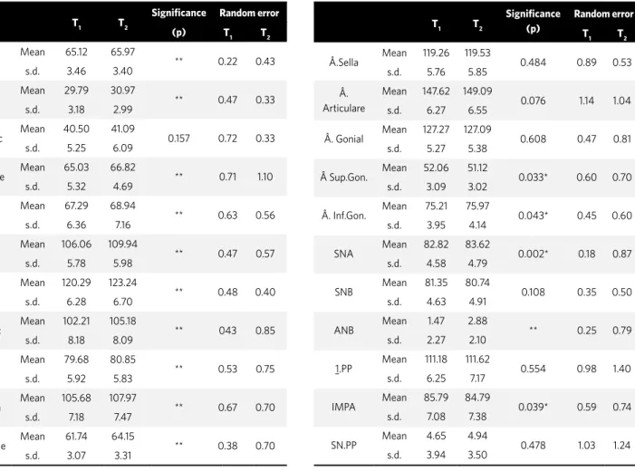
All cases evolved into a Class I correction or a class II overcorrection. Systematic error (bias) was not significant in any of the cases. Random er-ror is depicted in Tables 1 and 2. Ar-Goc was the only linear cephalometric variable that showed no statistically significant difference between T1 and T2 (Table 1). Among the angular variables, the su-perior and inferior Gonial angles SNA, ANB and IMPA showed statistically significant differences between T1 and T2. In the remaining angular mea-sures no significant changes occurred (Table 2).
At T1, 9 cases showed hypodivergent patterns
(52.94%), 5 cases neutral patterns (29.41%) and 3
cases hyperdivergent patterns (17.64%). In 12 cases (70.58%) there were no changes in facial pattern between T1 and T2. In 5 cases (29.41%) the follow-ing changes occurred: Case 2 displayed a hyperdi-vergent pattern, which became neutral, 2 cases (3 and 8) exhibited neutral patterns, which became hyperdivergent, and 2 cases (10 and 17) had hypodi-vergent patterns which ultimately became neutral.
DISCUSSION
Given the difficulty of restraining the man-dibular growth and the plasticity of the maxillary growth, the combination of RME and reverse pull maxillary headgear is a treatment protocol often used in the correction of Angle Class III malocclu-sion.3,6,13,18,21,27 Prognosis of this type of malocclusion
will depend on variables such as etiology and loca-tion of the skeletal problem.4 In this study, patients
were clinically evaluated and facially classified as
Table 1 - Mean and standard deviation (SD) of linear cephalometric
measure-ments (in mm) and random error at T1 and T2. Table 2 -surements (in degrees) and random error at T Mean and standard deviation (SD) of angular cephalometric mea-1 and T2.
T1 T2 Significance (p) Random error
T1 T2
Â.Sella Mean 119.26 119.53 0.484 0.89 0.53 s.d. 5.76 5.85
Â. Articulare
Mean 147.62 149.09
0.076 1.14 1.04 s.d. 6.27 6.55
Â. Gonial Mean 127.27 127.09 0.608 0.47 0.81 s.d. 5.27 5.38
 Sup.Gon. Mean 52.06 51.12 0.033* 0.60 0.70 s.d. 3.09 3.02
Â. Inf.Gon. Mean 75.21 75.97 0.043* 0.45 0.60 s.d. 3.95 4.14
SNA Mean 82.82 83.62 0.002* 0.18 0.87
s.d. 4.58 4.79
SNB Mean 81.35 80.74 0.108 0.35 0.50
s.d. 4.63 4.91
ANB Mean 1.47 2.88 ** 0.25 0.79
s.d. 2.27 2.10
1.PP Mean 111.18 111.62 0.554 0.98 1.40 s.d. 6.25 7.17
IMPA Mean 85.79 84.79 0.039* 0.59 0.74
s.d. 7.08 7.38
SN.PP Mean 4.65 4.94 0.478 1.03 1.24
s.d. 3.94 3.50
T1 T2 Significance Random error
(p) T1 T2
S-N Mean 65.12 65.97 ** 0.22 0.43
s.d. 3.46 3.40
S-Ar Mean 29.79 30.97 ** 0.47 0.33
s.d. 3.18 2.99
Ar-Goc Mean 40.50 41.09 0.157 0.72 0.33 s.d. 5.25 6.09
Goc-Me Mean 65.03 66.82 ** 0.71 1.10
s.d. 5.32 4.69
S-Goc Mean 67.29 68.94 ** 0.63 0.56
s.d. 6.36 7.16
N-Me Mean 106.06 109.94 ** 0.47 0.57
s.d. 5.78 5.98
S-Gnc Mean 120.29 123.24 ** 0.48 0.40
s.d. 6.28 6.70
N-Goc Mean 102.21 105.18 ** 043 0.85
s.d. 8.18 8.09
Co-A Mean 79.68 80.85 ** 0.53 0.75
s.d. 5.92 5.83
Co-Gn Mean 105.68 107.97 ** 0.67 0.70
s.d. 7.18 7.47
ANS-Me Mean 61.74 64.15 ** 0.38 0.70
Class III due to maxillary deficiency, mandibular excess or a combination of both factors. The mag-nitude of skeletal discrepancy was not taken into account as it can be seen in the wide variability ex-hibited by the ANB angle at T1 (mean 1.470 ± 2.270). The present study combined prior expansion with maxillary traction based on the fact that pro-traction in combination with an initial period of expansion may yield more significant skeletal re-sults7,13,18,27 even though expansion produces
unde-sirable dentoalveolar side effects, such as mandib-ular rotation.16 On the other hand, studies showed
that RME does not influence the correction of Class III with a face mask.7,20
A meta-analysis13 of clinical studies that used
face masks was undertaken to determine the most convenient time to employ this treatment meth-od. The authors found major orthopedic altera-tions in younger patients. In summary, maxillary protraction may be effective during the period in which the maxillary sutures are still open. Major orthopedic changes can be achieved and retained in permanent dentition as long as the face mask treatment happens in the deciduous or early mixed dentition.30 In this study the average chronological
age of patients was 8 years and 7 months (ranging from 6 years and 1 month to 11 years old at T1).
Although the treatment goal when using a face mask is to displace the maxilla forward by applying force to the circum-maxillary sutures, there are skeletal and dental changes with forward displace-ment of the maxilla (1-3 mm),2,19 maxillary incisors
flaring, downward and backward mandibular rota-tion and, finally, lingual inclinarota-tion of mandibular incisors.2,5,9,19,29 The orthopedic alterations are
re-sponsible for 75% of the correction (25% dental) with maxillary advancement representing 75% of the skeletal correction (25% due to downward and
backward mandibular rotation).27 In comparison
with the average, the results of this research are in agreement with other findings in the literature. There was an anterior displacement of the maxilla and the mean value of the SNB angle decreased, al-though this reduction was not statistically signifi-cant, suggesting that the downward and backward mandibular rotation increased the ANB angle. Interestingly, although the gonial angle did not
change, the upper and lower gonial angles changed significantly. This is due to the tendency of the mandible to rotate clockwise.
The patients in this study did not show any max-illary rotation. The direction of the force produced by the mask was more horizontal and parallel to the occlusal plane.11,27 The literature shows a high
incidence of anterior movement without rotation.3
The earlier the therapy is started the greater is the anterior displacement due to the release of the pterygomaxillary fissure.2
The anterior and posterior vertical dimensions of the face increased significantly between T1 and T2. When patients were evaluated separately, they showed no facial patterns changes between T1 and T2 in 12 cases (70.5%). The changes followed a more ver-tical pattern In four out of five cases (29.4%) whose facial patterns experienced modifications. In only one case there was a more horizontal pattern. In-creases were found in all linear values, although they were not significant at the level of the ramus. Angular measurements tended to worsen in the vertical di-rection. Overall, the changes may be considered min-imal in the vertical plane, with stability occurring in the facial growth pattern25 in 70.5% of the cases.
It is noteworthy that at T1, 9 cases showed hypo-divergent patterns (52.94%), 5 cases neutral pat-terns (29.41%) and 3 cases hyperdivergent patpat-terns (17.64%). Thus, regarding the absence of the pala-tal plane rotation, it can be speculated that most patients exhibited horizontal growth patterns, which helped to preserve the facial pattern.
Dentoalveolar compensation had a bearing on the process of malocclusion correction, although only the lower incisors changed significantly between T1 and T2. A non-significant change was found in upper incisor inclination, which may have been due to ex-pansion in all cases, with a consequent compensation caused by the uprighting of these teeth. A marked variability was observed in treatment time (6 to 18 months) with this type of protocol, which can be as-cribed to the severity of the malocclusion at T1 and patient cooperation in wearing the face mask.
(S-N) is 71 ± 3 mm. The patients in this study had an average chronological age of 8 years and 7 months with an average size of the anterior cranial base of 65.12 mm at T1. These results were in agree-ment with the findings of Jarabak, who noted a de-creased anterior cranial base in subjects with skel-etal Class III malocclusion. According to Jarabak26
the length of the mandibular body at that same age (11 years) is 71 ± 5 mm. A difference between 0 and 5 mm in favor of the anterior cranial base is usu-ally found in prepubertal ages. The mandibular body, therefore, is 5 mm shorter than the anterior cranial base in 8-year-old children. In this study, the subjects displayed a mean value of 65.03 mm
of mandibular length at T1, therefore nearly the
same size as the anterior cranial base, which char-acterized a Class III malocclusion. At T2, the aver-age size of the anterior cranial base was 65.97 mm,
showing an increase of 0.85 mm compared to T1
and growing less than 1 mm, what is considered the average standard for a 1-year assessment.26 In
pa-tients with a ratio of 1:1 (Goc-Me and S-N) at age 11 years the annual increment in mandibular growth is 1.5 mm per year, reaching 2 mm in Class III mal-occlusions. In this study, a mean increase of 1.8 mm was noted in the mandibular length between T1 and T2, showing increased mandibular growth.
According to Björk,26 the sella angle (Ar.S.N.)
displays a mean value of 123 ± 6°. The present study found a mean value of 119.26° at T1 and 119.53° at T2, whereas no significant change was noticed during treatment. A smaller angle lower than the norm, or a closed angle, indicates a more vertical position of the posterior cranial base (S-Ar). With growth, this
situation tends to favor the anterior projection of the mandible, usually found in Class III malocclu-sions and skeletal deep bite.
The clinical outcomes showed that malocclusions were overcorrected in compliant patients, achieving in some cases a Class II of 3 to 4 mm. A longitudinal follow-up of the treated cases is warranted before stability of the results can be ascertained. The long-term treatment prognosis of Angle’s Class III mal-occlusions tends to be better if the malocclusion is caused by maxillary deficiency rather than by man-dibular prognathism.28 New treatment protocols are
emerging for maxillary traction and research should be conducted alternating rapid expansion and con-striction of the maxilla, where previous studies14,15
reported an average protraction of 5.8 mm at point A. It was conducted a study24 using anchorage
im-plants in the search for a device capable of providing an extremely stable and secure anchorage in maxil-lary orthopedic treatments. A discrete anterior dis-placement of the jaw has also emerged as an alter-native treatment. Osseointegrated mini-implants have emerged which can also be used as anchorage for maxillary protraction.20 Thus, in a short term,
alternative evidence-based treatment protocols will afford more efficient orthopedic corrections that minimizes undesirable side effects.
CONCLUSIONS
1. Aidar LAA, Scanavini MA, Masi M, Luppi M, Scanavini C. Expansão rápida associada à tração extrabucal reversa da maxila e utilização do regulador de função de Fränkel (RF-3) como contenção. Ortodontia. 1998;31:72-82.
2. Baccetti T, McGill JS, Franchi L, McNamara JA Jr, Tollaro I. Skeletal effects of early treatment of Class III malocclusion with maxillary expansion and facemask therapy. Am J Orthod Dentofacial Orthop. 1998 Mar;113(3):333-43.
3. Buschang PH, Porter C, Genecov E, Genecov D, Sayler KE. Face mask therapy of preadolescents with unilateral cleft lip and palate. Angle Orthod. 1994;64(2):145-50.
4. Capelozza Filho L. Tratamento ortodôntico da Classe III: Revisando o método (ERM e tração) por meio de um caso clínico. R Dental Press Ortodon Ortop Facial. 2002;7:99-119.
5. Delaire J. Maxillary development revisited: relevance to the orthopedic treatment of Class III malocclusions. Eur J Orthod. 1997 Jun;19(3):289-311.
6. Gallagher RW, Miranda F, Buschang PH. Maxillary protraction: treatment and posttreatment effects. Am J Orthod Dentofacial Orthop. 1998 Jun;113(6):612-9.
7. Gautam P, Valiathan A, Adhikari R. Skeletal response to maxillary protraction with and without maxillary expansion: A inite element study. Am J Orthod Dentofacial Orthop. 2009 Jun;135(6):723-8. 8. Haas AJ. Rapid expansion of the maxillary dental arch and
nasal cavity by opening the midpalatal suture. Angle Orthod. 1961;31:73-90.
9. Hiyama S, Suda N, Ishii-Suzuki M, Tsuiki S, Ogawa M, Suzuki S, et al. Effects of maxillary protraction on craniofacial structures and upper-airway dimension. Angle Orthod. 2002 Feb;72(1):43-7.
10. Houston WJ. Analysis of errors in orthodontic measurements. Am J Orthod. 1983 May;83(5):382-90.
11. Itoh T, Chaconas SJ, Caputo AA, Matyas J. Photoelastic effects of maxillary protraction on craniofacial complex. Am J Orthod. 1985 Aug;88(2):117-24.
12. Janson GRP, Canto GL, Martins DR, Pinzan A, Vargas Neto J. Tratamento precoce da má oclusão de Classe III com a máscara individual individualizada. R Dental Press Ortodon Ortop Facial. 1998;3:41-51.
13. Kim JH, Viana MA, Graber TM, Omerza FF, BeGole EA. The effectiveness of protraction face mask therapy: a meta-analysis. Am J Orthod Dentofacial Orthop. 1999 Jun;115(6):675-85.
14. Liou EJ. Effective maxillary orthopedic protraction for growing Class III patients: a clinical application simulates distraction osteogenesis. Prog Orthod. 2005;6(2):154-71.
15. Liou EJ, Tsai WC. A new protocol for maxillary protraction in cleft patients: Repetitive weekly protocol of alternate rapid maxillary expansions and constrictions. Cleft Palate Craniofac J. 2005 Mar;42(2):121-7.
REFERENCES
16. Loh MK, Kerr WJ. The functional regulator III: effects and indications for use. Br J Orthod. 1985 Jul;12(3):153-7.
17. Matsui Y. Effect of chin cup on the growing mandible. Nihon Kyosei Shika Gakkai Zasshi. 1965;24(2):165-81.
18. McNamara JA Jr. An orthopedic approach to the treatment of Class III malocclusion in young patients. J Clin Orthod. 1987 Sep;21(9):598-608.
19. Ngan P, Yiu C, Hu A, Hägg U, Wei SH, Gunel E. Cephalometric and occlusal changes following maxillary expansion and protraction. Eur J Orthod. 1998 Jun;20(3):237-54.
20. Ngan PR. Entrevista. R Dental Press Ortodon Ortop Facial. 2008;13:24-33.
21. Ricketts RM. Entrevista. R Dental Press Ortodon Ortop Facial. 2003;8:7-22.
22. Silva Filho OG, Freitas SF, Cavassan A. Prevalência de oclusão normal e má-oclusão em escolares da cidade de Bauru- São Paulo. Parte I: relação sagital. R Odontol da Univ São Paulo. 1990;4:130-7. 23. Silva Filho OG, Magro AC, Capelozza Filho L. Early treatment
of the Class III malocclusion with rapid maxillary expansion and maxillary protraction. Am J Orthod Dentofacial Orthop. 1998 Feb;113(2):196-203.
24. Singer SL, Henry PJ, Rosenberg I. Osseointegrated implants as an adjunct to facemask therapy: a case report. Angle Orthod. 2000 Jun;70(3):253-62.
25. Siriwat PP, Jarabak JR. Malocclusion and facial morphology is there a relationship? Angle Orthod. 1985 Apr;55(2):127-38.
26. Suzuki H, Ayala J. Análise cefalométrica de Jarabak. In: Interlandi S. Ortodontia: bases para a Iniciação. 4a ed. São Paulo (SP): Artes Médicas; 1999. p. 451-76.
27. Turley PK. Orthopedic correction of Class III malocclusion with palatal expansion and custom protraction headgear. J Clin Orthod. 1988 May;22(5):314-25.
28. Van Der L. Entrevista. R Dental Press Ortodon Ortop Facial. 2003;8:7-15.
29. Vaughn GA, Mason B, Moon HB, Turley PK. The effects of maxillary protraction therapy with or without rapid palatal expansion: A prospective, randomized clinical trial. Am J Orthod Dentofacial Orthop. 2005 Sep;128(3):299-309.