Changes in skeletal and dental relationship in Class II
Division I malocclusion after rapid maxillary expansion:
a prospective study
Carolina Baratieri1, Matheus Alves Jr2, Ana Maria Bolognese3, Matilde C. G. Nojima4, Lincoln I. Nojima4
Objective:To assess skeletal and dental changes immediately after rapid maxillary expansion (RME) in Class II Divi-sion 1 maloccluDivi-sion patients and after a retention period, using cone beam computed tomography (CBCT) imaging. Methods: Seventeen children with Class II, Division 1 malocclusion and maxillary skeletal transverse deficiency un-derwent RME following the Haas protocol. CBCT were taken before treatment (T1), at the end of the active expan-sion phase (T2) and after a retention period of 6 months (T3). The scanned images were measured anteroposteriorly (SNA, SNB, ANB, overjet and MR) and vertically (N-ANS, ANS-Me, N-Me and overbite). Results: Significant differences were identified immediately after RME as the maxilla moved forward, the mandible moved downward, overjet increased and overbite decreased. During the retention period, the maxilla relapsed backwards and the mandi-ble was displaced forward, leaving patients with an overall increase in anterior facial height. Conclusion: RME treat-ment allowed more anterior than inferior positioning of the mandible during the retention period, thus significantly improving Class II dental relationship in 75% of the patients evaluated.
Keywords:Palatal expansion technique. Angle Class II malocclusion. Clinical trial. Orthodontics.
How to cite this article: Baratieri C, Alves Jr M, Bolognese AM, Nojima MCG, Nojima LI. Changes in skeletal and dental relationship in Class II Division I malocclusion after rapid maxillary expansion: a prospective study. Dental Press J Orthod. 2014 May-June;19(3):75-81. DOI: http://dx.doi.org/10.1590/2176-9451.19.3.075-081.oar
Submitted: January 29, 2013 - Revised and accepted: April 05, 2013
» The authors report no commercial, proprietary or financial interest in the products or companies described in this article.
Contact address: Lincoln Issamu Nojima
Avenida Professor Rodolpho Paulo Rocco, 325 – Ilha do Fundão
Rio de Janeiro/RJ — Brazil — CEP: 21941-617 – E-mail: [email protected] 1 Professor, Department of Orthodontics, Federal University of de Santa
Catarina, UFSC.
2 PhD resident inOrthodontics, Federal University of Rio de Janeiro.
3 Full professor, Department of Orthodontics, Federal University of Rio de
Janeiro.
4 Professor, Department of Orthodontics, Federal University of Rio de Janeiro,
UFRJ.
» Patients displayed in this article previously approved the use of their facial and in-traoral photographs.
DOI: http://dx.doi.org/10.1590/2176-9451.19.3.075-081.oar
Objetivo:avaliar, por meio de imagens de tomografia computadorizada de feixe cônico (TCFC), as mudanças es-queléticas e dentárias, imediatas e após 6 meses de contenção, causadas pela expansão rápida da maxila (ERM) em pacientes com má oclusão de Classe II, divisão 1. Métodos: dezessete crianças com má oclusão de Classe II, divisão 1, e deficiência transversal da maxila, foram submetidas a ERM, de acordo com o protocolo proposto por Haas. TCFC foram realizadas antes da ERM (T1), imediatamente após a fase ativa (T2) e após 6 meses de contenção (T3). Alterações anteroposteriores (SNA, SNB, ANB, overjet e RM) e verticais (N-ANS, ANS-Me, N-Me e overbite) foram analisadas. Resultados: imediatamente após a ERM, enquanto a maxila se deslocou para frente, a mandíbula se movimentou para frente e para baixo, aumentando o overjet e diminuindo o overbite. Durante o período de contenção, a maxila re-tornou para posterior e a mandíbula deslocou em direção anterior, aumentando a altura facial anterior. Conclusão: a realização da ERM permitiu que a mandíbula se posicionasse mais anteriormente do que inferior durante o período de contenção, melhorando a relação molar de Classe II em 75% dos pacientes avaliados.
INTRODUCTION
Angle1 defined Class II malocclusion as the distal
relationship of the lower first molar in relation to the upper first molar. Studies have recently shown that in addition to the anteroposterior and vertical problems related to Class II malocclusions, posterior transverse
discrepancy is also frequently associated with it.2
Diagnosis of posterior transverse discrepancy often passes unnoticed at clinical examination as this prob-lem is camouflaged by the Class II skeletal pattern. The characteristics of Class II malocclusion, in all three spatial planes, pre-exist in deciduous dentition
and persist into mixed dentition without correction.3
As soon as transverse maxillary deficiency is diag-nosed, rapid maxillary expansion (RME) should be implemented regardless of other skeletal alterations because transverse maxillary growth ends earlier than
growth in other directions.4
The majority of studies assessing RME outcomes showed that the mandible rotated downward and
backward,5 which is usually an unwanted effect in
Class II patients. Clinical observations and case re-ports reveal either an improvement or correction of the sagittal relationship in Class II patients during the
retention period following RME.6
Cone beam computed tomography (CBCT) allows a complete scan of the face within a few seconds, with
less ionizing irradiation than CT7 or full-mouth
ra-diographic survey for orthodontic diagnosis.8 Recent
technological advances in dental software allow ceph-alometric concepts and tools to be combined with CBCT advantages.
Despite a large number of studies reporting on the effects of RME, most of them failed to specify or dis-tinguish the type of malocclusion (Class I, II or III) in the subjects evaluated. Accordingly, there is a lack of information surrounding Class II malocclusion patients who underwent RME as the only treatment intervention. Therefore, the aim of this study is to use CBCT imaging to assess changes in dental and skeletal relationships in Class II, Division 1 maloc-clusion patients immediately after RME as well as af-ter a 6-month retention period.
MATERIAL AND METHODS
This prospective study was carried out in the De-partment of Orthodontics of the Federal University
of Rio de Janeiro with the approval of the Institute of Collective Health Studies Research Ethics Commit-tee (ref.128/2009-0052.0.239.000-09) and with an informed consent form signed by patients and parents.
Seventeen white Brazilian subjects (8 boys and 9 girls with mean age of 10.67 and 10.05 years old, respectively) presenting Class II Division 1 malocclusion and maxil-lary transverse skeletal deiciency were selected and di-agnosed to receive RME therapy. In addition, patients were followed for the following six months.
In selecting the sample, the following inclusion criteria were applied: Chronological age ranging from 7 to 12 years old; overjet greater than 3 mm; Class II
molar (unilateral or bilateral) and skeletal (ANB ≥ 4o)
relationship; maxillary skeletal transverse deficiency
(distance from J point to facial frontal line > 12 mm);9
skeletal maturation CS1 through CS3 as evaluated by the Cervical Vertebral Maturation method.
All patients were submitted to RME following
the Haas protocol.4 The appliances were
standard-ized with stainless steel wire, 0.047-in in diam-eter (Rocky Mountain Orthodontics) and expan-sion screw of 11 mm (Dentaurum, Magnum – 600.303.30). Upon insertion, the expansion screw was activated four turns (0.2 mm per turn) on the first day, and on the following days it was activated two turns per day, (0.4 mm daily). The active phase varied from 2 to 3 weeks, depending on the invidual maxillary transverse deficiency originally di-agnosed. Afterwards, the expander screw was stabi-lized with a 0.012-in double thread ligature and was passively kept in place for the following six months after which the appliance was removed.
CBTC scans were taken before treatment (T1), immediately after stabilization of the expansion
screw (T2), and after removal of the expander (T3).
The scans were performed with the same cone beam machine (i-CAT, Imaging Sciences International, Hatfield, Pennsylvania, USA), according to a stan-dard protocol (120 KVp, 3 mA, FOV 13x17 cm and
voxel 0.4 mm). Volume data at T1, T2, and T3 were
exported in DICOM (digital imaging and commu-nication in medicine) format into Dolphin Imaging
software® (Charsworth, Calif, USA).
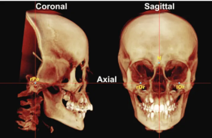
Once imported by means of speciic sotware tools, each 3D-volumetric data set was standardized using reference planes. The three planes are shown in Figure 1 and are deined by an axial plane passing through right and let infraorbitale points as well as right porion; a coronal plane passing through let and right porion perpendicular to the axial plane of choice; and a sagittal plane passing through the nasion point perpendicular to the axial and coronal planes of choice.
After standardization of head positioning, ana-tomical points (Sella, Nasion, A point, B point, Ante-rior Nasal Spine, and Menton) were analyzed through mid-sagittal slice images. Subsequently, landmarks 0.025 mm in diameter were identified (Table 1). The following measurements were performed (Fig 2): SNA (anteroposterior maxillary position), SNB (an-teroposterior mandibular position), ANB (antero-posterior maxillo-mandibular relationship), N-ANS (upper anterior facial height), ANS-Me (lower an-terior facial height), N-Me (anan-terior facial height), overjet, overbite, rMR (right molar relationship), and lMR (left molar relationship). Molar relationship
Figure 2 - Sagittal slice with landmarks and measurements. A) SNA, SNB, overbite and overjet; B) N-Me, N-ANS and ANS-Me; C. MR* (right molar relationship and left molar relationship).
Table 1 - Definition of landmarks
* Bilateral landmark
was determined as the perpendicular distance from the tip of mesiobuccal cusps of upper first permanent molar to the mesiobuccal sulcus of lower first perma-nent molar on the same side. Values of rMR and lMR
could not be obtained at T2 because of the artefacts
caused by orthodontic bands in these CBCT images. Measurements were performed separately at each
time (T1, T2 and T3) by the same examiner with a
one-week interval in between. Intraexaminer reli-ability values were determined by means of intraclass correlation coefficient (ICC), with 95% confidence interval. Fifteen CBCT scans were randomly selected and remeasured by the same examiner (CB) within 2 weeks, under the same conditions, and compared
Landmarks
(abbreviation)
Definition
Orbitale (Or)* Most inferior point on infraorbital rim Porion (Po)* Most superior point of anatomic external auditory
meatus
Nasion (N) Midsagittal point at junction of frontal and nasal bones at nasofrontal suture Sella (S) Midpoint of rim between anterior process at
mid-sagittal plane
A point (A) Deepest point of the maxillary alveolar bone concavity at mid-sagittal plane B point (B) Deepest point of the mandibular alveolar concavity
at mid-sagittal plane Anterior nasal spine
(ANS)
Most anterior limit of loor of nose, at tip of ANS at mid-sagittal plane
Posterior nasal spine (PNS)
Most posterior point along palate at mid-sagittal plane
Menton (Me) Most inferior point along curvature of chin at mid-sagittal plane
(-0.5 mm >and <0.5 mm); and decreased measure-ment (≤ –0.5 mm). Statistical analysis was carried out using the SPSS software version 16.0 (SPSS Inc., Chicago, IL, USA).
RESULTS
Separation of the mid-palatal suture was clinically confirmed in all patients with increased opening of inter-incisor diastema or within 3-5 days following expander activation. These data were confirmed on
the CBCT image at T2. During the retention period,
one of the patients returned without the expander,
thus, his data were not computed at T3. Transverse
deficiency was corrected in all patients. Data of RME
transverse effects were previously published.10
Table 2 shows the descriptive analysis (minimum, maximum and standard deviation) of measurements with the first measurements. All measurement error
coefficients were found to be close to 1.00 and within acceptable limits (higher than 0.95, except for MR measurement that was 0.91). The mean measurement
difference obtained was less than 0.4 mm and 0.3o,
which was considered not significant.
Means, standard deviations, minimum and maxi-mum values were calculated for each measurement. After finding normal data distribution by means of the Kolmogorov-Smirnov non-parametric test, sta-tistically significant differences were identified using paired Student’s t-test (P < 0.05 - 95% interval
confi-dence) between T2 and T1, T3 and T2, and T3 and T1.
The percentage of patients who had the same
qualita-tive mean changes during the interval T1-T3 was also
calculated. Patients were considered to have increased measurement (mean difference ≥ 0.5 mm); no change
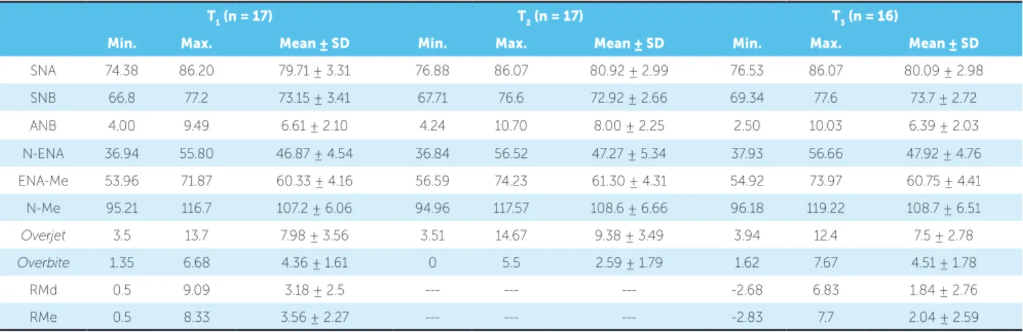
T1 (n = 17) T2 (n = 17) T3 (n = 16)
Min. Max. Mean ± SD Min. Max. Mean ± SD Min. Max. Mean ± SD
SNA 74.38 86.20 79.71 ± 3.31 76.88 86.07 80.92 ± 2.99 76.53 86.07 80.09 ± 2.98 SNB 66.8 77.2 73.15 ± 3.41 67.71 76.6 72.92 ± 2.66 69.34 77.6 73.7 ± 2.72 ANB 4.00 9.49 6.61 ± 2.10 4.24 10.70 8.00 ± 2.25 2.50 10.03 6.39 ± 2.03 N-ENA 36.94 55.80 46.87 ± 4.54 36.84 56.52 47.27 ± 5.34 37.93 56.66 47.92 ± 4.76 ENA-Me 53.96 71.87 60.33 ± 4.16 56.59 74.23 61.30 ± 4.31 54.92 73.97 60.75 ± 4.41 N-Me 95.21 116.7 107.2 ± 6.06 94.96 117.57 108.6 ± 6.66 96.18 119.22 108.7 ± 6.51
Overjet 3.5 13.7 7.98 ± 3.56 3.51 14.67 9.38 ± 3.49 3.94 12.4 7.5 ± 2.78
Overbite 1.35 6.68 4.36 ± 1.61 0 5.5 2.59 ± 1.79 1.62 7.67 4.51 ± 1.78
RMd 0.5 9.09 3.18 ± 2.5 --- --- --- -2.68 6.83 1.84 ± 2.76
RMe 0.5 8.33 3.56 ± 2.27 --- --- --- -2.83 7.7 2.04 ± 2.59
Table 2 - Descriptive analysis of measurements obtained in before treatment onset (T1), immediately after expansion (T2) and after retention (T3).
n = number of patients; Min = minimum; Max = maximum; SD = standard deviation.
Table 3 - Results regarding skeletal and dental changes between pre-treatment and post-expansion (T2 – T1), post-retention and post-expansion (T3 – T2), and post-retention and initial (T3 – T1).
n=number of patients; SD= Standard Deviation; Level of significance = * P < 0.05;**P < 0.01; ***P < 0.001.
T2-T1 (n = 17) T3-T2 (n = 16) T3-T1 (n = 16)
Mean ± SD Mean ± SD Mean ± SD
SNA 1.21* ± 1.96 -0.83* ± 1.28 0.38 ± 1.32
SNB -0.23 ± 2.05 0.78* ± 1.26 0.55 ± 1.76
ANB 1.39*** ± 1.09 -1.61*** ± 1.32 0.22 ± 0.84
N-ENA 0.40 ± 1.88 0.65 ± 1.31 1.06* ± 1.45
ENA-Me 0.97* ± 1.40 -0.55* ± 0.90 0.42 ± 1.40
N-Me 1.44*** ± 1.82 0.02 ± 1.18 1.46*** ± 1.42
Overjet 1.4* ± 1.96 -1.87*** ± 1.50 -0.47 ± 1.33
Overbite -1.76*** ± 0.72 1.91*** ± 0.92 0.15 ± 0.56
RMd --- --- -1.33** ± 1.23
RMe --- --- -1.55** ± 1.55
whereas the mandible moved backward to a lesser de-gree. Skeletal changes were previously reported by
Haas12 and have been recently confirmed by
meta-analysis13 and systematic reviews.14-16 Dental changes
mirrored skeletal changes by showing significant in-crease in overjet and dein-crease in overbite. Changes in dental and skeletal relationships were more likely to be associated with premature contacts involving pal-atal cusps and dental-alveolar inclination caused by
RME17 than to inferior displacement of the maxilla.
This effect was confirmed by the significant increase
in buccal inclination (7.31°/6.46°)10 found in upper
first molars immediately after RME.
The 6-month retention period with the Haas ex-pander did not only maintain the new skeletal, al-veolar and dental transverse dimensions, (1.66 mm,
4.69 mm and 5.89 mm, respectively, P < 0.001),10 but
also resulted in significant decrease in dentoalveolar angulation of original levels. The wider maxilla al-lowed mandible to shift forward more than upward, therefore improving skeletal and dental relationships. This was revealed by overjet decrease, overbite in-crease and MR improvement.
By the end of the assessment period, sagittal skel-etal changes were not significantly different when compared with initial data, except for patient’s vertical dimension. However, Class II dental relationship sig-nificantly improved in 75% of patients. Studies assess-ing untreated Class II malocclusions determined that
dental and skeletal patterns were not self-corrected,3,18
but became even worse.19 Wendling et al20 observed
obtained before treatment onset (T1), immediately
ater expansion (T2) and ater retention (T3). Table 3
shows Student’s t-test results yielded between the
fol-lowing intervals: T2-T1, T3-T2 and T3-T1. Signiicant
diferences were identiied immediately ater RME
(T2-T1) as the maxilla moved forward (SNA mean
increase was 1.21o), the mandible moved downward
(ANS-Me mean increase was 0.97 mm and N-Me mean increase was 1.44 mm), overjet increased in 1.4 mm and overbite decreased in 1.76 mm. During the
retention period (T3-T2), the maxilla relapsed
back-ward (SNA mean decrease was 0.83o) and the
man-dible was displaced forward (SNB mean increase was
0.78o), improving Class II ANB relationship (mean
decrease of 1.61o), although patients were let with an
overall increase in anterior facial height.
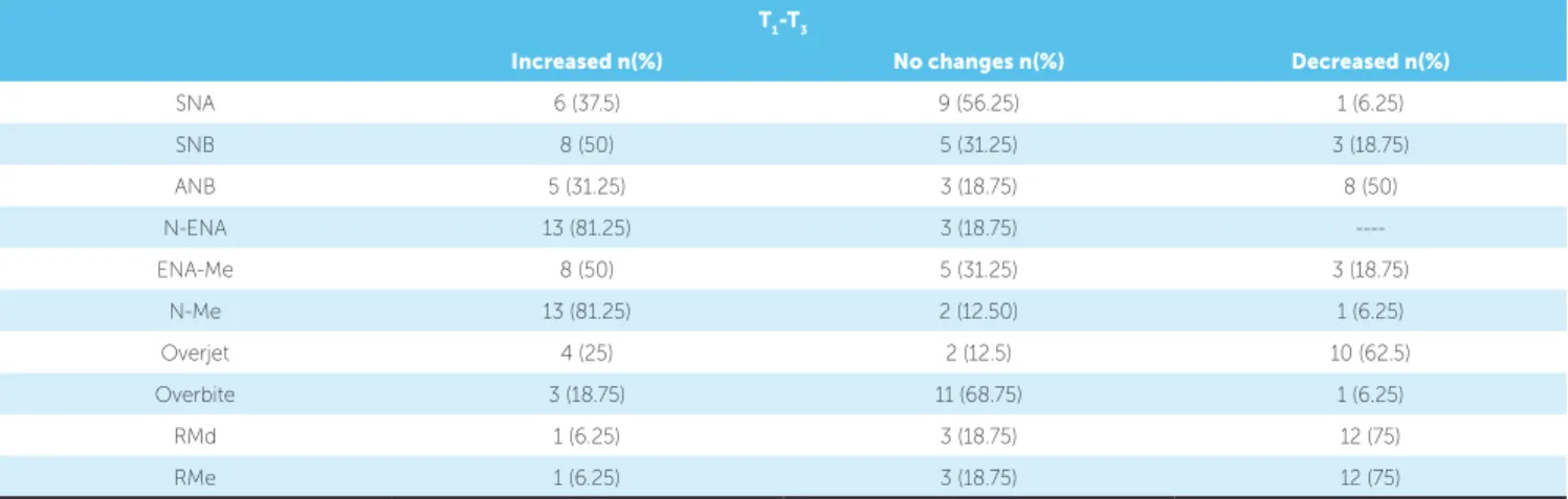
Table 4 shows a qualitative description of changes
found within T1-T3. Class II dental relationship (rMR
and lMR) improved in 75% of patients.
DISCUSSION
This study is part of a long-term prospective clinical investigation into the effects of RME on
Class II malocclusions using CBCT imaging.10,11
Understanding the effects of RME on Class II, Di-vision 1 patients is of paramount importance, since transverse maxillary deficiency is often associated with this malocclusion.
Immediately after RME therapy, Class II relation-ship was worse in the anteroposterior and vertical di-mensions. The maxilla significantly moved forward,
Table 4 - Number and percentage of patients with increased (≥ 0.5), no changes (- 0.5 > and < 0.5) or decreased (≤ - 0.5) measurements during the interval T1-T3.
n = number of patients.
T1-T3
Increased n(%) No changes n(%) Decreased n(%)
SNA 6 (37.5) 9 (56.25) 1 (6.25)
SNB 8 (50) 5 (31.25) 3 (18.75)
ANB 5 (31.25) 3 (18.75) 8 (50)
N-ENA 13 (81.25) 3 (18.75)
----ENA-Me 8 (50) 5 (31.25) 3 (18.75)
N-Me 13 (81.25) 2 (12.50) 1 (6.25)
Overjet 4 (25) 2 (12.5) 10 (62.5)
Overbite 3 (18.75) 11 (68.75) 1 (6.25)
RMd 1 (6.25) 3 (18.75) 12 (75)
that some patients had spontaneous Class II correc-tion after RME during the retencorrec-tion period (6-12 months) in cases of moderate Class II malocclusions.
McNamara et al21 recently observed great
improve-ment (1.8 mm) in MR after RME therapy in 81% of Class II patients when compared to non-treated controls (0.3 mm).
No statistically signiicant vertical changes were iden-tiied immediately ater RME. This difers from
previ-ous studies that used cephalometric imaging5,22-25 and
re-ported downward displacement of the maxilla. However, ater the retention period, a signiicant increase in the superior anterior facial height was observed in 81.25% of patients examined herein (N-ANS increased 1.06 mm). In contrast to RME active phase, the retention pe-riod was longer which could possibly explain the vertical
growth of the maxilla over this period.26,27 It is expected
that untreated 9-year-old subjects would undergo verti-cal growth of 1.5 mm per year for boys and 1.2 mm for
girls.26 Mc Namara et al21 observed a facial height increase
of 3.4 mm in a RME group and 4.2 mm in the control group over a mean observation period of 3.7 years.
Despite the fact that the present study only assessed Class II Division 1 malocclusion patients, the severity of malocclusion was not considered (Table 2). The large variability of skeletal involvement may precipitate dif-ferent responses to the same therapy. Vertical changes, resulting either from RME or growth, may limit hori-zontal mandibular changes and hinder forward
posi-tioning of the menton.28 Vertical maxillary control
dur-ing the active phase and the retention period would al-low further anterior repositioning of the mandible.
The number of patients included in the present study, although suicient to detect statistically signii-cant changes, is likely insuicient to generalize the re-sults to all Class II malocclusions. The lack of a control group was a limitation of the present study; however, a control group was unfeasible for the present study due to ethical reasons, since it is impossible not to intervene when a diagnosed transverse discrepancy is present.
The routine use of CBCT is not recommended for orthodontic procedures, given that conventional images emit lower radiation doses. However, some orthodontic patients require temporomandibular im-ages, frontal and lateral cephalograms, panoramic, periapical, occlusal or bite-wing radiographs. It is worth noting that the effective dose related to a
full-mouth radiographic survey, as reported by Gibbs,8
and the sum of the effective doses for panoramic, lateral cephalometric and periapical images are simi-lar, if not higher than that of CBCT without a 3D evaluation. This study used CBCT images because a 3D evaluation had also been carried out for other analyses and some data had already been previously
reported.10,11
CONCLUSIONS
1. Angle EH. Treatment of malocclusion of the teeth. Philadelphia: SS White; 1899.
2. Tollaro I, Baccetti T, Franchi L, Tanasescu CD. Role of posterior transverse interarch discrepancy in Class II, Division 1 malocclusion during the mixed dentition phase. Am J Orthod Dentofacial Orthop. 1996;110(4):417-22.
3. Baccetti T, Franchi L, McNamara JA Jr, Tollaro I. Early dentofacial features of Class II malocclusion: a longitudinal study from the deciduous through the mixed dentition. Am J Orthod Dentofacial Orthop. 1997;111(5):502-9. 4. Haas AJ. Long-term posttreatment evaluation of rapid palatal expansion.
Angle Orthod. 1980;50(3):189-217.
5. Silva Filho OG, Boas CV, Capelozza LFO. Rapid maxillary expansion in the primary and mixed dentitions: a cephalometric evaluation. Am J Orthod Dentofacial Orthop. 1991;100(2):171-9.
6. Lima Filho RMA, Lima AC, Ruellas ACO. Spontaneous correction of Class II malocclusion after rapid palatal expansion. Angle Orthod. 2003;73(6):745-52.
7. Silva MAG, Wolf U, Heinicke F, Bumann A, Visser H, Hirsch E. Cone-beam computed tomography for routine orthodontic treatment planning: a radiation dose evaluation. Am J Orthod Dentofacial Orthop. 2008;133(5):640.e1-5.
8. Gibbs SJ. Efective dose equivalent and efective dose: comparison for common projections in oral and maxillofacial radiology. Oral Surg Oral Med Oral Pathol Oral Radiol Endod. 2000;90(4):538-45.
9. Ricketts RM. Perspectives in the clinical application of cephalometrics. Angle Orthod. 1981;51(2):115-50.
10. Baratieri C, Nojima LI, Alves Jr M, Souza MMGd, Nojima MG. Transverse efects of rapid maxillary expansion in Class II malocclusion patients: a cone-beam computed tomography study. Dental Press J Orthod. 2010;15(5):89-97.
11. Baratieri C, Alves Jr M, Sant’Anna EF, Nojima MdCG, Nojima LI. 3D Mandibular positioning after rapid maxillary expansion in Class II malocclusion Braz Dent J. 2011;22(5):428-34.
12. Haas AJ. The treatment of maxillary deiciency by opening the midpalatal suture. Angle Orthod. 1965;35(3):200-17.
13. Lagravére MO, Heo G, Major PW, Flores-Mir C. Meta-analysis of immediate changes with rapid maxxillary expansion treatment. J Am Dent Assoc. 2006;137(1):44-53.
14. Baratieri C, Alves Jr M, Souza MMG, Araújo MTS, Maia LC. Does rapid maxillary expansion have long-term efects on airway dimensions and breathing? Am J Orthod Dentofacial Orthop. 2011;140(2):146-56.
REFERENCES
15. Lagravere MO, Major PW, Flores-Mir C. Long-term dental arch changes after rapid maxillary expansion treatment: a systematic review. Angle Orthod. 2005;75(2):155-61.
16. Lagravere MO, Major PW, Flores-Mir C. Long-term skeletal changes with rapid maxillary expansion: a systematic review. Angle Orthod. 2005;75(6):1046-52.
17. Wertz R. Skeletal and dental changes accompanying rapid midpalatal suture opening. Am J Orthod. 1970;58(1):41-66.
18. You Z-H, Fishman LS, Rosenblum RE, Subtelny JD. Dentoalveolar changes related to mandibular forward growth in untreated Class II persons. Am J Orthod Dentofacial Orthop. 2001;120(6):598-607. 19. Fröhlich FJ. Changes in untreated Class II type malocclusions. Angle
Orthod. 1962;32(3):167-79.
20. Wendling LK, McNamara JA, Franchi L, Baccetti T. A prospective study of the short-term treatment efects of the acrylic-splint rapid maxillary expander combined with the lower Schwarz Appliance. Angle Orthod. 2004;75(1):7-14.
21. McNamara JA, Sigler LM, Franchi L, Guest SS, Baccetti T. Changes in Occlusal Relationships in mixed dentition patients treated with rapid maxillary expansion. Angle Orthod. 2010;80(2):230-8.
22. Haas AJ. Rapid expansion of the maxillary dental arch and nasal cavity by opening the midpalatal suture. Angle Orthod. 1961;31(2):73-90. 23. Akkaya S, Lorenzon S, Üçem TTA. A comparison of sagittal and vertical
efects between bonded rapid and slow maxillary expansion procedures. Eur J Orthod. 1999;21(2):175-80.
24. Chung C-H, Font B. Skeletal and dental changes in the sagittal, vertical, and transverse dimensions after rapid palatal expansion. Am J Orthod Dentofacial Orthop. 2004;126(5):569-75.
25. Akkaya S, Lorenzon S, Üçem TTA. A comparison of sagittal and vertical efects between bonded rapid and slow maxillary expansion procedures. Eur J Orthod. 1999;21(2):175-80.
26. Riolo ML, Moyers RE, McNamara JA, Hunter W. An atlas of craniofacial growth -Cephalometric standards from the University School Growth Study. Michigan: University of Michigan-Monograph Craniofacial Series; 1974.
27. Wendling LK, McNamara JA, Franchi L, Baccetti T. A Prospective study of the short-term treatment efects of the acrylic-splint rapid maxillary expander combined with the lower Schwarz Appliance. Angle Orthod. 2005;75(1):7-14.