Evaluation of cytotoxicity and
corrosion resistance of orthodontic mini-implants
Celha Borges Costa Alves1, Márcio Nunes Segurado2, Miriam Cristina Leandro Dorta3, Fátima Ribeiro Dias3, Maurício Guilherme Lenza1, Marcos Augusto Lenza4
Objective: To evaluate and compare in vitro cytotoxicity and corrosion resistance of mini-implants from three different commercial brands used for orthodontic anchorage. Methods: Six mini-implants (Conexão™, Neodent™ and SIN™) were separately immersed in artificial saliva (pH 6.76) for 30 and 60 days. The cytotoxicity of the corrosion extracts was assessed in L929 cell cultures using the violet crystal and MTT assays, as well as cell morphology under light microscopy. Metal surface characteristics before and after immersion in artificial saliva were assessed by means of scanning electron microscopy (SEM). The samples underwent atomic absorption spectrophotometry to determine the concentrations of aluminum and vanadium ions, constituent elements of the alloy that present potential toxicity. For statistical analysis, one-way ANOVA/Bonferroni tests were used for comparisons among groups with p < 0.05 considered significant. Sta-tistical analysis was carried out with Graph Pad PRISM software Version 4.0. Results: No changes in cell viability or morphology were observed. Mini-implants SEM images revealed smooth surfaces with no obvious traces of corrosion. The extracts assessed by means of atomic absorption spectrophotometry presented concentrations of aluminum and va-nadium ions below 1.0 µg/mL and 0.5 µg/mL, respectively. Conclusion: Orthodontic mini-implants manufactured by Conexão™, Neodent™ and SIN™ present high corrosion resistance and are not cytotoxic.
Keywords: Mini-implants. Corrosion. Cytotoxicity.
1 PhD student, Universidade Federal de Goiás, School of Dentistry, Department of Clinical Dentistry, Graduate Program in Dentistry, Goiânia, Goiás, Brazil. 2 Professor, Universidade Paulista (UNIP), School of Dentistry, Department of
Orthodontics, Goiânia, Goiás, Brazil.
3 Associate Professor, Universidade Federal de Goiás (UFG), Institute of Tropical Pathology and Public Health, Immunology Section, Department of Microbiology, Immunology, Parasitology and Patology, Goiânia. Goiás, Brazil. 4 Full Professor and Coordinator, Universidade Federal de Goiás (UFG), School of
Dentistry, Specialization Course in Orthodontics, Goiânia, Goiás, Brazil).
DOI: http://dx.doi.org/10.1590/2177-6709.21.5.039-046.oar
How to cite this article: Alves CBC, Segurado MN, Dorta MCL, Dias FR, Lenza MG, Lenza MA. Evaluation of cytotoxicity and corrosion resistance of orthodontic mini-implants. Dental Press J Orthod. 2016 Sep-Oct;21(5):39-46. DOI: http://dx.doi.org/10.1590/2177-6709.21.5.039-046.oar
Submitted: August 01, 2015 - Revised and accepted: August 26, 2016
» The authors report no commercial, proprietary or financial interest in the products or companies described in this article.
Contact address: Celha Borges Costa Alves E-mail: [email protected]
Objetivo: avaliar, in vitro, e comparar a citotoxicidade e a resistência à corrosão de mini-implantes de três marcas comerciais diferentes, utilizados para ancoragem ortodôntica. Métodos: seis mini-implantes fabricados pelas empresas Conexão®, Neo-dent® e SIN® foram imersos, separadamente, em saliva artificial (pH = 6,76), por 30 e 60 dias, de forma a obter os extratos da corrosão. A citotoxicidade dos extratos foi avaliada em cultura de células L929, empregando-se a análise de ensaios do cristal violeta e MTT, bem como da morfologia celular sob microscopia óptica. As características da superfície do metal antes e após a imersão em saliva artificial foram avaliadas usando microscopia eletrônica de varredura. Os extratos foram submetidos a espec-trofotometria de absorção atômica, para determinar as concentrações dos íons alumínio e vanádio, elementos constituintes da liga e que apresentam toxicidade em potencial. Para análise estatística, os testes one-way ANOVA/Bonferroni foram usados para comparação entre os grupos, com p < 0,05 sendo considerado significativo. A análise estatística foi realizada com o programa Graph Pad PRISM v. 4.0. Resultados: não foi observada alteração na viabilidade ou morfologia celular após a exposição dos mini-implantes aos extratos. A análise dos mini-implantes por microscopia eletrônica de varredura revelou superfícies lisas e sem traços evidentes de corrosão. Os extratos analisados usando espectrofotometria de absorção atômica apresentaram concen-trações de íons alumínio e vanádio inferiores a 1,0 µg/ml e 0,5 µg/ml, respectivamente. Conclusão: os mini-implantes fabrica-dos pelas empresas Conexão®, Neodent® e SIN® apresentam alta resistência à corrosão e não são citotóxicos.
INTRODUCTION
Understanding each patient’s anchorage require-ments is extremely important and ensures high-qual-ity orthodontic treatment. If anchorage is lost, it will undoubtedly result in compromised results. Relying on patient’s compliance to obtain the desirable force sys-tem will also increase the risk of not achieving the de-sirable inishing results. Today, mini-implants provide the much-desired absolute anchorage and, more impor-tantly, the use of these devices does not rely on patient’s compliance. They are used primarily as direct or indi-rect anchorage — a biomechanical setup in which force is directly or indirectly applied from the mini-implant to a tooth or a group of teeth that needs to be orthodon-tically moved. Therefore, in the last few years, mini-implants have been extensively used for anchorage, thus simplifying orthodontic mechanics and minimizing side efects during orthodontic treatment.1-5
The ongoing and continuous use of metal material in Orthodontics has led to a large number of labora-tory and clinical studies on the detrimental efects of corrosion products to one’s general health. The oral cavity is not only extremely aggressive, but also a po-tential corrosive environment. Corrosion resistance of orthodontic alloys depends on the oral environment which is inluenced by several variables, such as the amount and quality of saliva, pH of food and beverages, among others.6 The release of metal ions from
orth-odontic devices is a genuine concern.
Although all types of metallic material are subject to corrosion, titanium is widely used in orthopedic com-ponents because of its attractive characteristics, such as high corrosion resistance and excellent biocompatibil-ity. Additionally, it presents excellent mechanical prop-erties and provides resistance to stress and strain. It is, therefore, considered an ideal material.7-11 However,
pure titanium has less fatigue strength than titanium alloys. Orthodontic mini-implants should withstand
high orthodontic loads for tooth movement. In order to overcome potential fractures of commercially pure titanium during mini-implant placement and removal, aluminum and vanadium have been added to the alloy for greater strength and fatigue resistance.12,13
Titanium alloy (Ti6Al4V) is now most often used in Dentistry to overcome this disadvantage. Howev-er, this alloy may undergo corrosion in the oral en-vironment due to its low corrosion resistance. Tita-nium, aluminum and vanadium ions can be released to local and remote tissues and have been associated with side effects in the human body, particularly aluminum and vanadium.14-18
Although in vitro studies do not reproduce the complex oral environment, standard assays are useful to evaluate the cytotoxicity and biocompatibility of temporary an-chorage devices, such as mini-implants. ISO 10271:2011 provides test methods to determine the corrosion behav-ior of metallic material used in the oral cavity.19
The aim of this in vitro study was to compare the cytotoxicity and corrosion resistance of mini-implants from three diferent commercial brands used for orth-odontic anchorage.
MATERIAL AND METHODS Samples
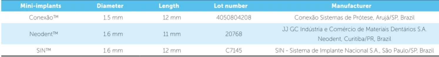
This study investigated metal mini-implants used for orthodontic anchorage fabricated by three com-mercial manufacturers: ConexãoTM, São Paulo, Brazil;
NeodentTM, Curitiba, Brazil and SINTM, São Paulo,
Brazil — respectively with mini-implants head diam-eter and total length of 1.5 x 12 mm, 1.6 x 11 mm and 1.6 x 12 mm (Table 1). Although the exact mini-im-plant chemical composition was not provided by the manufacturers, they followed the standard speciica-tion for Titanium-6Aluminum-4Vanadium ELI (extra low interstitial) alloy for surgical implant applications (ASTM F136-08e1 – UNSR 56401).
Mini-implants Diameter Length Lot number Manufacturer
Conexão™ 1.5 mm 12 mm 4050804208 Conexão Sistemas de Prótese, Arujá/SP, Brazil
Neodent™ 1.6 mm 11 mm 20768 JJ GC Indústria e Comércio de Materiais Dentários S.A. Neodent, Curitiba/PR, Brazil
Six samples of each orthodontic mini-implant man-ufacturer were individually weighed with the aid of an analytical balance (model 410 - Kern & Sohn GmbH, Balingen, Germany) and autoclaved at 120 oC for
30 minutes. Subsequently, each sample was transferred to individual sterile BD VacutainerTM glass tubes
(Bec-ton Dickinson Indústrias Cirúrgicas Ltda, Juiz de Fora, MG, Brazil) and immersed in artiicial saliva for 30 and 60 periods. The number of samples and methods used are in accordance to corrosion test methods for metal material speciied in ISO 10271.19 The procedures were
carried out in a laminar low hood, with ultraviolet ra-diation used to obtain an aseptic ield.
The artiicial saliva chemical composition used in this study was a modiication of Meyer’s solution20,21
which has been shown to present corrosion activity and chloride concentration similar to natural saliva. It was composed of 0.40 mg/L of NaCl, 0.40 mg/L of KCl, 0.80 mg/L of CaCl2.H2O, 1.0 mg/L of CO(NH2)2 in
distilled water with a pH adjusted and controlled with a 10-N NaOH solution. The performance of any mate-rial placed into the oral environment should be assessed with artiicial saliva of a known composition, since natural saliva varies widely.22
The amount of saliva was calculated by the ratio of 1 mL of artiicial saliva for 0.2 g of mini-implant weight, according to ISO 1027119 and ISO 10993-15.20
Mini-im-plants were maintained in immersion and stored at 37 oC
under stationary conditions. Tubes containing only artii-cial saliva, without the mini-implant extract, were stored under the same conditions as negative control.
Ater the immersion periods, mini-implants were re-moved from the tubes, washed in deionized water, dried and stored in new sterile airtight plastic tubes, and saliva with the mini-implant corrosive product extracts was stored in 1.5-mL tubes at 4 oC for further analysis. The methods
used in this study have already been described.24
L929 cell culture
Murine ibroblast L929 cells were cultured in 75-cm2
culture lasks (Corning Costar Corporation, Cambridge, MA, USA) containing RPMI 1640 culture medium (Sigma-Aldrich Co. LLC, St. Louis, MO, USA), buf-ered with 10-mM HEPES and supplemented with 10% fetal bovine serum (FBS) (Gibco/BRL Division, Grand Island, NY, USA), 2-mM L-glutamine, 11-mM so-dium bicarbonate, 100-U/mL penicillin, and 100-g/mL
streptomycin (Sigma-Aldrich Co. LLC, St. Louis, MO, USA), herein named complete medium. Ater L929 cell monolayer formation, the culture medium was removed and the cells washed with 1 mL of incomplete medium (RPMI 1640 without FBS). The cells were detached from the culture lasks with 0.025% trypsin (Sigma-Al-drich Co. LLC, St. Louis, MO, USA).
Ater trypsinization, cultured cells were resuspend-ed in 5 mL of culture mresuspend-edium, transferrresuspend-ed to 50-mL plastic tubes (Corning Costar Corporation, Cambridge, MA, USA) and centrifuged at 2,000 rpm for 10 min-utes at 15 oC. For culture maintenance, cells were
cul-tivated again in complete medium (@ 1 × 10⁵ cells/mL).
For cytotoxicity assays, cells were resuspended in 1 mL of complete medium. Viable cells were counted by try-pan blue dye exclusion test (in 0.1% phosphate bufered saline) using a hemacytometer adjusted to a concentra-tion of 3.5 × 10⁵ cells/mL by adding 0.9% NaCl.
Artiicial saliva was used as negative control and as a medium to obtain mini-implant extracts, since it is not cytotoxic to cell-culture. Tumor necrosis factor (TNF, Sigma-Aldrich Co. LLC, St. Louis, MO, USA), a cytokine capable of destroying L929 cells ater approxi-mately 20 hours of culture, was used as positive control.
Cytotoxicity assays
Aliquots of 100 µL of L929 cell suspension were pi-petted into 96-well lat bottom plates (Corning Costar Corporation, Cambridge, MA, USA). External wells were half illed and the plates incubated for 48 hours at 37 oC in a humidiied atmosphere with 5% CO
2 to
obtain a cell monolayer. Ater this period, monolayer growth was conirmed by inverted light microscope and 20-µL aliquots (20%) of mini-implant extracts or 20-µL (20%) of artiicial saliva (used as negative con-trol) were added to the correspondent well. Mini-im-plant extract solution was tested in triplicate on every plate and incubated for 48 hours at 37 oC in a
humidi-ied atmosphere with 5% CO2.
Determination of cell viability by means of the crystal violet colorimetric assay
Ater a 48-hour incubation period, 10-µL aliquots of 0.5% crystal violet in 30% acetic acid were added to each well to ix the living cells to the bottom of the plate. Ater 10 minutes, the plates were washed to have dead cells re-moved and, ater complete drying in a bacteriological in-cubator at 37 oC, 100 mL of absolute methanol (Synth,
Di-adema, SP, Brazil) was added to dissolve the stained cells. The resulting stained solution, corresponding to the total number of viable cells retained on the plates, was placed in a microplate spectrophotometer (Multiskan Original, Model 352, Thermo Labsystems, China, ilter 620 nm) and optical density (OD) was read. Culture medium with-out cells was the blank. Control wells absorbance (cells cultured in complete medium) was considered as 100% cell viability. Results were expressed as OD.
Determination of cellular metabolism by means of the MTT colorimetric assay
For the MTT assay, L929 cells were grown and, ater 48 hours of incubation, 10-µL aliquots of MTT solution (5 mg/mL phosphate-bufered solution, PBS) were added to each well and incubated for 3 hours at 37 oC in a humidiied atmosphere with 5% CO
2.
Af-ter this period, 100-mL aliquots of a sodium dodecyl
sulfate (SDS) solution in 10% 0.01-N hydrochloric acid (HCl) were added to each well to dissolve the crystals, and the plate was incubated again for 24 hours at 35 oC
for further OD readings.25,26,27 The OD was measured
in a Thermo Labsystems 352 Multiskan MS micro-plate reader (Labsystems Oy, Helsinki, Finnland) with a 550-nm ilter. Culture medium without cells was the blank. Control wells absorbance (cells cultured in com-plete medium) was considered as 100% cell viability. Results were expressed as OD.
Mini-implant surface scanning electron microscopy (SEM)
In order evaluate qualitatively mini-implants sur-face characteristics as to the presence of any imperfec-tion and corrosion areas, a sample of each artiicial saliva immersion group and a sterile packaged control sample from the same lot were chosen randomly and examined by means of scanning electron microscope (JEOL Mod-el JSM5410, Jeol Ltd, Japan) equipped with energy dis-persive spectroscopy (EDS) to analyze surface element composition. Surface topography of the mini-implant head, normally exposed to the oral environment, was examined under 35x and 1000x magniication.
Atomic absorption spectrophotometry analysis (AAS) of artificial saliva mini-implant corrosion products
Mini-implant extract solutions obtained ater 30- and 60-day immersion periods in artiicial saliva were analyzed with the aid of an atomic absorption spectro-photometer (AanalystTM 200, PerkinElmer, Waltham,
MA, USA) to determine and quantify the amount of aluminum and vanadium ions released due to corro-sion and oxidation. Artiicial saliva incubated for 30 and 60 days was used as a control solution (blank). The gas mixture used was air/acetylene. The wavelengths em-ployed were 309.3 nm for aluminum and 313.3 nm for vanadium. The limits of sensitivity for aluminum and vanadium were 1.0 µg/mL and 0.5 µg/mL, respectively.
Statistical analysis
Results were expressed as a mean ± SEM (standard error of the mean). One-way ANOVA/Bonferroni’s post-tests were performed with GraphPad PRISM sotware (Graph-Pad Sotware, Inc., San Diego, CA, USA). Statistical sig-niicance was determined at the level of p < 0.05.
RESULTS
Cytotoxicity assays
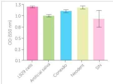
L929 cell morphological analysis under light micros-copy showed no cell monolayer destruction. Similarly, the crystal violet assay indicated absence of cell death. A certain optical density decrease was registered for the L929 cell samples incubated with Conexão™ and Neo-dent™ mini-implant extract solution, but this decrease was similar to negative control (artiicial saliva) and no statistical diference was found among them (p = 0.781 and p = 0.514 for 30 and 60 days, respectively) (Fig 2).
The MTT colorimetric assay demonstrated no cell metabolic activity inhibition for the three mini-implant extract solutions tested, particularly in the 30-day samples. Although SIN™ mini-implants led to more cell metabolism alteration than the others in the 60-day period, the diference was not statistically signiicant (p = 0.125 and p = 0.273 for 30 and 60 days, respectively) (Fig 3).
Analysis of mini-implant surfaces by means of SEM
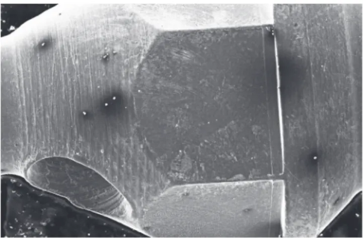
Micro analysis of Neodent™ mini-implants demon-strated more adhered particles and a higher number of
darkened spots on their surfaces, especially samples im-mersed for a longer period, as compared to the control group, although these surfaces revealed to be smooth and regular (Fig 4).
SIN™ mini-implant analysis of the control group revealed a smooth surface, but with adhered parti-cles in some darkened areas. The artificial saliva im-mersed sample demonstrated a rough area between the screw body itself and the head, thus suggesting corrosion (Fig 5).
Conexão™ mini-implant analysis of the control group demonstrated a very smooth surface without sig-niicant roughness, without adhered particles or dark-ened spots. The artiicial-saliva-immersed samples re-mained smooth and free from corrosion, presenting only small amounts of adhered particles and darkened areas, especially ater 30 days. The 60-day samples pre-sented some whitish spots, characteristic of calcium buildup (Fig 6).
The energy-dispersive X-ray spectrometer (EDS) revealed the presence of titanium, aluminum and vana-dium, as well as traces of calcium, silicon, potassium, chloride, magnesium and carbon, thus relecting the artiicial saliva composition in all mini-implants tested.
Figure 2 - Crystal violet colorimetric assay for L929 cell samples incubated with extract solutions of mini-implants obtained after 30 days of immersion in artificial saliva.
Figure 3 - MTT colorimetric assay for L929 cell samples incubated with ex-tract solutions of mini-implants obtained after 60 days of immersion in arti-ficial saliva.
OD (620 nm)
0.7
0.5
0.3
0.1 0.6
0.4
0.2
0
1.3
0.8
0.3 1.0
0.5
0.1
OD (550 nm)
L929 c ells
Conexão Neodent
SIN
Artificial saliva Células L929
Conexão Neodent
SIN
Measurement of aluminum and vanadium ions in mini-implant extract solutions by means of atomic absorption spectrophotometry (AAS)
The artiicial saliva used solely as control, in both pe-riods, showed no sign of aluminum or vanadium. Simi-larly, the concentration of Al and V ions in the artiicial saliva mini-implant extract solution was below the sen-sitivity threshold of the equipment, thus demonstrating that whatever amount is released, it is so minimal that it is not detrimental to an individual’s health.
DISCUSSION
In this study, orthodontic mini-implants ready for clinical use as anchorage devices were tested for their potential toxic efect. These devices are manufactured almost exclusively from a titanium alloy (Ti6Al4V) with
Figure 4 - Photomicroscopy of Neodent mini-implant after 60 days of im-mersion in artificial saliva (50x).
Figure 6 - Photomicroscopy of Conexão mini-implant after 60 days of im-mersion in artificial saliva, evidencing the presence of whitish spots.
Figure 5 - Photomicroscopy of SIN mini-implant after 30 days of immersion in artificial saliva (150x).
the addition of aluminum and vanadium for greater strength and fatigue resistance12,13 to withstand
orth-odontic forces for tooth movement. However, alumi-num and vanadium have been associated with side ef-fects in the human body.
Results yielded by the present study demonstrated that Conexão™, Neodent™ and SIN™ mini-implant extract solutions obtained ater 30- and 60-day immer-sion periods did not afect cell viability or decreased cell metabolism, thus demonstrating that none of them are cytotoxic. There was no statistical diference among groups (p > 0.05). This inding is in agreement with several studies that support the high biocompat-ibility of titanium and its alloys.4,8,10,11 One of the main
requirements for a metal or alloy to be biocompatible is the lack of release of corrosion products, which may lead to adverse efects.
According to SEM mini-implant surface analysis, there was no signiicant corrosion. This result conirms the high corrosion resistance of these mini-implants, even if they are composed of a less resistant alloy com-pared with other devices, which do not have aluminum and vanadium in their composition. However, all mini-implants immersed for 60 days showed darkened spots and more adhered particles suggestive of decreased cor-rosion resistance.
worth mentioning that, despite the evidence of good corrosion resistance and biocompatibility of all mini-implants tested, SIN™ mini-implant presented rough areas that suggest corrosion or manufacturing defects. The 60-day samples exhibited the greatest alteration in the MTT assay, which is more sensitive than the crys-tal violet assay.26 The combination of these two results
calls attention to the corrosion potential of this mini-implant, although the results demonstrated that they were not statistically diferent.
The other elements also detected in the alloy were contaminants, such as calcium, potassium, chloride, ox-ygen, silicon and magnesium; they were probably from artiicial saliva or were incorporated during the clean-ing and passivation protocols in industrial handlclean-ing of all mini-implants tested.
Recent studies28,29,30 have demonstrated that although
titanium alloys are considered highly corrosion-resistant because of the stable passive titanium oxide layer on their surface, they are not inert to corrosive attack. Retrieved mini-implants showed considerable surface and structural alterations, such as dullness, corrosion, and blunting of threads and tips. Their surfaces showed interactions and adsorption of several elements, such as calcium, at the body region.
In the present study, taking into consideration that 60 days was the maximum period that the mini-im-plants were exposed to artiicial saliva, a time in which all samples remained static, not submitted to any orth-odontic force in which the results demonstrate no signs of corrosion in the mini-implants from all manufactur-ers, the presence of manufacturing/corrosion defects on the SIN™ mini-implants surface causes concern. In studies employing longer immersion periods and fric-tion simulafric-tion, these mini-implants most probably would release greater amounts of corrosive products, which could be harmful because the protective oxide layer would be removed from certain areas and, there-fore, would not prevent corrosion.9,13,29
Although the corrosion resistance of titanium is well documented in the literature, a gap regarding this mat-ter in mini-implants commonly used in Orthodontics still remains. Therefore, further studies should be per-formed to clarify corrosion resistance and cytotoxicity of these devices, testing longer periods of immersion, harder wear simulation conditions, pH alterations, and the presence of luoride ions in the corrosive medium.
CONCLUSION
Mini-implants of three commercial brands (Conexão™, Neodent™ and SIN™) exhibited good corrosion resistance ater 30- and 60-day immersion periods in artiicial saliva. The release of aluminum and vanadium ions was not detected in the extract so-lutions analyzed, within the limits of the AAS tech-nique used. No cytotoxicity was observed in L929 cell morphological evaluation, growth inhibition, cell damage, and/or alteration of cellular metabolism.
1. Cousley RR, Sandler PJ. Advances in orthodontic anchorage with the use of mini-implant techniques. Br Dent J. 2015 Feb 16;218(3):e4.
2. Chang HP, Tseng YC. Miniscrew implant applications in contemporary orthodontics. Kaohsiung J Med Sci. 2014 Mar;30(3):111-5.
3. Rodriguez JC, Suarez F, Chan HL, Padial-Molina M, Wang HL. Implants for orthodontic anchorage: success rates and reasons of failures. Implant Dent. 2014 Apr;23(2):155-61.
4. Jasoria G, Shamim W, Rathore S, Kalra A, Manchanda M, Jaggi N. Miniscrew implants as temporary anchorage devices in orthodontics: a comprehensive review. J Contemp Dent Pract. 2013 Sept 1;14(5):993-9.
5. Bae SM, Park HS, Kyung HM, Kwon OW, Sung JH. Clinical application of micro-implant anchorage. J Clin Orthod. 2002 May;36(5):298-302.
6. Eliades T, Bourauel C. Intraoral aging of orthodontic materials: the picture we miss and its clinical relevance. Am J Orthod Dentofacial Orthop. 2005 Apr;127(4):403-12.
7. Burmann PF, Ruschel HC, Vargas IA, de Verney JC, Kramer PF. Titanium alloy orthodontic mini-implants: scanning electron microscopic and metallographic analyses. Acta Odontol Latinoam. 2015 Apr;28(1):42-7.
8. Huang LH, Shotwell JL, Wang HL. Dental implants for orthodontic anchorage. Am J Orthod Dentofacial Orthop. 2005 Jun;127(6):713-22.
9. Chen G, Wen X, Zhang N. Corrosion resistance and ion dissolution of titanium with diferent surface microroughness. Biomed Mater Eng. 1998;8(2):61-74. 10. Lautenschlager EP, Monaghan P. Titanium and titanium alloys as dental materials.
Int Dent J. 1993 Jun;43(3):245-53.
11. Strietzel R, Hösch A, Kalbleisch H, Buch D. In vitro corrosion of titanium. Biomaterials. 1998 Aug;19(16):1495-9.
12. Morais LS, Serra GG, Muller CA, Andrade LR, Palermo EF, Elias CN, Meyers M. Titanium alloy mini-implants for orthodontic anchorage: immediate loading and metal ion release. Acta Biomater. 2007 May;3(3):331-9.
13. Morais LS, Serra GG, Albuquerque Palermo EF, Andrade LR, Müller CA, Meyers MA, Elias CN. Systemic levels of metallic ions released from orthodontic mini-implants. Am J Orthod Dentofacial Orthop. 2009 Apr;135(4):522-9. 14. Frisken KW, Dandie GW, Lugowski S, Jordan G. A study of titanium release into
body organs following the insertion of single threaded screw implants into the mandibles of sheep. Aust Dent J. 2002 Sept;47(3):214-7.
15. Martín-Cameán A, Jos A, Puerto M, Calleja A, Iglesias-Linares A, Solano E, et al. In vivo determination of aluminum, cobalt, chromium, copper, nickel, titanium and vanadium in oral mucosa cells from orthodontic patients with mini-implants by Inductively coupled plasma-mass spectrometry (ICP-MS). J Trace Elem Med Biol. 2015 Oct;32:13-20.
REFERÊNCIAS
16. Barceloux DG. Vanadium. J Toxicol Clin Toxicol. 1999;37(2):265-78. 17. Chen WJ, Monnat RJ Jr, Chen M, Mottet NK. Aluminum induced pulmonary
granulomatosis. Hum Pathol. 1978 Nov;9(6):705-11.
18. Marquis JK. Aluminum neurotoxicity: an experimental perspective. Bull Environ Contam Toxicol. 1982 July;29(1):43-9.
19. International Standard Organization. Dentistry: corrosion test methods for metallic materials. Geneva: ISO; 2011. ISO Document 10271.
20. International Standard Organization. Biological evaluation of medical devices — Part 15: Identiication and quantiication of degradation products from metals and alloys. Geneva: ISO; 2000. ISO Document 10993-15.
21. Park HY, Shearer TR. In vitro release of nickel and chromium from simulated orthodontic appliances. Am J Orthod. 1983 Aug;84(2):156-9.
22. Hwang CJ, Shin JS, Cha JY. Metal release from simulated ixed orthodontic appliances. Am J Orthod Dentofacial Orthop. 2001 Oct;120(4):383-91. 23. Leung VW, Darvell BW. Artiicial salivas for in vitro studies of dental materials.
J Dent. 1997 Nov;25(6):475-84.
24. Costa MT, Lenza MA, Gosch CS, Costa I, Ribeiro-Dias F. In vitro evaluation of corrosion and cytotoxicity of orthodontic brackets. J Dent Res. 2007 May;86(5):441-5.
25. Flick DA, Giford GE. Comparison of in vitro cell cytotoxic assays for tumor necrosis factor. J Immunol Methods. 1984 Mar 30;68(1-2):167-75.
26. Mosmann T. Rapid colorimetric assay for cellular growth and survival: application to proliferation and cytotoxicity assays. J Immunol Methods. 1983 Dec 16; 65(1-2):55-63.
27. Gerlier D, Thomasset N. Use of MTT colorimetric assay to measure cell activation. J Immunol Methods. 1986 Nov 20;94(1-2):57-63.
28. Chaddad K, Ferreira AF, Geurs N, Reddy MS. Inluence of surface characteristics on survival rates of mini-implants. Angle Orthod. 2008 Jan;78(1):107-13. 29. Patil P, Kharbanda OP, Duggal R, Das TK, Kalyanasundaram D. Surface
deterioration and elemental composition of retrieved orthodontic miniscrews. Am J Orthod Dentofacial Orthop. 2015 Apr;147(4 Suppl):S88-100.