Reliability of overbite depth indicator (ODI) and
anteroposterior dysplasia indicator (APDI) in the assessment of
different vertical and sagittal dental malocclusions: a receiver
operating characteristic (ROC) analysis
Farheen Fatima1, Mubassar Fida2, Attiya Shaikh3
Introduction: Differential diagnosis of skeletal and dental relationships is crucial for planning orthodontic treatment. Overbite depth
indicator (ODI) and anteroposterior dysplasia indicator (APDI) had been introduced in the past for assessment of vertical and sagittal jaw relationships, respectively. Objective: The objectives of this study were to evaluate the reliability of ODI and APDI in overbite and Angle malocclusions, as well as assess their diagnostic reliability among males and females of different age groups. Material and
Methods: This study was conducted using pretreatment dental casts and lateral cephalograms of 90 subjects. For ODI, subjects were divided
into three groups based on overbite (normal overbite, open bite and deep bite). Likewise, the same subjects were divided for APDI into three groups, based on Angle’s malocclusion classification (dental Class I, II and III malocclusions). Mann-Whitney U test was applied for compari-son of study parameters regarding sex and different age groups. The mean values of ODI and APDI were compared among study groups by means of Kruskal-Wallis and post-hoc Dunnet T3 tests. The receiver operating characteristic (ROC) curve was applied to test diagnostic reli-ability. Results: Insignificant differences were found for ODI and APDI angles, particularly in regards to sex and age. Significant inter-group differences were found in different overbite inter-groups and Angle’s classification for ODI and APDI, respectively (p < 0.001). ROC showed 91% and 88% constancy with dental pattern in ODI and APDI, respectively. Conclusions: ODI can reliably differentiate deep bite versus normal overbite and deep bite versus open bite. APDI can reliably differentiate dental Class I, II and III malocclusions.
Keywords: Malocclusion. Overbite. Cephalometry. Receiver operating characteristic curve.
1 Resident in Orthodontics, The Aga Khan University Hospital, Section of
Dentistry, Department of Surgery, Karachi Pakistan.
2 Consultant Orthodontist/Associate Professor, The Aga Khan University
Hospital, Program Director Orthodontics Residency Program, Section of Dentistry, Department of Surgery, Karachi, Pakistan.
3 Consultant Orthodontist/ Assistant Professor, The Aga Khan University
Hospital, Program Coordinator, Orthodontics Residency Program, Section of Dentistry, Department of Surgery, Karachi, Pakistan.
Submitted: December 07, 2015 - Revised and accepted: February 28, 2016 DOI: http://dx.doi.org/10.1590/2177-6709.21.5.075-081.oar
How to cite this article: Fatima F, Fida M, Shaikh A. Reliability of overbite depth indicator (ODI) and anteroposterior dysplasia indicator (APDI) in the as-sessment of different vertical and sagittal dental malocclusions: a receiver operating characteristic (ROC) analysis. Dental Press J Orthod. 2016 Sept-Oct;21(5):75-81. DOI: http://dx.doi.org/10.1590/2177-6709.21.5.075-081.oar
» The authors report no commercial, proprietary or financial interest in the products or companies described in this article.
Contact address: Farheen Fatima E-mail: [email protected]
Introdução: o diagnóstico diferencial das relações esqueléticas e dentárias é essencial para o plano de tratamento ortodôntico. O indicador de
profundidade da sobremordida (ODI)) e o indicador de displasia anteroposterior (APDI) foram desenvolvidos, no passado, para avaliação das relações verticais e sagitais dos maxilares, respectivamente. Objetivo: o objetivo desse estudo foi avaliar a confiabilidade do ODI e do APDI em diferentes sobremordidas e más oclusões de Angle, bem como verificar sua confiabilidade diagnóstica em homens e mulheres de diferentes grupos etários. Métodos: esse estudo foi conduzido utilizando-se modelos de estudo e radiografias cefalométricas laterais pré-tratamento de 90 indivíduos. Para o ODI, os indivíduos foram divididos em três grupos, com base na sobremordida (sobremordida normal, mordida aberta, mordida profunda). Ainda, para avaliação do APDI, esses mesmos indivíduos foram divididos em três grupos baseados na classificação de Angle para as más oclusões (Classes I, II e III dentárias). O teste U de Mann-Whitney foi aplicado para comparar os parâmetros estudados, quanto ao sexo e diferentes grupos etários. Os valores médios do ODI e do APDI foram comparados entre os grupos estudados por meio dos testes de Kruskal-Wallis e post-hoc T3 de Dunnett. A curva ROC (receiver operating characteristic) foi aplicada para testar a confiabilidade do diagnóstico. Resultados: diferenças não significativas foram encontradas para os ângulos ODI e APDI, particularmente em relação ao sexo e à idade. Diferenças significativas foram encontradas entre os grupos com diferentes sobremordidas e diferentes más oclusões de Angle, para o ODI e para o APDI, respectivamente (p < 0,001). A ROC mostrou 91% e 88% de concordância com o padrão dentário, para o ODI e APDI, respectivamente. Conclusões: o ODI mostrou-se confiável para diferenciar entre a mordida profunda e a sobremordida normal, e entre a mordida profunda e a mordida aberta. O APDI é confiável para se diferenciar entre as más oclusões dentárias de Classes I, II e III.
INTRODUCTION
Malocclusions are classiied on the basis of skeletal discrepancies and occlusal disharmonies. In clinical practice, a dental malocclusion is usually found with a corresponding skeletal discrepancy. However, in several cases, dental and skeletal malocclusions may not follow an analogous pattern. This might be due to variations in dental malocclusion which are more amenable to en-vironmental inluences.1 Hence, diferential diagnosis is crucial for planning the treatment of complex orth-odontic problems.
Identiication of dentoalveolar and skeletal relationships in the vertical and sagittal planes can be achieved by vari-ous cephalometric analyses.2-6 Skeletal relationship in the vertical plane is commonly assessed by Downs mandibular plane angle (FMA), Y-axis, Steiner mandibular plane angle (SNMP), facial angle and several others. In 1948, Downs4 introduced FMA, Y-axis and facial angle, using Frankfort horizontal plane as the reference plane.The problem re-garding these analyses was related to diiculty identifying the landmarks. Additionally, the mandibular plane used in FMA was drawn as a tangent to the lower border of the body of the mandible, which is not very reliable and may lead to measurement error.5 To overcome this deiciency and facilitate diagnosis, Kim7 studied cephalograms of 119 subjects with ideal occlusion and 500 subjects with difer-ent malocclusions, and introduced the overbite depth indi-cator (ODI) to assess the skeletal relationship in the vertical plane. The ODI is the sum of two interplaner angles that showed the highest correlation with incisor overbite. It de-scribes the skeletal trends towards open bite or deep bite.
Assessment of sagittal skeletal relationship is most com-monly performed by ANB angle, Wits appraisal, McNa-mara analysis and several others.2,3,8,9 Riedel2 introduced the ANB angle in 1952. It estimates the discrepancy of maxilla and mandible in reference to the anterior cranial base.Various studies have reported that the values of the ANB angle are afected by steepness of the S-N plane, variation in the position of point A due to root position, exceptionally long or short mandible, and excessively long or short face.3,6 To overcome these problems, Jacob-son,3 in 1975, proposed a simple method to measure the degree of anteroposterior dysplasia: “Wits appraisal.” In this method, perpendicular lines were drawn from points A and B on the occlusal plane. However, the value of Wits appraisal was afected by occlusal plane angle and incisor angulations.6 Moreover, these analyses do not
describe the relationship between dental and skeletal pat-terns. Hence, the diagnosis drawn from the most com-monly used analyses is still questionable.2,3,8,9,10 In order to overcome these shortcomings, Kim and Vietas11 studied cephalograms of 102 subjects with normal occlusion and 874 subjects with diferent dental malocclusions based on Angle’s classiication, and proposed the anteroposte-rior dysplasia indicator (APDI) which scores the sagittal skeletal relationship. The APDI is the sum of three in-terplaner angles that showed the highest correspondence with Angle’s classiication.12,13,14
Similarly, a few studies have been conducted to test the reliability of ODI and APDI in Caucasian and Japa-nese populations.7,11,14 However, to date, no study has been conducted in a Pakistani population. Therefore, the aim of our study was to determine and compare the mean ODI and APDI values in various overbite and Angle’s classiication groups, respectively. In addition, we aimed to assess the diagnostic validity of ODI and APDI and compare them among diferent sex and age groups.
MATERIAL AND METHODS
Data were collected retrospectively from the pretreatment orthodontic records of patients presenting to our dental clinics during 2006-2015. Sample size was calculated using the values of ODI in three overbite groups, as reported by Freudanthaler et al.12 Alpha was set as 0.05 and the power of study as 80% for sample size calculation which showed that a sample of 16 was neces-sary in each group. However, to ensure the validity of comparison among diferent study groups, sample size was increased to 30 subjects in each of the three groups. Subjects with good-quality pretreatment lateral cephalograms and dental casts with well-established molar and incisor relationship were included in the study. A digital vernier caliper (0-150 mm ME00183, Dentaurum, Pforzheim, Germany) with accuracy of 0.02 mm and reliability of 0.01 mm (manufacturer’s speciication) was used to record overbite on dental casts. Subjects having subdivision malocclusion and those with anterior teeth showing combined character-istics of open and deep bite were excluded.
A sample of 90 subjects was divided into three groups for ODI on the basis of overbite:
» Normal overbite group: overbite 1-3 mm (30 subjects);
» Open bite group: overbite < 0 mm (30 subjects);
For APDI, 90 subjects were equally divided into three groups on the basis of Angle’s classiication of malocclusion8 :
» Dental Class I (30 subjects); » Dental Class II (30 subjects); » Dental Class III (30 subjects).
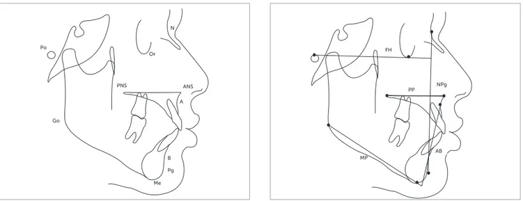
Each study group was further divided into adolescent group (10-18 years old) and adult group (19-30 years old). Each group included 14 males and 16 females, except for the open bite group that had an equal number of male and female subjects. Lateral cephalograms of these subjects were traced manually on acetate paper, with a 0.5-mm lead pencil in a dark room by the main investigator. Spe-ciic landmarks were identiied (N, Or, Po, ANS, PNS, A, B, Pg, Me, Go) and angular measurements were de-termined with the aid of a protractor (Fig 1).
The ODI was measured as the sum of two angles (AB-MP and PP-FH), as described by Kim7 (Fig 2).
The APDI was measured as the sum of three angles (FH-NPg, PP-FH and AB-NPg), as described by Kim and Vietas11 (Fig 2).
Statistical analysis of data was carried out by means of SPSS for Windows (version 20.0, SPSS Inc. Chicago, USA). Shapiro-Wilk test was used to test for normality of data and revealed normal distribution; hence, non-parametric tests were applied. Mann-Whitney-U test was used to compare the study parameters between males and females as well as adolescent and adult groups. The mean values of ODI and APDI angles were compared among study groups by means of Kruskal-Wallis test. Multiple
comparisons for ODI and APDI among study groups were carried out by means of post-hoc Dunnet T3 test. A p ≤ 0.05 was consigned as statistically signiicant. The reliability of ODI and APDI as diagnostic analyses was tested by means of the receiver operating characteristic curve (ROC).
RESULTS
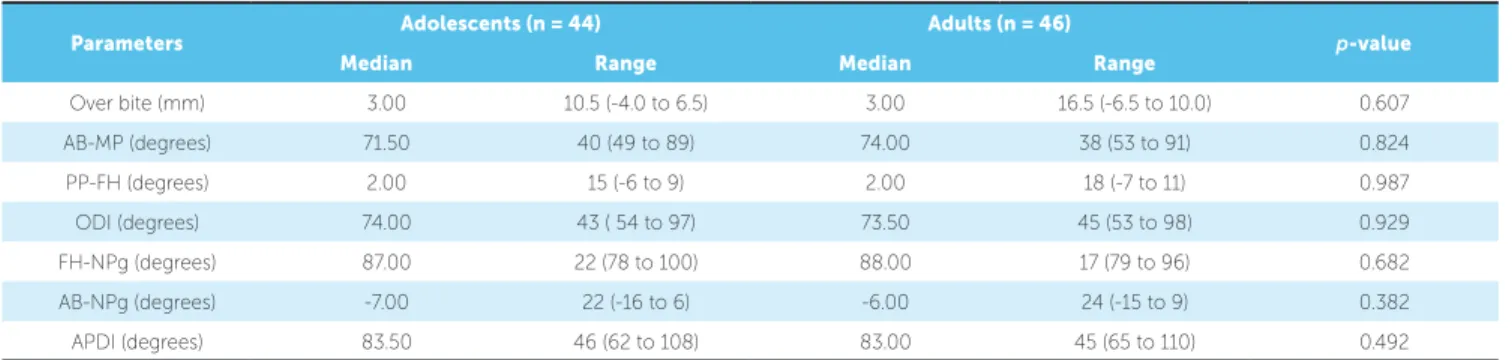
The study parameters were compared between males and females as well as between adolescents and adults. Results showed insigniicant diferences. Hence, to conserve the power of study, data were not stratiied according to sex and age (Tables 1 and 2).
Comparison among overbite groups showed signiicant diferences for AB-MP angle (p < 0.001) and ODI (p < 0.001). However, insigniicant diference was found for the palatal plane angle among the three over-bite groups (p = 0.775) (Table 3).
Comparison among Angle’s classiication groups showed signiicant diferences for the facial plane angle (p < 0.001), denture base to facial plane angle (p < 0.001) and APDI (p < 0.001). However, insigniicant diference was found for the palatal plane angle among Classes I, II and III (p = 0.214) (Table 4).
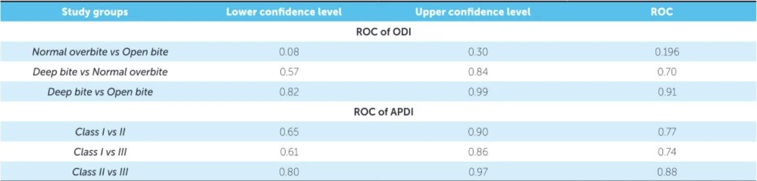
ROC plot comparing overbite groups for ODI showed an area under curve with a value equal to 0.196 between normal overbite and open bite groups; 0.70 between deep bite and normal overbite groups; and 0.91 between deep bite and open bite groups. The calculated values of ODI were consistent with incisor overbite in 91% of subjects (Table 5, Fig 3).
Figure 1 - Landmarks for ODI and APDI. Figure 2 - ODI and APDI parameters.
FH
PP NPg
AB MP
Me Pg B
A ANS PNS
Or N
Po
Parameters Adolescents (n = 44) Adults (n = 46) p-value
Median Range Median Range
Over bite (mm) 3.00 10.5 (-4.0 to 6.5) 3.00 16.5 (-6.5 to 10.0) 0.607
AB-MP (degrees) 71.50 40 (49 to 89) 74.00 38 (53 to 91) 0.824
PP-FH (degrees) 2.00 15 (-6 to 9) 2.00 18 (-7 to 11) 0.987
ODI (degrees) 74.00 43 ( 54 to 97) 73.50 45 (53 to 98) 0.929
FH-NPg (degrees) 87.00 22 (78 to 100) 88.00 17 (79 to 96) 0.682
AB-NPg (degrees) -7.00 22 (-16 to 6) -6.00 24 (-15 to 9) 0.382
APDI (degrees) 83.50 46 (62 to 108) 83.00 45 (65 to 110) 0.492
Table 1 - Comparison of study parameters between adolescent and adult groups.
n = 90. Mann-Whitney U Test. p ≤ 0.05.
Table 2 - Comparison of study parameters between male and female groups.
n = 90. Mann-Whitney U Test. p ≤ 0.05.
Parameters Male (n = 44) Female (n = 46) p-value
Median Range Median Range
Over bite (mm) 2.75 15.5 (-5.5 to 10.0) 3.00 14.0 (-6.5 to 7.5) 0.786
AB-MP (degrees) 74.00 42 (49 to 91) 72.00 32 (55 to 87) 0.759
PP-FH (degrees) 2.00 15 (-7 to 8) 3.00 16 (-5 to 11) 0.065
ODI (degrees) 73.50 44 (53 to 97) 74.50 41 (57 to 98) 0.288
FH-NPg (degrees) 87.50 22 (78 to 100) 87.50 17 (70 to 96) 0.761
AB-NPg (degrees) -7.00 25 (-16 to 9) -6.50 19 (-14 to 5) 0.557
APDI (degrees) 83.00 45 (65 to 110) 83.00 4162 to 103) 0.965
Table 3 - Comparison of ODI among overbite groups.
n = 90, SD – Standard Deviation. Kruskal-Wallis Test. Post hoc-Dunnet T3. *p ≤ 0.05, **p < 0.01. Variables
ODI (degrees)
P
Post hoc Dunnet T3
Open bite (n = 30) Normal overbite (n = 30) Deep bite (n = 30) Open vs
Deep bite (p)
Deep vs
Normal overbite (p)
Open vs
Normal overbite (p) Median Range Median Range Median Range
AB-MP 62.00 31 (49 to 80) 74.50 30 (54 to 84) 81.50 31 (60 to 91) <0.001** <0.001** 0.012* <0.001**
PP-FH -1.00 14 (-6 to 8) 2.00 16 (-7 to 9) 2.00 18 (-7 to 11) 0.775 0.738 0.963 0.931
ODI 64.50 24 (53 to 77) 74.50 32 (57 to 89) 83.00 42 (56 to 98) <0.001** <0.001** 0.022* <0.001**
Table 4 - Comparison of APDI among Angle’s molar classes groups.
n = 90, SD – Standard Deviation. Kruskal-Wallis Test. Post hoc-Dunnet T3. *p ≤ 0.05, **p < 0.01. Variables
APDI (degrees)
P
Post hoc Dunnet T3
Dental Class I (n = 30) Dental Class II (n = 30) Dental Class III (n = 30)
Dental
Class I vs II
Dental
Class II vs III
Dental
Class I vs III Median Range Median Range Median Range
FH-NPg 88.00 11 (82 to 93) 85.00 22 (78 to 100) 85.50 18 (79 to 97) < 0.001** 0.085 0.001* 0.109
PP-FH 3.00 15 (-7 to 8) -1.00 18 (-7 to 11) -1.00 15 (-6 to 9) 0.214 0.236 0.999 0.271
AB-NPg -7.00 13 (-14 to -1) -9.00 17 (-16 to 1) 1.50 18 (-9 to 9) <0.001** 0.025* <0.001** <0.001**
Table 5 - ROC of ODI and APDI among overbite and Angle’s classes, respectively.
ROC = Reciever Operating Characteristic. ROC > 0.6 is significantly reliable.
Study groups Lower confidence level Upper confidence level ROC
ROC of ODI
Normal overbite vs Open bite 0.08 0.30 0.196
Deep bite vs Normal overbite 0.57 0.84 0.70
Deep bite vs Open bite 0.82 0.99 0.91
ROC of APDI
Class I vs II 0.65 0.90 0.77
Class I vs III 0.61 0.86 0.74
Class II vs III 0.80 0.97 0.88
Figure 3 - ROC of ODI amongst vertical groups: (A) Normal overbite vs Open bite; (B) Deep bite vs Normal overbite; (C) Deep bite vs Open bite.
Figure 4 - ROC of APDI amongst sagittal groups: (A) Dental Class I vs II; (B) Dental Class I vs III; (C) Dental Class II vs III.
Normal overbite vs Open bite Dental Class I vs Dental Class II
Deep bite vs Normal overbite Dental Class I vs Dental Class III
Deep bite vs Open bite Dental Class II vs Dental Class III
A A
B B
C C
ROC Curve
ROC Curve ROC Curve
ROC Curve ROC Curve
ROC Curve
se
n
sit
iv
it
y
se
n
sit
iv
it
y
se
n
sit
iv
it
y
se
n
sit
iv
it
y
se
n
sit
iv
it
y
se
n
sit
iv
it
y
1 - Specificity
1 - Specificity 1 - Specificity
1 - Specificity 1 - Specificity
1 - Specificity
1.25
1.25 1.25
1.0 1.2 1.0
1.0 0.75
0.75 0.75
0.6 0.6
0.6 0.25
0.25 0.25
0.2 0.0 0.2
0.2 1.00
1.00 1.00
0.8 0.8
0.8 0.50
0.50 0.50
0.4 0.4
0.4 0.00
0.00 0.00
0.0 0.0
0.0
0.0
0.0 0.2
0.2 0.2
0.2 0.2
0.2 0.4
0.4 0.4
0.4 0.4
0.4 0.6
0.6 0.6
0.6 0.6
0.6 1.0
1.0 1.0
1.0 1.0
1.0
0.8
0.8 0.8
0.8 0.8
ROC plot comparing Angle’s classiication groups for APDI showed an area under curve with a value equal to 0.77 between dental Classes I and II; 0.74 between dental Class I and III; and 0.88 between dental Classes II and III. The calculated values of APDI were consistent with Angle’s classiication in 88% of subjects (Table 5, Fig 4).
DISCUSSION
Cephalometric analysis is an essential clinical tool in orthodontic diagnosis and treatment planning. To this end, several cephalometric analyses have been intro-duced by researchers, but none of them provides de-tailed information regarding dental malocclusion and their corresponding skeletal discrepancy.6 Hence, the objective of the current study was to identify whether the skeletal and dental components of malocclusion can be clearly identiied by ODI and APDI.
To evaluate ODI, subjects were divided into three equal groups on the basis of overbite. The present study showed signiicant diferences in ODI among open bite, normal overbite and deep bite groups. Our results were in accordance with the study conducted by Kim.7 Another study conducted by Freudenthaler et al12 found signii-cant diferences between deep bite and open bite groups as well as normal overbite and deep bite groups. Howev-er, insigniicant diferences were reported between nor-mal overbite and open bite groups. The reason behind the diferences in results may be due to the stratiication of subjects on the basis of incisor overbite.
The ODI is the sum of the AB-MP angle and the pal-atal plane angle. Considering these components of ODI independently, the AB-MP angle value showed signiicant intergroup diferences. A lower value of AB-MP angle was observed in the horizontal growth pattern, while an in-creased value was observed in the vertical growth pattern. However, the palatal plane angle did not show signiicant diferences among the three overbite groups. The inclina-tion of palatal plane upward and forward to the Frankfort horizontal plane results in decreased value of ODI. This indicates a tendency towards skeletal open bite. Therefore, it showed that the primary determinant of ODI is AB-MP angle, while the palatal plane angle does not play any sig-niicant role in the value of ODI. These results were simi-lar to those reported by other studies.7,12,14
In the assessment of APDI, our study showed significant intergroup differences regarding Angle’s classification. Analogous results were found in the
previous studies.11,12,15 In evaluating each compo-nent individually, the facial plane angle showed sig-nificant differences between dental Classes II and III. The lowest values were presented in cases of man-dibular retrognathism, while the highest values were found in mandibular excess, indicating skeletal Class III pattern. A higher value of the mean palatal plane angle was observed in dental Class I pattern, but statis-tical analysis showed insignificant differences among the three Angle’s classification groups. In contrast, the third component of APDI, denture base to facial plane angle, showed significant intergroup differences among all three sagittal groups. Clockwise rotation of this angle led to a decrease in the APDI value, which expressed clinically as dental Class II pattern. On the other hand, an increased value of APDI and dental Class III pattern was observed with counter clock-wise rotation of this angle.11 Hence, facial plane angle and denture base to facial plane angle were the deci-sive factors for APDI to determine various Angle’s classification groups.
The reliability of diagnostic information provided by the analyses plays a vital role in treatment success. Re-liability could be assessed by means of the ROC curve which describes eicacy in terms of sensitivity and speci-icity.15 An ideal test shows a value of 1, while a test re-sult of 0.5 or less indicates no diagnostic value.16-21 In our study, assessment of ROC demonstrated that ODI yielded the highest diagnostic value for deep bite and open bite groups. These results were 91% correspondent with incisor overbite. In contrast, a study conducted by Freudenthaler et al12 reported a value of 81%. Wardlaw et al20 showed a high diagnostic value between open bite and positive overbite groups, using a modiication of ODI. They used palatal plane to cranial base plane angle (PP-SN) instead of Frankfort horizontal plane to palatal plane angle (PP-FH). Although landmark identiication is diicult with the use of the Frankfort horizontal plane, the latter provides more accurate information regarding jaw position. Moreover, a true horizontal plane provides better information in terms of ODI.7,22
However, there are cases that present diferent Angle’s malocclusions with variable skeletal patterns, i.e., mo-lar Class II could present with skeletal Class III pat-tern, showing a higher APDI value; or molar Class I with skeletal Class II pattern. Such cases require care-ful treatment planning and cautious use of biomechan-ics, since the dental decompensation occurring during orthodontic treatment may result in the expression of underlying skeletal discrepancy. 23-26
CLINICAL IMPLICATION
The results of the present study indicate that ODI and APDI can be reliably used to assess the nature of dental malocclusion. However, there may be cases in which skeletal and dental malocclusions are not in correspondence with each other and for which cautious treatment planning would be required.
CONCLUSIONS
The following conclusions could be drawn from this study:
1. No diference was found in ODI and APDI val-ues between males and females, as well as between adolescents and adults.
2. ODI can signiicantly diferentiate between bite groups and was consistent with patient’s over-bite in 91% of the cases.
3. APDI can signiicantly difer between Angle’s malocclusions and was consistent with the dental classiication in 88% of the cases.
1. Graber TM. Orthodontics: principles and practice. 2nd ed. Philadelphia: WB Saunders; 1966.
2. Riedel RA. The relation of maxillary structures to cranium in malocclusion and in normal occlusion. Angle Orthod. 1952 July;22(3):142-45. 3. Jacobson A. The “Wits” appraisal of jaw disharmony. Am J Orthod. 1975
Feb;67(2):125-38.
4. Downs WB. Variations in facial relationships; their signiicance in treatment and prognosis. Am J Orthod. 1948 Oct;34(10):812-40.
5. Isaacson JR, Isaacson RJ, Speidel TM, Worms FW. Extreme variation in vertical facial growth and associated variation in skeletal and dental relations. Angle Orthod. 1971 July;41(3):219-29.
6. Freeman RS. Adjusting A-N-B angles to relect the efect of maxillary position. Angle Orthod. 1981 Apr;51(2):162-71.
7. Kim YH. Overbite depth indicator with particular reference to anterior open-bite. Am J Orthod. 1974 Jun;65(6):586-611.
8. Angle EH. Treatment of malocclusion of the teeth and fractures of the maxillae. 6th ed. Philadelphia: SS. White Dental; 1990.
9. Singh AK, Ganeshkar SV, Mehrotra P, Bhagchandani J. Comparison of
diferent parameters for recording sagittal maxillomandibular relation using natural head posture: a cephalometric study. J Orthod Sci.
2013 Jan-Mar;2(1):16-22.
10. Veeranarayana D, Rajani, Vani SVK, Rao GS, Raju KS, Pullaparaju G. Reliability, accuracy and interchangeability of diferent cephalometric methods for assessing sagittal jaw relationships. J Res Adv Dent. 2014;3:232-8.
11. Kim YH, Vietas JJ. Anteroposterior dysplasia indicator: an adjunct to cephalometric diferential diagnosis. Am J Orthod. 1978 Jun;73(6):619-33. 12. Freudenthaler JW, Celar AG, Schneider B. Overbite depth and
anteroposterior dysplasia indicators: the relationship between occlusal and skeletal patterns using the receiver operating characteristic (ROC) analysis. Eur J Orthod. 2000 Feb;22(1):75-83.
13. Kim YH. A comparative cephalometric study of Class II, Division 1 nonextraction and extraction cases. Angle Orthod. 1979 Apr;49(2):77-84. 14. Freudenthaler JW, Celar AG, Kubota M, Akimoto S, Sato S, Schneider B.
Comparison of Japanese and European overbite depth indicator and anteroposterior dysplasia indicator values. Eur J Orthod. 2011 Feb;33(1):1-5. 15. Han UK, Kim YH. Determination of Class II and Class III skeletal patterns:
Receiver operating characteristic (ROC) analysis on various cephalometric measurements. Am J Orthod Dentofacial Orthop. 1998 May;113(5):538-45. 16. Weinstein MC, Fineberg HV. Clinical decision analysis. Philadelphia:
WB Saunders; 1980.
17. Douglass CW. Evaluating diagnostic tests. J Eval Clin Pract. 2016 Aug;22(4):575-9.
18. Centor RM. Signal detectability: the use of ROC curves and their analyses. Med Decis Making. 1991 Apr-Jun;11(2):102-6.
19. Berkey CS, Douglass CW, Valachovic RW, Chauncey HH, McNeil BJ. Statistical methods for comparing dental diagnostic procedures. Community Dent Oral Epidemiol. 1990 Aug;18(4):169-76.
20. Wardlaw DW, Smith RJ, Hertweck DW, Hildebolt CF. Cephalometrics of anterior open bite: a receiver operating characteristic (ROC) analysis. Am J Orthod Dentofacial Orthop. 1992 Mar;101(3):234-43.
21. Newhauser D, Yin XP. Deciding whether a new test/measure is useful. Med Care. 1991 Aug;29(8):685-9.
22. Jacobson A. Radiographic cephalometry. How reliable is cephalometric prediction? São Paulo: Quintessence; 1995.
23. Meyers DM. A multivariate analysis of the relations between craniofacial structure and occlusion with DiPaolo’s quadrilateral measures. Am J Orthod Dentofacial Orthop. 1992 July;102(1):52-61.
24. Dibbets JM. Morphological associations between the Angle classes. Eur J Orthod. 1996 Apr;18(2):111-8.
25. Harris EF, Johnson MG. Heritability of craniometric and occlusal variables: a longitudinal sib analysis. Am J Orthod Dentofacial Orthop. 1991 Mar;99(3):258-68.
26. Bishara SE, Jakobsen JR, Vorhies B, Bayati P. Changes in dentofacial structures in untreated Class II division 1 and normal subjects: a longitudinal study. Angle Orthod. 1997;67(1):55-66.