The duration of pubertal growth peak among
three skeletal classes
Waqar Jeelani1, Mubassar Fida2, Attiya Shaikh3
Introduction: Pubertal growth peak is closely associated with a rapid increase in mandibular length and offers a wide range of therapeutic modifiability. Objective: The aim of the present study was to determine and compare the mean ages of onset and duration of pubertal growth peak among three skeletal classes. Methods: A retrospective cross-sectional study was con-ducted using lateral cephalograms of 230 subjects with growth potential (110 males, 120 females). Subjects were categorized into three classes (Class I = 81, Class II = 82, Class III = 67), according to the sagittal relationship established between the maxilla and the mandible. The cervical vertebral maturation stage was recorded by means of Baccetti’s method. The mean ages at CS3 and CS4 and the CS3-CS4 age interval were compared between boys and girls and among three skeletal classes.
Results: Pubertal growth peak occurred on average four months earlier in girls than boys (p = 0.050). The average duration of pubertal growth peak was 11 months in Class I, seven months in Class II and 17 months in Class III subjects. Interclass differences were highly significant (Cohen’s d > 0.08). However, no significant difference was found in the timing of pubertal growth peak onset among three skeletal classes (p = 0.126 in boys, p = 0.262 in girls). Conclusions: Girls enter pubertal growth peak on average four months earlier than boys. Moreover, the duration of pubertal growth peak is on average four months shorter in Class II and six months longer in Class III subjects as compared to Class I subjects.
Keywords: Puberty. Age of onset. Cervical vertebrae. Cephalometry.
1 Resident Orthodontist, The Aga Khan University Hospital, Section of
Dentistry, Department of Surgery, Karachi, Pakistan.
2 Consultant Orthodontist/Associate Professor, The Aga Khan University
Hospital, Program Director, Orthodontics Residency Program Section of Dentistry, Department of Surgery, Karachi, Pakistan.
3 Consultant Orthodontist/ Assistant Professor, The Aga Khan University
Hospital, Program Coordinator, Orthodontics Residency Program Section of Dentistry, Department of Surgery, Karachi, Pakistan.
DOI: http://dx.doi.org/10.1590/2177-6709.21.5.067-074.oar
How to cite this article: Jeelani W, Fida M, Shaikh A. The duration of pubertal growth peak among three skeletal classes. Dental Press J Orthod. 2016 Sept-Oct;21(5):67-74. DOI: http://dx.doi.org/10.1590/2177-6709.21.5.067-074.oar
Submitted: December 05, 2015 - Revised and accepted: May 30, 2016
» The authors report no commercial, proprietary or financial interest in the products or companies described in this article.
Contact address: Waqar Jeelani E-mail: [email protected]
Introdução: o pico de crescimento puberal está intimamente relacionado a um rápido aumento no comprimento da mandíbula e propicia uma larga gama de abordagens terapêuticas. Objetivos: o objetivo do presente estudo foi deter-minar e comparar as idades médias ao começo do pico de crescimento puberal, bem como sua duração, nas três dife-rentes classes esqueléticas. Métodos: esse estudo retrospectivo transversal foi conduzido usando radiografias celafométricas laterais de 230 indivíduos com potencial de crescimento (110 homens, 120 mulheres). Os indivíduos foram categorizados em 3 classes (Classe I = 81, Classe II = 82, Classe III = 67), conforme a relação sagital presente entre a maxila e a mandíbula. O estágio de maturação cervical foi registrado por meio do método de Baccetti. As idades médias em EMVC3 e EMVC4, bem como o intervalo de idade EMVC3-EMVC4 foram comparados entre meninos e meninas e entre as três classes es-queléticas. Resultados: o pico de crescimento puberal ocorreu, em média, quatro meses mais cedo nas meninas do que nos meninos (p = 0,050). A duração média do pico de crescimento puberal foi de 11 meses nos indivíduos Classe I, 7 meses nos indivíduos Classe II e de 17 meses nos indivíduos Classe III. As diferenças interclasses foram altamente significativas (d de Cohen > 0,08). Porém, não foram identificadas diferenças significativas quanto à época de início do pico de crescimento puberal entre as três classes esqueléticas (p = 0,126 nos garotos, p = 0,262 nas garotas). Conclu-sões: as meninas entram no pico de crescimento puberal, em média, quatro meses antes dos meninos. Além disso, a duração do pico de crescimento puberal é, em média, quatro meses menor em indivíduos Classe II e seis meses maior nos indivíduos Classe III, em comparação aos indivíduos Classe I.
INTRODUCTION
Modiication of children’s facial growth to achieve a more harmonious relationship between diferent fa-cial structures is oten part of orthodontic treatment.1,2,3 Normal human development is constituted of certain periods of growth accelerations and decelerations.4-7 The periods of rapid growth are of particular interest to orthodontists, as growth modiications are best achieved during the adolescent growth spurt when diferent facial bones are growing at a favourable rate.4,5 By initiating treatment at patient’s optimal skeletal maturational stage, a favorable outcome with minimum risk of un-wanted efects can be expected.4
Longitudinal studies based on lateral cephalograms have identiied wide individual variations in the time of pubertal growth spurt onset and duration.8 In this context, identiication of patient’s maturation stage be-comes a critical component of orthodontic diagnosis, helping to identify children of the same chronological age, but with diferent degrees of skeletal maturation.
Individual patient’s skeletal maturity can be assessed by means of diferent biological indicators, for example, increase in body weight and height,9-12 skeletal matu-ration of the hand and wrist,6,13 dental development,14 sexual changes,15,16 and cervical vertebral matura-tion.17,18,19 Franchi et al20 reported several advantages of using the cervical vertebral maturation (CVM) meth-od in assessing the skeletal maturity of an individual. These advantages include: straightforward appraisal of cervical vertebrae shape; more than 98% interexaminer reliability; and no need for second radiation exposure to determine patient’s skeletal age.20,21
Several studies4,19-24 and a systematic review25 have established the CVM method as a highly reliable ap-proach of assessing diferent stages of adolescent growth spurt. Current studies26,27 continue to establish that the CVM method can be used as an alternative to the hand and wrist radiographs to assess skeletal matu-rity. Cervical stage 3 (CS3) and cervical stage 4 (CS4) of the CVM method correspond to the initial and inal stages of the accelerative portion of the pubertal growth peak, respectively.4,24 Longitudinal studies by Gu and McNamara28 as well as Perinetti et al29 report that the maximum increment in mandibular growth occurs between CS3 and CS4. The age interval be-tween these two stages is regarded as the duration of the pubertal growth peak.28-32
A rapid increase in mandibular length during pubertal growth peak highlights the potential impact of variations in the time of pubertal growth peak onset and duration on the inal size of the mandible.28-33 Thus, evaluation of such aberrations at the time of pubertal growth peak onset and duration may provide a better understanding of the development of diferent skeletal malocclusions and subsequently facilitate treatment of skeletal problems during this period of rapid growth.
The timing of pubertal growth peak varies signifi-cantly between males and females; thus, a separate analysis for girls and boys is highly desirable. How-ever, previous studiesfailed to provide a comprehen-sive analysis of pubertal growth peak duration among three skeletal classes and reported combined results for male and female samples.30,31,32
In this context, this study was designed to de-termine and compare the mean ages of pubertal growth peak onset and duration among children with different skeletal classes.
MATERIAL AND METHODS
A cross-sectional study was conducted at The Aga Khan University Hospital, Karachi. Ethical approval was obtained from the institutional Ethics Committee prior to data col-lection (3503-Sur-ERC-15). Sample size for three skeletal classes was calculated by taking α = 0.05 and keeping a pow-er of study of 80%. Findings by Kuc-Michalska and Bac-cetti30 were used for sample size calculation, showing that a sample size of 63 in each group was suicient in order to detect a clinically signiicant diference of 0.50 + 1.00 year in the mean age at CS4 between Class I and Class III sub-jects. In order to increase the power of study, the maximum number of available subjects was included in the study, which resulted in a total sample of 230 subjects.
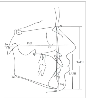
Patients’ age was recorded to the nearest month and converted into decimal expression for further use in statisti-cal analyses. Lateral cephalograms of all patients were traced manually on acetate paper by the main investigator, and the skeletal class of each subject was determined based on the ANB angle and Downs facial angle. The vertical facial pat-tern was assessed from the anterior cranial base to the man-dibular plane angle (SNMP angle), and lower anterior facial height to total anterior facial height ratio (LAFH/TAFH) (Fig 1).34,35 Dental malocclusion was assessed on pretreat-ment dental casts. Subjects were divided into three groups, according to the following criteria:
» Skeletal Class I: subjects with ANB angle > 0° and < 5°; Downs facial angle > 83° and < 91°; and Class I molar relationship (81 subjects).
» Skeletal Class II: subjects with ANB angle > 5°; Downs facial angle < 83°; and more than half unit Class II molar relationship (82 subjects).
» Skeletal Class III: subjects with ANB angle < 0°; Downs facial angle > 91°; and more than half unit Class III molar relationship (67 subjects).
Cervical vertebral maturation stages were assessed on the lateral cephalograms by means of Baccetti’s method4 (Fig 2). The age interval between CS3 and CS4 stages was regarded as the duration of pubertal growth peak.28-32
Data were analyzed in SPSS for Windows (version 20.0, SPSS Inc. Chicago). The normality of variable age was as-sessed by means of Shapiro-Wilk test that showed normal distribution of data. The mean ages at CS3 and CS4 and the age interval between these two stages were compared between boys and girls by means of independent t-test. The mean ages at CS3 and CS4 and the age intervals be-tween them were compared among three skeletal classes by one-way ANOVA and post-hoc Tukey tests. Efect sizes were calculated by means of Cohen’s d and the recom-mended interpretations were used to describe the results.36 A p < 0.05 was taken as statistically signiicant, but this value was adjusted to the appropriate level when Bonferroni cor-rections were employed for multiple comparisons to mini-mize the chance of type I error.
To test interexaminer reliability, 30 lateral cephalograms were randomly selected, and steps of tracing, landmark identiication and measurement were repeated by the main investigator and a second observer. Kappa statistics was em-ployed and showed that the values of coeicients of reliabil-ity were greater than 0.892 for the identiication of skeletal class and the CVM stage.
Figure 1 - ANB angle and Downs facial angle (angle formed between FHP and NPog) were used to classify subjects into three skeletal classes. The SNMP angle (angle formed between SN plane and GoGn plane) and LAFH/TAFH ratio were used to determine the vertical growth pattern of a subject.
RESULTS
A total of 230 subjects (110 males and 120 females) were included in this study. The mean SNMP angle of the total sample was 31.28 ± 4.53°, and no signiicant diference (p = 0.065) was found among three classes. Similarly, the mean LAFH/TAFH of the sample was 55.37 ± 3.02%, and no signiicant diference was found among three skeletal classes (p = 0.125).
The mean ages at CS3 and CS4 were compared between the male and female samples by means of an independent sample t-test (Table 1). Significant sex-based differences in the mean ages at CS3 (p = 0.050) showed that the pubertal growth peak oc-curred around four months (0.33 years) earlier in girls, as compared to boys. The mean duration of pubertal growth peak was 11.7 months in girls and 13.3 months in boys. This sex-related difference in the duration of pubertal growth peak was found to be statistically significant, but had a small effect (Cohen’s d = 0.13) (Table 2).
Comparison of mean ages at CS3 and CS4 among three skeletal classes was performed by means of one-way ANOVA and post-hoc Tukey tests separately for the male and female samples (Table 3). No signiicant diference was found in the mean ages at CS3 in boys (p = 0.126) or girls (p = 0.262). However, highly signiicant diferences (p = < 0.001) were present in the mean ages at CS4 among three skeletal classes for both males and females.
The mean duration of pubertal growth peak was 0.95 ± 0.20 years (11.4 months) in Class I; 0.60 ± 0.15 years (7.2 months) in Class II; and 1.44 ± 0.16 years (17.3 months) in Class III children. The durations of pubertal growth peak were compared among various skeletal class-es and showed that Class II subjects had on average 4.2 months shorter duration of pubertal growth peak, as com-pared to Class I subjects. On the other hand, the duration of pubertal growth peak was on average 5.9 months longer in Class III subjects, as compared to Class I counterparts. These interclass diferences were characterized by highly signiicant efect (Cohen’s d efect size > 0.08) (Table 4).
Table 1 - A comparison of mean ages between boys and girls at CS3 and CS4.
Table 2 - Mean duration of pubertal growth peak in boys and girls. n = 230; SD: Standard Deviation; Independent sample t-test. *p < 0.05.
n = 230; SD: Standard Deviation; CI: Confidence Interval;
Cohen’s d effect size: *low significance (0.2 – 0.5), **Moderate significance (0.5 – 0.8), ***High significance ( > 0.8).
Cervical stage Boys (n = 110) Girls (n = 120) Mean diference p value Mean ± SD (Years) Mean ± SD (Years) (Years)
CS3 12.18 ± 0.81 11.85 ± 0.85 0.33 (3.9 months) 0.050*
CS4 13.30 ± 0.78 12.84 ± 0.92 0.46 (5.5 months) 0.004*
CS3 CS4 Duration of pubertal growth peak
CI inferior limit
CI Superior limit
Cohen’s d p value Mean ± SD
(Years)
Mean ± SD (Years)
CS3 – CS4
(Years) (Years) (Years)
Boys 12.18 + 0.81 13.30 + 0.78 1.11 ± 0.15 (13.3 months)
0.08 0.17 0.13 < 0.001
DISCUSSION
Variations in pubertal growth spurt onset and du-ration may afect the inal size of diferent craniofacial structures.10,37 Longitudinal studies have shown that growth changes during adolescent growth spurt are more pronounced in the mandible, as compared to the maxilla.10,38,39 The current study reports that the onset of pubertal growth peak occurs around four months earlier in girls, as compared to boys. However, the diference in the overall duration of pubertal growth peak between males and females was only of one and a half month. A literature review reveals insigniicant diferences in the duration of pubertal growth peak between boys and girls.31 Late onset of adolescent growth spurt accompa-nied by continued post-pubertal increase in mandibu-lar length in boys help explaining mandibu-large mandibumandibu-lar size and more prevalent Class III jaw relationship in males, as compared to females.40,41
Table 3 - Comparison of mean ages at CS3 and CS4 among three skeletal classes.
Table 4 - Comparison of mean duration (in years) of pubertal growth peak among three skeletal classes.
n = 230; SD: Standard Deviation; One-way ANOVA test. After applying Bonferroni correction for multiple testing. a p-value of <0.025 was taken as statistically significant. *p < 0.025.
Cohen’s d effect size: *low significance (0.2 – 0.5), **Moderate significance (0.5 – 0.8), *** High significance (> 0.8).
Cervical stage
Age (Years ± SD)
p value Post-hoc Tukey HSD Class I Class II Class III I vs II I vs III II vs III
Girls CS3 11.94 + 0.99 11.57 + 0.70 12.08 + 0.75 0.262 0.431 0.904 0.274
CS4 12.95 + 1.04 12.26 + 0.56 13.62 + 0.43 <0.001* 0.003* 0.014* <0.001*
Boys CS3 12.22 + 0.83 11.93 + 0.89 12.47 + 0.61 0.126 0.492 0.613 0.103
CS4 13.25 + 0.36 13.62 + 0.52 13.85 + 0.75 <0.001* 0.018* 0.015* <0.001*
Duration of pubertal growth peak (CS3 – CS4 interval)
CI inferior limit
CI superior limit
Interclass
diference Cohen’s d p value
Class I (n = 81) 0.95 ± 0.20 (11.4 months)
-0.40 -0.29 -0.35
(4.2 months) 1.98*** < 0.001 Class II (n = 82) 0.60 ± 0.15 (7.2 months)
Class I (n = 81) 0.95 ± 0.20 (11.4 months)
0.43 0.54 0.49
(5.9 months) 2.67*** < 0.001 Class III (n = 67) 1.44 ± 0.16 (17.3 months)
Class II (n = 82) 0.60 ± 0.15 (7.2 months)
0.78 0.89 0.82
(9.8 months) 5.40*** < 0.001 Class III (n = 67) 1.44 ± 0.16 (17.3 months)
Our results show that the duration of puber-tal growth peak was on average 4.2 months shorter in Class II subjects, as compared to Class I sub-jects. Salazar-Lazo et al32 also showed this differ-ence to be of four months. On the other hand, we found, on average, a 5.9-month longer duration of pubertal growth peak in Class III subjects, as com-pared to Class I subjects. Studies conducted on South American and Caucasian subjects showed this differ-ence to be of 4.8 and five months, respectively.30,31 These findings are suggestive that a longer duration of pubertal growth peak may be related to a larger size of the mandible.30,31,33,37 On the other hand, a shorter duration of pubertal growth peak may result in early deceleration of mandibular lengthening; thus, result-ing in a smaller final size of the mandible.33,37
The current recommendations endorse CS3 as the ideal time for the initiation of functional jaw ortho-pedics for the treatment of mandibular deficiency.4 Functional appliance therapy is less likely to be suc-cessful if commenced in the prepubertal period in-stead of pubertal growth peak.5 In the context of our results and the findings of previous studies,10,33,37,42 Class II subjects and girls can be regarded as early maturers, for which commencement of functional jaw orthopedics should be started earlier than usual. Simi-larly, Class III subjects and boys may be considered late maturers; therefore, they may require treatment with Class III orthopedic appliances to be carried out for a longer period of time until the accelerated phase of adolescent growth spurt is over.
Despite some recent investigations43,44 showing a weak correlation between CVM and mandibular growth spurt, strong evidence is available in favor of CVM as a good predictor of mandibular growth peak.4,5,18-20,24-27,45,46 Different studies reported variable levels of validity and reliability of the CVM meth-od ranging from below average to excellent.47,48,49 However, Santiago et al47 showed a moderate to high-level of reproducibility of the CVM method in their systematic review using Kappa statistics. A high de-gree of intra- and interexaminer reliability was found in the current study. Though a recent meta-analysis25 shows that the CVM method of Hassel and Farman
performs better than Baccetti’s method in predicting the overall status of pubertal growth spurt, the later was used because of its proven efficiency in assessing the pubertal growth peak, as shown by longitudinal studies.28,29 Assessment of craniofacial growth asks for a longitudinal study design as an essential method for reliable results. Longitudinal studies require repeated exposure to X-ray radiations, which has certain ethi-cal limitations. Moreover, a few studies report that variations in skeletal and dental maturation may be re-lated to the vertical facial pattern of the individual.50,51 In this context, subjects were matched according to the vertical facial pattern by means of SNMP angle and LAFH/TAFH ratio which have been shown to be the most reliable indicators of vertical growth pat-tern.52 Moreover, separate analyses were performed for boys and girls, as required.
CONCLUSIONS
There is no signiicant diference in the duration of pubertal growth peak between girls and boys. The aver-age duration of pubertal growth peak was found to be 11 months in Class I, seven months in Class II and 17 months in Class III subjects. However, no signiicant interclass diferences were found in the time of pubertal growth peak onset among three skeletal classes.
A 4-month shorter duration of pubertal growth peak in Class II subjects and a 6-month longer duration of pubertal growth peak in Class III subjects, as com-pared to Class I subjects, may explain a smaller and a larger increment in mandibular length during pubertal growth peak in Class II and Class III subjects, respec-tively. However, the validity of these results needs to be endorsed by indings of longitudinal studies.
1. Chen L, Liu J, Xu T, Lin J. Longitudinal study of relative growth rates of the maxilla and the mandible according to quantitative cervical vertebral maturation. Am J Orthod Dentofacial Orthop. 2010 Jun;137(6):736.e1-8; discussion 736-7.
2. Singer J. Physiologic timing of orthodontic treatment. Angle Orthod. 1980 Oct;50(4):322-33.
3. O’Reilly MT, Yanniello GJ. Mandibular growth changes and maturation of cervical vertebrae--a longitudinal cephalometric study. Angle Orthod. 1988 Apr;58(2):179-84.
4. Baccetti T, Franchi L, McNamara JA. The Cervical Vertebral Maturation (CVM) method for the assessment of optimal treatment timing in dentofacial orthopedics. Semin Orthod. 2005 Sept;11(3):119-29. 5. Perinetti G, Primožič J, Franchi L, Contardo L. Treatment efects of
removable functional appliances in pre-pubertal and pubertal Class II patients: a systematic review and meta-analysis of controlled studies. PLoS One. 2015 Oct 28;10(10):e0141198.
6. Fishman L. Chronological versus skeletal age, an evaluation of craniofacial growth. Angle Orthod. 1979 July;49(3):181-9.
7. San Román P, Palma JC, Oteo MD, Nevado E. Skeletal maturation determined by cervical vertebrae development. Eur J Orthod. 2002 Jun;24(3):303-11.
8. Fishman LS. Can cephalometric x-rays of the cervical column be used instead of hand-wrist x-rays to determine patient’s maturational age? Am J Orthod Dentofacial Orthop. 2002 July;122(1):18A-9A.
9. Nanda RS. The rates of growth of several facial components measured from serial cephalometric roentgenograms. Am J Orthod. 1955 Sept;41(9):658-73.
10. Bambha JK. Longitudinal cephalometric roentgenographic study of face and cranium in relation to body height. J Am Dent Assoc. 1961 Dec;63:776-99.
11. Hunter WS. The correlation of facial growth with body height and skeletal maturation at adolescence. Angle Orthod. 1966 Jan;36(1):44-54. 12. Green LJ. The interrelationships among height, weight and chronological,
dental and skeletal ages. Angle Orthod. 1961 July;31(3):189-93. 13. Hägg U, Taranger J. Skeletal stages of the hand and wrist as indicators of
the pubertal growth spurt. Acta Odontol Scand. 1980;38(3):187-200. 14. Hägg U, Matsson L. Dental maturity as an indicator of chronological age: the
accuracy and precision of three methods. Eur J Orthod. 1985 Feb;7(1):25-34.
REFERENCES
15. Hägg U, Taranger J. Menarche and voice change as indicators of the pubertal growth spurt. Acta Odontol Scand. 1980;38(3):179-86. 16. Hägg U, Taranger J. Maturation indicators and the pubertal growth
spurt. Am J Orthod. 1982 Oct;82(4):299-309.
17. Lamparski DG. Skeletal age assessment utilizing cervical vertebrae [thesis]. Pittsburgh, (Penn): University of Pittsburgh; 1972. 18. Hassel B, Farman AG. Skeletal maturation evaluation using cervical
vertebrae. Am J Orthod Dentofacial Orthop. 1995 Jan;107(1):58-66. 19. Baccetti T, Franchi L, McNamara JA Jr. An improved version of the cervical vertebral maturation (CVM) method for the assessment of mandibular growth. Angle Orthod. 2002 Aug;72(4):316-23.
20. Franchi L, Baccetti T, McNamara JA Jr. Mandibular growth as related to cervical vertebral maturation and body height. Am J Orthod Dentofacial Orthop. 2000 Sept;118(3):335-40.
21. Pasciuti E, Franchi L, Baccetti T, Milani S, Farronato G. Comparison of three methods to assess individual skeletal maturity. J Orofac Orthop. 2013 Sept;74(5):397-408.
22. Mito T, Sato K, Mitani H. Cervical vertebral bone age in girls. Am J Orthod Dentofacial Orthop. 2002 Oct;122(4):380-5.
23. García-Fernandez P, Torre H, Flores L, Rea J. The cervical vertebrae as maturational indicators. J Clin Orthod. 1998 Apr;32(4):221-5.
24. Baccetti T, Franchi L, McNamara JA Jr. The cervical vertebral maturation method: some need for clarification. Am J Orthod Dentofacial Orthop. 2003 Jan;123(1):19A-20A.
25. Cericato GO, Bittencourt MA, Paranhos LR. Validity of the assessment method of skeletal maturation by cervical vertebrae: a systematic review and meta-analysis. Dentomaxillofac Radiol. 2015;44(4):20140270.
26. Perinetti G, Perillo L, Franchi L, Di Lenarda R, Contardo L. Maturation of the middle phalanx of the third finger and cervical vertebrae: a comparative and diagnostic agreement study. Orthod Craniofac Res. 2014 Nov;17(4):270-9.
27. Uysal T, Ramoglu SI, Basciftci FA, Sari Z. Chronologic age and skeletal maturation of the cervical vertebrae and hand-wrist: is there a relationship? Am J Orthod Dentofacial Orthop. 2006 Nov;130(5):622-8. 28. Gu Y, McNamara JA. Mandibular growth changes and cervical vertebral
29. Perinetti G, Contardo L, Castaldo A, McNamara JA Jr, Franchi L. Diagnostic reliability of the cervical vertebral maturation method and standing height in the identiication of the mandibular growth spurt. Angle Orthod. 2016 July;86(4):599-609.
30. Kuc-Michalska M, Baccetti T. Duration of the pubertal peak in skeletal Class I and Class III subjects. Angle Orthod. 2010 Jan;80(1):54-7.
31. García-Drago AG, Arriola-Guillén LE. Duration of the peak of growth in Class I and III subjects using the Baccetti’s cervical vertebrae maturation analysis on lateral cephalometric radiographs. Oral Health Dent Manag. 2014 Dec;13(4):963-6.
32. Salazar-Lazo R, Arriola-Guillén LE, Flores-Mir C. Duration of the peak of adolescent growth spurt in class i and ii malocclusion subjects using a cervical vertebrae maturation analysis. Acta Odontol Latinoam. 2014;27(2):96-101.
33. Reyes BC, Baccetti T, McNamara JA Jr. An estimate of craniofacial growth in Class III malocclusion. Angle Orthod. 2006 July;76(4):577-84. 34. Steiner CC. Cephalometrics for you and me. Am J Orthod.
1953;39(10):720-55.
35. Downs WB. Analysis of the dentofacial proile. Angle Orthod. 1956 Oct;26(4):191-212.
36. Cohen J. Statistical power analysis for the behavioral sciences. 2nd ed. Mahwah: Ed. Lawrence Erlbaum; 1988.
37. Silveira AM, Fishman LS, Subtelny JD, Kassebaum DK. Facial growth during adolescence in early, average and late maturers. Angle Orthod. 1992 Fall;62(3):185-90.
38. Linder-Aronson S, Woodside DG, Daigle DJ. A longitudinal study of the growth in length of the maxilla in boys between ages 6-20 years. Trans Eur Orthod Soc. 1975:169-79.
39. Jamison JE, Bishara SE, Peterson LC, DeKock WH, Kremenak CR. Longitudinal changes in the maxilla and the maxillary-mandibular relationship between 8 and 17 years of age. Am J Orthod. 1982 Sept;82(3):217-30.
40. Love RJ, Murray JM, Mamandras AH. Facial growth in males 16 to 20 years of age. Am J Orthod Dentofacial Orthop. 1990 Mar;97(3):200-6. 41. Mitani H, Sato K, Sugawara J. Growth of mandibular prognathism
after pubertal growth peak. Am J Orthod Dentofacial Orthop. 1993 Oct;104(4):330-6.
42. Armond MC, Generoso R, Falci SG, Ramos-Jorge ML, Marques LS. Skeletal maturation of the cervical vertebrae: association with various types of malocclusion. Braz Oral Res. 2012 Mar-Apr;26(2):145-50.
43. Ball G, Woodside D, Tompson B, Hunter WS, Posluns J. Relationship between cervical vertebral maturation and mandibular growth. Am J Orthod Dentofacial Orthop. 2011 May;139(5):e455-61.
44. Gray S, Bennani H, Kieser JA, Farella M. Morphometric analysis of cervical vertebrae in relation to mandibular growth. Am J Orthod Dentofacial Orthop. 2016 Jan;149(1):92-8.
45. Perinetti G, Contardo L, Castaldo A, McNamara JA Jr, Franchi L. Diagnostic reliability of the cervical vertebral maturation method and standing height in the identiication of the mandibular growth spurt. Angle Orthod. 2016 July;86(4):599-609.
46. Moshfeghi M, Rahimi H, Rahimi H, Nouri M, Bagheban AA. Predicting mandibular growth increment on the basis of cervical vertebral dimensions in Iranian girls. Prog Orthod. 2013;14(1):3.
47. Santiago RC, de Miranda Costa LF, Vitral RW, Fraga MR, Bolognese AM, Maia LC. Cervical vertebral maturation as a biologic indicator of skeletal maturity. Angle Orthod. 2012 Nov;82(6):1123-31.
48. Gabriel DB, Southard KA, Qian F, Marshall SD, Franciscus RG, Southard TE. Cervical vertebrae maturation method: poor reproducibility. Am J Orthod Dentofacial Orthop. 2009 Oct;136(4):478.e1-7; discussion 478-80. 49. Nestman TS, Marshall SD, Qian F, Holton N, Franciscus RG, Southard
TE. Cervical vertebrae maturation method morphologic criteria: poor reproducibility. Am J Orthod Dentofacial Orthop. 2011 Aug;140(2):182-8. 50. Gottimukkala P, Gandikota CS, Challa PL, Perumalla K, Palla Y, Juvvadi
SR. Assessment of skeletal and dental maturation of short and long-face children of South Indian Population. J Ind Orthod Soc. 2012;46(3):148-53. 51. Jamroz GM, Kuijpers-Jagtman AM, van’t Hof MA, Katsaros C. Dental
maturation in short and long facial types. Is there a diference? Angle Orthod. 2006 Sept;76(5):768-72.
52. Rizwan M, Mascarenhas R, Hussain A. Reliability of the existing vertical dysplasia indicators in assessing a deinitive growth pattern. Rev Latinoam Ortod Odontopediatría 2011;1-7.