Periodontics
Roberto Andrade(a) Manuel Espinoza(a) Elena Maria Gómez(a) José Rolando Espinoza(a) Elizabeth Cruz(a)
(a)Private practice. San Salvador, El Salvador.
Corresponding author: Roberto Andrade E-mail:
Received for publication on Aug 16, 2011 Accepted for publication on Nov 16, 2011
Intra- and inter-examiner
reproducibility of manual probing
depth
Abstract: The periodontal probe remains the best clinical diagnostic tool for the collection of information regarding the health status and the at-tachment level of periodontal tissues. The aim of this study was to evalu-ate intra- and inter-examiner reproducibility of probing depth (PD) mea-surements made with a manual probe. With the approval of an Ethics Committee, 20 individuals without periodontal disease were selected if they presented at least 6 teeth per quadrant. Using a Williams periodon-tal probe, three calibrated thesis-level students (k > 0.6) assessed PD at 6 sites per tooth, from the gingival margin to the bottom of the periodon-tal sulcus (rounded to the next 0.5 mm). Initial and repeated measure-ments were performed by the same three examiners. The intra-examiner agreement (± 1 mm > 90%) was 99.85%, 100%, and 100% for the three examiners, respectively. When the variables vestibular/lingual surfaces, mesial/distal surfaces, or superior/inferior jaws were evaluated, no sig-niicant differences in reproducibility were detected at the inter-examiner level (p < 0.05). At this level, the only signiicant differences observed were in the three examiners’ measurements of the anterior and poste-rior sites. While high intra-examiner reproducibility was detected, inter-examiner level proved to be low. We can conclude that measurement of PD with a manual periodontal probe produced high reproducibility in healthy individuals. The operator’s position can affect the reproducibility of repeated measures of PD. Calibration and operator training, rather than operator experience, were fundamental for reproducibility. Other factors, such as individual technique and probing depth force, can affect inter-examiner reproducibility.
Descriptors: Dental Instruments; Reproducibility of Results; Diagnostic Errors; Calibration.
Introduction
Traditionally, investigators have used the periodontal probe to de-tect the presence and progression of some periodontal diseases, such as chronic periodontitis, by evaluating numerical data and/or clinical signs of inlammation.1-3 Typically evaluated parameters include probing depth,
gingival levels, presence or absence of bleeding, and clinical attachment levels.2,4-6 The more exact the measuring tools are, the more reproducible
the measurements will be; additionally, a higher level of control of the variables that affect probing will be achieved. Furthermore, an accuracy Declaration of Interests: The authors
of the measuring tools will provide more precise in-formation for diagnosis and earlier detection of dis-ease, which, in turn, can foster proper and timely treatment, as well as long-term periodontal con-trol. Several factors affect periodontal parameters recorded through clinical measurements, and these can lead to a misinterpretation of an individual’s true periodontal condition, resulting in a lack or an excess of treatment. Some of these factors are: pres-ence of swollen gingival tissue, dental prosthetics, dental calculus, and diameter of and/or variations in the standardization of probe marks, as well as probing force and operator errors. The aim of this study was to compare inter- and intra-examiner re-producibility using a manual probe to obtain a clini-cal record of probing depth, with different variables considered.
Methodology
Before beginning the study, three examiners were trained in a calibration process. In a calibration group (not part of the experimental group), study participants were probed by each examiner, using a Williams SE manual probe (Hu-Friedy Co., Chicago, USA), once a week over a period of 4 months. The probing process was repeated until each examiner had substantial correlation as measured by Cohen’s Kappa (k ≥ 0.6). ln addition to the Kappa agreement, the measurements had to show a 90% agreement for ± 1 mm, as well as an exact agreement in 75% of the PD repeated measurements. Once the exam-iners were trained, the study was initiated. Twenty students (mean age, 21 years) from the Dentistry Program at the Evangelica University of El Salvador were selected (Institutional Ethics Committee Cer-tiication No. 9). Exclusion criteria for individuals participating in the study included: loss of clinical at-tachment levels, the presence of ixed or partial pros-thetics, individual crowns, or orthodontics, as well as surgical or non-surgical periodontal treatments in the preceding 8 months, and pregnancy. The trained examiners measured PD in the study participants, to determine the intra- and inter-examiner reproduc-ibility. Probing depth was deined as the distance between the gingival margin and the bottom of the sulcus/pocket.5 The examiners used a Williams SE
manual periodontal probe (1, 2, 3, 5, 7, 8, 9, and 10 mm), which was introduced into the interior of the gingival sulcus following the length of the tooth until resistance was felt by the penetrating probe. Each participant had to have a minimum of 6 teeth per quadrant to be included in the study, and 6 sites per tooth were probed (mesiobuccal, buccal, distal buccal, mesiolingual, lingual, and distolingual), ex-cluding third molars. The PD measurements were done according to the following criteria: All mea-surements were rounded to the closest 0.5 mm (up or down), and when the PD measurement was halfway between 2 marks on the probe, the closest millimeter immediately above the mark was recorded. The ini-tial probing depths (baseline) were taken as follows: On day 1, examiner 1 did an initial PD; two days later (day 3), examiner 2 did an initial PD; and on day 5, examiner 3 did an initial PD (two days’ dif-ference between measurements). On day 7, examiner 1 began the second round (repeated measurements) of PD. Examiners 2 and 3 probed on days 9 and 11, respectively. All these procedures were conducted on participant number 1. This same methodology was used on all 20 participants until complete data were recorded for all of them. Each individual was probed a maximum of 6 times (2 probes per exam-iner: the initial and the repeated probing, with two days’ difference between measurements). Therefore, the 20 participants were probed twice in different appointments by the three examiners. The sequence of the examiners was random. The results were ana-lyzed statistically by a t-test for independent samples (intra-examiner reproducibility) and an analysis of variance (ANOVA) for inter-examiner reproducibil-ity (p = 0.01). By evaluating the results from the irst PD (initial) and the repeated PD measurements tak-en by the three trained examiners, we could calculate the Kappa value and create mathematical measure-ments for subsequent statistical analyses. The statis-tical package used was SPSS for Windows 13.0 (IBM Corporation, Chicago, USA).
Results
Intra-examiner reproducibility
Based on a criterion of an exact agreement > 75% between the initial and repeated probes per exam-iner, it was shown that examiner 1 had an exact agreement of 85.47%; examiner 2, 93.97%; and ex-aminer 3, 90.03% (Figure 1).
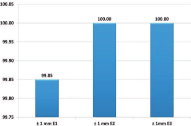
Based on the criterion of exact agreement > 90% for ± 1 mm between the initial and repeated PD per examiner, it was shown that examiner 1 had an agreement of 99.85%, while examiners 2 and 3 had 100% agreement (Figure 2).
Analysis of intra-examiner variables
A statistical t-test for independent samples was used to determine if there was a signiicant differ-ence between the initial PD measurements and the repeated measurements for each examiner, by ana-lyzing the following variables:
a) PD of mesial surfaces versus distal surfaces;
b) PD of anterior area versus posterior area;
c) PD of buccal surfaces versus lingual surfaces; and
d) PD of superior versus inferior arches.
For examiner 1, the t-test for independent sam-ples (p < 0.01) results showed no statistically signii-cant differences between each probe for the above-mentioned variables (Table 1).
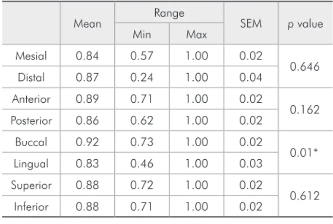
For examiner 2, the results of the t-test for inde-pendent samples (p < 0.01) showed no statistically signiicant differences between the initial and
re-peated probes for the following variables:
• mesial surfaces versus distal surfaces;
• buccal surfaces versus lingual surfaces; and
• superior versus inferior arch PD.
A statistically signiicant difference did appear between the initial and repeated probes for the PD of anterior versus posterior areas (Table 2).
For examiner 3, the results of the t-test for inde-pendent samples (p < 0.01) showed no statistically signiicant differences between the initial and re-peated probes for the variables:
• mesial surfaces versus distal surfaces;
• buccal surfaces versus lingual surfaces; and
• superior versus inferior arch PD. Figure 1 - Percentage of exact intra-examiner
correla-tion > 75% between the initial and repeated probes. Figure 2 -for ± 1 mm between the initial and repeated probes. Percentage of intra-examiner correlation > 90%
Table 1 - Results from the t-test for independent samples on the probe depth from examiner 1 for dental surface, area, and arch variables.
Mean Range SEM p value
Min Max
Mesial 0.6 0.19 0.94 0.04
0.413 Distal 0.72 0.27 1.00 0.05
Anterior 0.72 0.31 1.00 0.04
0.280 Posterior 0.62 0.17 0.91 0.04
Buccal 0.71 0.40 1.00 0.03
0.400 Lingual 0.66 0.26 0.85 0.04
Superior 0.68 0.26 0.90 0.03
0.489 Inferior 0.7 0.24 1.00 0.04
A statistically signiicant difference did appear between the initial and repeated probes for the ante-rior versus posteante-rior areas (Table 3).
Inter-examiner reproducibility
By evaluating the probe results from the three trained examiners, we could calculate the Kappa value and create mathematical measurements for subsequent application of the t-test. An analysis of variance (ANOVA) (p = 0.01, p = 0.05) showed that there were differences between the groups of ex-aminers (Table 4). For detection of the differences (examiner 1 versus examiner 2, examiner 1 versus examiner 3, examiner 2 versus examiner 3), the Scheffé method was applied a posteriori (p = 0.01).
The Scheffé method showed that examiners 1 and 2 differed statistically signiicantly (p < 0.01). Examiner 2 and examiner 3 differed statistically signiicantly, with p < 0.05, while examiner 3 and examiner 1 also differed at the same level (p < 0.05) (Figure 3).
Discussion
This study evaluated intra- and inter-examiner
reproducibility among three trained and calibrated examiners as they used a manual probe for clini-cal recording of periodontal probing depth (PD) in healthy individuals. The large number of probing sites by examiner (n = 6,564) and rigorous opera-tor calibration were demonstrated as necessary for obtaining reliable records, which can be used for appropriate decision-making. In our opinion, the high intra-examiner reproducibility observed was the result of the calibration and training program, and was not related to the operators’ experience, as have stated other authors,7-9 who believe that
experi-ence is the most important factor in measurement reproducibility. When comparing experienced and Figure 3 - Scheffé method for the
evaluation of inter-examiner probes showing statistically significant differences between and among the examiners.
Table 3 - Results from the t-test for independent samples on the probe depth from examiner 3 for dental surface, area, and arch variables.
Mean Range SEM p value
Min Max
Mesial 0.77 0.31 0.96 0.04
0.198 Distal 0.85 0.28 1.00 0.04
Anterior 0.8 0.46 0.97 0.03
0.916 Posterior 0.81 0.51 0.98 0.03
Buccal 0.87 0.59 1.00 0.03
0.013* Lingual 0.76 0.39 0.94 0.03
Superior 0.81 0.58 0.96 0.03
0.419 Inferior 0.79 0.37 0.93 0.03
*p < 0.01.
Table 4 - Analysis of Variance (ANOVA) for inter-examiner reproducibility.
Source of variation DF SS MS F
Between 2 0.38 0.19 19*
Within 57 0.82 0.01
Total 59 1.20
* There are statistically significant differences between the groups. DF: De-gree of Freedom; SS: Sum of Squares; MS: Mean Squares; F: Critical value.
Table 2 - Results from the t-test for independent samples on the probe depth from examiner 2 for dental surface, area, and arch variables.
Mean Range SEM p value
Min Max
Mesial 0.84 0.57 1.00 0.02
0.646 Distal 0.87 0.24 1.00 0.04
Anterior 0.89 0.71 1.00 0.02
0.162 Posterior 0.86 0.62 1.00 0.02
Buccal 0.92 0.73 1.00 0.02
0.01* Lingual 0.83 0.46 1.00 0.03
Superior 0.88 0.72 1.00 0.02
0.612 Inferior 0.88 0.71 1.00 0.02
inexperienced examiners, Samuel and co-workers10
showed that experienced examiners reproduced their probe measurements; however, they also showed that inexperienced examiners had signiicantly more reproducibility with manual probes (Williams probe), as was also shown in this study. Despite the fact that the examiners in this study were students, their measurements were highly reproducible. As the calibration process progressed (calibration group), the intra-examiner agreement gradually increased as well, a fact that permitted highly reproducible repeated measurements for the experimental group. This speaks positively of the calibration process, as presented by Grossi et al.11 When the results of the
intra-examiner measurements were compared with the Kappa test, exact agreement > 75% and > 90% for ± 1 mm was found between the two12,13 (repeated
measures), and the three examiners were positioned above benchmark parameters. These are favorable results in terms of diagnosis and periodontal con-trol, as suggested by Listgarten,4 who stated that a
measurement error of 2 mm or more in PD could lead to problems in the interpretation of results, thus leading to the provision of inappropriate treat-ment. There are many factors that could cause er-rors in clinical probing records.7,12-23 Among the
studied variables, our results conirmed the indings presented by Mullally and Linden12 and Mayield et al.,24 inding differences in probing measurements
between anterior and posterior areas (the only vari-ables with statistically signiicant differences) (Ta-bles 2 and 3). These differences can be explained by better access, probe position, and visibility of ante-rior areas or by the unconsciously high force that can be applied when posterior areas are probed. It should be noted that the results of examiners 2 and 3 were different compared with those of examiner 1. This could be due to the fact that the irst two exam-iners changed their working position when appro-priate, while examiner 1 did not. Working position directly inluences the PD record when a manual probe is being used to perform a periodontal probe, especially when the examiner does not use an angle periodontal probe. This has not been reported in the literature. As other authors have conirmed,8,9,24-26
our results showed greater reproducibility for
mea-surements with a manual probe, contrary to List-garten,4 who stated that reproducibility for manual
probes was low. We found that probing a healthy periodontium with a shallow sulcus, which offered greater resistance to probe penetration compared with a diseased sulcus,27,28 also contributed to high
reproducibility.27,28 This could be considered a
limi-tation of our study, since it was demonstrated that when calculus, inlammation, and bleeding are pres-ent, the clinically recorded data can be affected.29
Despite a high intra-examiner correlation, statisti-cally signiicant differences were found among the three examiners in terms of inter-examiner repro-ducibility (Figure 3). Our indings were similar to those presented by other authors, who had shown that differences (inter-examiner reproducibility) were more evident when manual probes were used.13
It can be estimated that these differences are related to individual differences in probing technique and force, even when high intra-examiner reproducibil-ity is demonstrated. To control the above-mentioned factors, it is recommended that pressure control probes2 be used to calibrate the operator’s probing
force, or even an acrylic stent to standardize the op-erator’s technique. We suggest that future research should evaluate other variables that affect peri-odontal clinical probing19,21,28,30 and the effects of
those variables over the resolution, reproducibility, and accuracy of periodontal probes,23 not only on
healthy tissues but mainly in the presence of peri-odontal disease. Finally, the methodology was care-fully designed to avoid irreversible damage of peri-odontal tissues as a result of repeated probing. This was avoided by providing suficient time between the initial and repeated measures and preserving tis-sue health, thus controlling the likelihood that an operator’s memory could induce a bias.13,25 Greater
emphasis should be placed in dental school class-rooms on how to take periodontal clinical records, since they are extremely relevant to diagnosis, prog-nosis, and treatment, and they are highly sensitive to error.
Conclusions
us-ing a manual periodontal probe produced high re-producibility in healthy individuals. The operator’s position can affect the reproducibility of repeated measures. Calibration and operator training, rather than operator experience, were fundamental to
re-producibility. However, since this methodology can-not demonstrate the reasons for low inter-examiner reproducibility, we can assume that there are other mitigating factors, such as individual technique and probing depth force.
References
1. Chu SJ, Hochman MN, Fletcher P. A biometric approach to aesthetic crown lengthening: Part II. Pract Proced Aesthet Dent. 2008 Oct;20(9):529-36.
2. Ramachandra SS, Mehta DS, Sandesh N, Baliga V, Amarnath J. Periodontal probing systems: a review of available equip-ment. Compend Contin Educ Dent. 2011 Mar;32(2):71-7. 3. Hourdin S, Glez D, Sorel O. [Periodontal diagnosis in
ortho-dontics]. Orthod Fr. 2010 Mar;81(1):9-17. French.
4. Listgarten MA. Periodontal probing: what does it mean? J Clin Periodontol. 1980 Jun;7(3):165-76.
5. Mombelli A. Clinical parameters: biological validity and clini-cal utility. Periodontol 2000. 2005;39(1):30-9.
6. Armitage GC. The complete periodontal examination. Peri-odontol 2000. 2004;34(1):22-33.
7. Seabra RC, Costa FO, Costa JE, Van Dyke T, Soares RV. Impact of clinical experience on the accuracy of probing depth measurements. Quintessence Int. 2008 Jul-Aug;39(7):559-65. 8. Niederman R. Manual and electronic probes have similar
reliability in the measurement of untreated periodontitis. Evid Based Dent. 2009 Jun;10(2):34-61.
9. Silva-Boghossian CM, Amaral CS, Maia LC, Luiz RR, Co-lombo AP. Manual and electronic probing of the periodontal attachment level in untreated periodontitis: a systematic re-view. J Dent. 2008 Aug;36(8):651-7. Epub 2008 Jun 5. 10. Samuel ED, Griffiths GS, Petrie A. In vitro accuracy and
reproducibility of automated and conventional periodontal probes. J Clin Periodontol. 1997 May;24(5):340-5.
11. Grossi SG, Dunford RG, Ho A, Koch G, Machtei EE, Genco RJ. Sources of error for periodontal probing measurements. J Periodontal Res. 1996 Jul;31(5):330-6.
12. Mullally BH, Linden GJ. Comparative reproducibility of proximal probing depth using electronic pressure-controlled and hand probing. J Clin Periodontol. 1994 Apr;21(4):284-8. 13. Buduneli E, Aksoy O, Köse T, Atilla G. Accuracy and repro-ducibility of two manual periodontal probes; an in-vitro study. J Clin Periodontol. 2004 Oct;31(10):815-9.
14. Barendregt DS, Van der Velden U, Timmerman MF, Van Der Weijden GA. Comparison of two automated periodontal probes and two probes with a conventional readout in peri-odontal maintenance. J Clin Periodontol. 2006 Apr;33(4):276-82.
15. Bulthuis HM, Barendregt DS, Timmerman MF, Loos BG, van der Velden U. Probe penetration in relation to the connective
tissue attachment level: influence of tine shape and probing force. J Clin Periodontol. 1998 May;25(5):417-23.
16. Araujo MW, Hovey KM, Benedek JR, Grossi SG, Dorn J, Wactawski-Wende J, et al. Reproducibility of probing depth measurements using a constant-force electronic probe: analysis of inter- and intra-examiner variability. J Periodontol. 2003 Dec;74(12):1736-40.
17. Garnick JJ, Silverstein L. Periodontal probing: probe tip di-ameter. J Periodontol. 2000 Jan;71(1):96-103.
18. Van Weringh M, Barendregt DS, Rosema NAM, Timmerman MF, Van Der Weijden GA. A thin or thick probe handle: does it make the difference? Int J Dent Hyg. 2006 Aug;4(3):140-4. 19. Hassan MA, Bogle G, Quishenbery M, Stephens D, Riggs M,
Egelberg J. Pain experienced by patients during periodontal recall examination using thinner versus thicker probes. J Peri-odontol. 2005 Jun;76(6):980-4.
20. Karpinia K, Magnusson I, Gibbs C, Yang MC. Accuracy of probing attachment levels using a CEJ probe versus traditional probes. J Clin Periodontol. 2004 Mar;31(3):173-6.
21. Neto JB, Filho GR, Tramontina VA, Sallum EA, Nociti FH Jr, Sallum AW. Millimeter marks and probe tip diameter standar-disation from commercially available periodontal probes. A comparative study. J Int Acad Periodontol. 2001 Jul;3(3):57-60.
22. Eickholz P, Grotkamp FL, Steveling H, Mühling J, Staehle HJ. Reproducibility of peri-implant probing using a force-con-trolled probe. Clin Oral Implants Res. 2001 Apr;12(2):153-8. 23. Bergenholtz A, al-Harbi N, al-Hummayani FM, Anton P,
al-Kahtani S. The accuracy of the Vivacare true pressure-sensitive periodontal probe system in terms of probing force. J Clin Periodontol. 2000 Feb;27(2):93-8.
24. Mayfield L, Bratthall G, Attström R. Periodontal probe preci-sion using 4 different periodontal probes. J Clin Periodontol. 1996 Feb;23(2):76-82.
25. Alves R de V, Machion L, Andia DC, Casati MZ, Sallum AW, Sallum EA. Reproducibility of clinical attachment level and probing depth of a manual probe and a computerized electronic probe. J Int Acad Periodontol. 2005 Jan;7(1):27-30. 26. Ishihata K, Wakabayashi N, Wadachi J, Akizuki T, Izumi
27. Khan S, Cabanilla LL. Periodontal probing depth measure-ment: a review. Compend Contin Educ Dent. 2009 Jan-Feb;30(1):12-21.
28. Hatakeyama Y, Uzel MI, Santana RB, Ruben MP. Relation-ship between position of probe tip and periodontal tissues after periodontal surgery in dogs. Int J Periodontics Restorative Dent. 2005 Jun;25(3):247-55.
29. Oringer RJ, Fiorellini JP, Koch GG, Sharp TJ, Nevins ML, Davis GH, et al. Comparison of manual and automated prob-ing in an untreated periodontitis population. J Periodontol. 1997 Dec;68(12):1156-62.