PB 41
Hand and ultrasonic instrumentation in the treatment of chronic
periodontitis after supragingival plaque control
Instrumentação manual e ultra-sônica no tratamento da
periodontite crônica após controle de placa supragengival
Ana Chapper*
Viviane Vasconcelos Catão** Rui Vicente Oppermann***
ABSTRACT: This study compared the clinical effects of hand or ultrasonic scaling and root planing on the treat-ment of chronic periodontitis. After supragingival plaque control, twenty patients were examined by a blinded and calibrated examiner for probing pocket depth (PPD), clinical attachment level (CAL) and bleeding on probing (BOP). Experimental teeth were allocated to the following subgingival treatment groups according to PPD: 1) hand instru-mentation; 2) hand instrumentation with irrigation; 3) ultrasonic instruinstru-mentation; 4) ultrasonic followed by hand instrumentation. Time used in the procedures was recorded. Follow-up examinations were performed at 30 and 90 days after treatment. Each patient’s individual BOP, PPD and CAL means were analyzed with repeated-measures ANOVA. Differences in the instrumentation time were analyzed with 1-way ANOVA. Significance level was estab-lished at 5%. All treatments produced significant changes in the clinical parameters. BOP reduced, in proximal surfaces, from 67.21-79.17% at baseline to 45.75-51.54% at 90 days. Significant reductions were also found for PPD and CAL in all groups, both in proximal and free surfaces. Reductions in mean PPD at 90 days ranged from 0.92 to 1.14 mm for the free surfaces and from 1.01 to 1.35 mm for proximal surfaces, whereas reductions in CAL ranged from 0.43 to 0.82 mm and from 0.60 to 0.73 mm for free and proximal surfaces, respectively. Mean instrumentation time ranged from 4.77 to 5.30 minutes. No statistically significant differences were found among the four study groups. It can be concluded that the four methods of subgingival instrumentation were equally ef-ficacious in the improvement of the studied clinical parameters.
DESCRIPTORS: Periodontitis, therapy; Dental scaling; Dental plaque, prevention & control; Ultrasonics; Root plan-ing.
RESUMO: Este estudo comparou, clinicamente, o efeito de raspagem e alisamento radicular por instrumentações manual e ultra-sônica no tratamento da periodontite crônica, após o controle de placa supragengival em 20 pa-cientes. Os parâmetros clínicos profundidade de sondagem (PS), nível de inserção clínica (NIC) e sangramento à sondagem (SS) foram avaliados por um examinador calibrado e cego. Os dentes experimentais foram alocados para uma das seguintes abordagens subgengivais, de acordo com a profundidade de sondagem: 1) manual, 2) manual associada à irrigação, 3) ultra-sônica, 4) ultra-sônica previamente à manual. O tempo usado nos proce-dimentos foi registrado. Avaliações foram feitas 30 e 90 dias após os tratamentos. Médias individuais de SS, PS e NIC foram analisadas pelo teste ANOVA de medidas repetidas (p < 0,05). Diferenças no tempo de instrumentação foram analisadas por meio do teste ANOVA para um critério de classificação (p < 0,05). Os resultados revelaram que os tratamentos produziram alterações significativas nos parâmetros clínicos sem diferenças estatisticamente significantes entre os quatro grupos. Observaram-se reduções no SS, que, nas faces proximais, variou de 67,21 a 79,17%, no início, para 45,75 a 51,54% no final. Também foram observadas reduções significativas na PS e no NIC em todos os grupos, tanto para faces livres quanto proximais: as médias da PS aos 90 dias reduziram entre 0,92 e 1,14 mm nas faces livres e entre 1,01 e 1,35 mm nas proximais, enquanto para NIC as reduções foram de 0,43 a 0,82 mm e 0,60 a 0,73 mm, para as faces livres e proximais respectivamente. A média do tempo operatório variou de 4,77 a 5,30 minutos. Pôde-se concluir que as quatro modalidades terapêuticas de instrumentação subgengival foram igualmente eficazes na melhora dos parâmetros clínicos estudados.
DESCRITORES: Periodontite, terapia; Raspagem subgengival; Placa dentária, prevenção & controle; Ultra-som; Aplainamento radicular.
* Master in Periodontology.
** Professor, Discipline of Periodontics, Gama Filho University.
42 43
42 43
INTRODUCTION
Different methodologies have been used to compare the effects of hand and ultrasonic in-strumentation. In vitro studies have shown that hand instrumentation tends to result in smoother root surfaces than ultrasonic instrumentation. However, remaining calculi and plaque have been found on the roots of teeth in studies conducted with either method12,13,17. Although these are
im-portant findings, in vitro studies are limited due to the need to extract the instrumented tooth for posterior analysis. The differences between in vitro and in vivo conditions make it difficult to interpret the clinical significance of such results. Moreover, the importance of a smooth root surface for suc-cessful healing has been questioned11,16.
Clinical studies have not demonstrated sig-nificant differences between the two subgingival instrumentation methods discussed here1,2,14.
However, the results of these studies might have been affected by a therapeutic model that sees scaling and root planing as a basic procedure in the preparation for a surgical approach considered as the definitive treatment. In addition, although hand instrumentation has traditionally been used to scale the subgingival biofilm and calculus off the pathologically exposed root surface, greater em-phasis has been given to nonsurgical subgingival scaling therapy performed with machine-driven instruments5,15.
For the treatment of periodontitis, procedures aiming at the subgingival dental area should be performed only when clinical signs of inflamma-tion associated with the presence of supragingival plaque are previously controlled. Most of the stud-ies comparing manual and ultrasonic instrumen-tation have not made such consideration and this may cause bias due to non controlled determinants such as established gingivitis8.
Therefore, the purpose of this study was to compare the clinical effect of subgingival scaling and root planing performed with hand and ul-trasonic instruments in the treatment of chronic periodontitis in patients showing adequate supra-gingival plaque control.
MATERIALS AND METHODS
Patients
Participants in this study were selected from a group of patients referred to the program of Peri-odontology, School of Dentistry, ULBRA (Lutheran
University of Brazil) for periodontal treatment. In-clusion criteria were: a) patients with chronic peri-odontitis aged 35 years or older; b) patients with at least four single-rooted teeth with one or more sites showing pocket probing depth ≥ 6 mm and periodontal bleeding after supragingival plaque control, c) patients who consented in participating in the study by signing an informed consent form for minimal risk procedures in adults, d) clinical and radiographic examination should demonstrate that selected teeth were free of endoperiodontal lesions. Patients with systemic diseases, cardiac pacemakers or hormonal disorders were excluded, as well as patients that had made use of antimicro-bial drugs in the preceding six months or that were taking immunosuppressive drugs. According to these criteria, 20 patients (13 males) were selected, with a median age of 42.50 years (38.50-47.75).
This study is in accordance with Resolution 196/96 of the Brazilian National Health Commit-tee (Conselho Nacional de Saúde) and its amend-ments, and with the Helsinki Declaration of 1975 as revised in 1983. This study was approved by the Ethics Committee of the Lutheran University of Brazil (protocol 2003-147H), and all patients signed an informed consent form to participate in the study.
Examination parameters
• Calibration procedures: The patients were amined by only one blinded and calibrated ex-aminer. The following clinical parameters were recorded: pocket probing depth (PPD), clinical attachment level (CAL) and bleeding on prob-ing (BOP). Two examinations with a one week interval were performed. The results showed agreement for PPD and CAL measurements of 94.01% and 89.08%, considering a difference of up to 1 mm between examinations.
• Clinical evaluation: Recordings were performed only in the single rooted teeth, although peri-odontal treatment included multirooted teeth as well. Clinical examinations were performed before the experimental period, and at 30 and 90 days. Efficiency was assessed by recording the mean time (in minutes) needed to treat each tooth with the aid of a chronometer.
Experimental procedures
42 43
42 43
hygiene instructions. The single-rooted teeth se-lected in each patient were distributed into groups to be treated, according to PPD, with one of the following types of subgingival instrumentation: hand instrumentation (H); hand instrumentation and subgingival irrigation with distilled water (H+I); ultrasonic instrumentation (U); ultrasonic instrumentation followed by hand instrumenta-tion (U+H). Teeth allocainstrumenta-tion to the different groups followed a systematic distribution pattern in such way that all patients underwent all types of treat-ment in a number of teeth as similar as possible. A total of 293 teeth were included in the study.
Hand instrumentation consisted of subgingi-val scaling and root planing with Hirschfeld files (Newmar, São Paulo, Brazil) and Gracey curettes (Newmar, São Paulo, Brazil) according to the op-erator’s criteria. Ultrasonic instrumentation was performed with an ultrasonic scaler with a piezo-ceramic transducer (Profi I Ceramic®, Dabi Atlante
SA – Indústrias Médico Odontológicas, São Paulo, Brazil). All clinical procedures were performed un-der local anesthesia (2% lidocaine hydrochloride + norepinephrine, Probem, Manaus, Brazil) by only one trained operator. After each subgingival in-strumentation session, patients received profes-sional supragingival plaque removal and home care was reinforced.
Treatment was completed in a maximum of four weekly sessions. Patient plaque control was monitored and reinforced during follow-up exam-inations, but no professional cleaning was per-formed.
Statistical analysis
The patient was used as the unit of analy-sis. Each patient’s individual BOP, PPD and CAL means were analyzed with repeated-measures ANOVA. Differences in the instrumentation time were analyzed with 1-way ANOVA. Significance level was established at 5%.
RESULTS
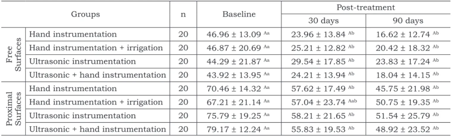
Significant reductions in the BOP between baseline and post-treatment measurements were observed for all experimental groups (Table 1). In the free surfaces, baseline mean values ranged from 43.92% to 46.96%, and were reduced to val-ues from 16.62% to 23.83% at 90 days. The valval-ues for bleeding on probing in the proximal surfaces ranged from 67.21% to 79.17% at baseline.
Signifi-cant reductions were already present at 30 days and after 90 days values ranged from 45.75% to 51.54%. No statistically significant differences be-tween treatments were observed.
The PPD mean in the free surfaces ranged from 2.60 to 2.85 mm. Significant reductions were found already at 30 days (Table 2). At 90 days, PPD values ranged from 1.64 to 1.85 mm and were significantly lower than baseline values, but simi-lar to the values observed after 30 days. A simisimi-lar pattern was observed for the proximal surfaces. Mean PPD values found for the different groups were not significantly different when compared at different time points.
Table 3 shows that CAL means were also low-er at 30 and 90 days for both free and proximal surfaces. Mean CAL values for the free surfaces ranged from 3.40 to 3.65 mm. Reductions were already found at 30 days. At 90 days, CAL values were significantly lower than baseline values, rang-ing from 2.80 to 2.97 mm. A similar result was observed for the proximal surfaces. No statistically significant differences were found among the four groups at different time points.
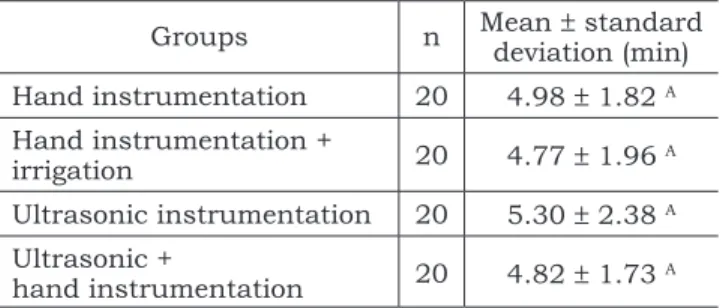
Mean instrumentation time for each tooth (Ta-ble 4) ranged from 4.77 to 5.30 minutes. No statis-tically significant differences were found in instru-mentation time among the four study groups.
DISCUSSION
The results of the present study showed that hand and ultrasonic treatments determined sig-nificant changes in the clinical parameters related to chronic periodontitis. Moreover, no statistical differences between treatment modalities could be observed.
PPD measured after supragingival plaque con-trol was established as an inclusion criterion. In fact, 8 patients were excluded due to pocket reduc-tions related to the supragingival plaque control. Similar results have been reported by Fernandes et al.8 (1989) and Catão3 (1999). Their studies showed
that important changes in subgingival parameters such as PPD and BOP occur associated with the reduction of inflammation in the marginal gingiva following supragingival plaque control.
44 45
44 45
TABLE 1 - Means and standard deviations for periodontal bleeding on probing (%) on free and proximal surfaces at baseline, 30 and 90 days.
Groups n Baseline Post-treatment
30 days 90 days
Fr
ee
S
u
rf
aces
Hand instrumentation 20 46.96 ± 13.09 Aa 23.96 ± 13.84 Ab 16.62 ± 12.74 Ab Hand instrumentation + irrigation 20 46.87 ± 20.69 Aa 25.21 ± 12.82 Ab 20.42 ± 18.32 Ab Ultrasonic instrumentation 20 44.29 ± 21.87 Aa 29.54 ± 17.85 Ab 23.83 ± 17.24 Ab Ultrasonic + hand instrumentation 20 43.92 ± 13.95 Aa 24.21 ± 13.94 Ab 18.04 ± 14.15 Ab
Pr
ox
im
al
S
u
rf
aces
Hand instrumentation 20 70.46 ± 14.32 Aa 57.62 ± 17.49 Ab 45.75 ± 21.98 Ab Hand instrumentation + irrigation 20 67.21 ± 21.14 Aa 57.04 ± 23.74 Aab 50.75 ± 19.35 Ab Ultrasonic instrumentation 20 75.79 ± 19.25 Aa 58.21 ± 21.65 Ab 51.54 ± 25.79 Ab Ultrasonic + hand instrumentation 20 79.17 ± 12.24 Aa 55.83 ± 19.53 Ab 48.92 ± 23.52 Ab Results followed by the same uppercase letter in the column do not differ statistically (p > 0.05). Results followed by the same lowercase letter in the line do not differ statistically (p > 0.05).
TABLE 2 - Means and standard deviations (mm) for pocket probing depth in free and proximal surfaces at baseline, 30 and 90 days.
Groups n Baseline Post-treatment
30 days 90 days
Fr
ee
S
u
rf
aces
Hand instrumentation 20 2.71 ± 0.73 Aa 2.06 ± 0.64 Ab 1.79 ± 0.59 Ab Hand instrumentation + irrigation 20 2.79 ± 0.83 Aa 1.85 ± 0.53 Ab 1.65 ± 0.50 Ab Ultrasonic instrumentation 20 2.85 ± 1.06 Aa 2.05 ± 0.75 Ab 1.85 ± 0.58 Ab Ultrasonic + hand instrumentation 20 2.60 ± 0.58 Aa 1.82 ± 0.46 Ab 1.64 ± 0.36 Ab
Pr
ox
im
al
S
u
rf
aces
Hand instrumentation 20 4.56 ± 0.83 Aa 3.43 ± 0.58 Ab 3.55 ± 1.14 Ab Hand instrumentation + irrigation 20 4.33 ± 1.00 Aa 3.14 ± 0.64 Ab 3.18 ± 0.61 Ab Ultrasonic instrumentation 20 4.47 ± 0.89 Aa 3.51 ± 0.73 Ab 3.21 ± 0.83 Ab Ultrasonic + hand instrumentation 20 4.58 ± 0.97 Aa 3.36 ± 0.63 Ab 3.23 ± 0.63 Ab Results followed by the same uppercase letter in the column do not differ statistically (p > 0.05). Results followed by the same lowercase letter in the line do not differ statistically (p > 0.05).
TABLE 3 - Means and standard deviations (mm) of clinical attachment level in free and proximal surfaces at base-line, 30 and 90 days.
Groups n Baseline Post-treatment
30 days 90 days
Fr
ee
S
u
rf
aces
Hand instrumentation 20 3.65 ± 1.09 Aa 3.08 ± 1.14 Ab 2.83 ± 1.14 Ab Hand instrumentation + irrigation 20 3.62 ± 1.26 Aa 2.95 ± 1.24 Ab 2.83 ± 1.14 Ab Ultrasonic instrumentation 20 3.40 ± 1.59 Aa 3.16 ± 1.52 Aab 2.97 ± 1.29 Ab Ultrasonic + hand instrumentation 20 3.46 ± 1.07 Aa 2.97 ± 1.15 Ab 2.80 ± 1.08 Ab
Pr
ox
im
al
S
u
rf
aces
44 45
44 45
It has been shown that bleeding may occur in what appears to be clinically healthy conditions6.
Con-versely, at sites with apparent histological evidence of chronic inflammation, bleeding may not occur6.
Thus, these BOP results do not indicate that the periodontal therapy was not effective. They suggest the need for short observational intervals to main-tain an adequate supragingival plaque control. Poor plaque control may lead to gingival bleeding, which may become a misleading factor when as-sessing the eficacy of the periodontal therapy.
Pocket probing depths and loss of attachment were significantly reduced in all types of treatment. This confirmed previous findings reported in stud-ies with different experimental conditions1,2,14. In
fact, it is reasonable to expect that greater initial depths lead to greater reductions after treatment6.
The changes in PPD and CAL, observed already at 30 days, were partially a consequence of the reduction in inflammation resulting from subgin-gival plaque removal. Changes in PPD and CAL are expected to occur as a consequence of edema reduction, increased tissue tonus, or formation of a long junctional epithelium7. The results of
clinical studies suggest that a 3-month post-treat-ment interval is suitable for re-evaluation6. Most
of the clinical healing has usually occurred at this time, even in areas with initial deep lesions1,2. In
the present study, edema reduction was probably primarily related to the reduction of inflamma-tion in the subgingival area, since the reducinflamma-tion of inflammation in the marginal area, which fol-lowed supragingival plaque control, had already occurred.
The findings reported in the present study are in agreement with the results reported in a number of similar studies15. In the present study,
it was observed 90 days following instrumentations that the mean reduction of attachment loss ranged
from 0.43 to 0.82 mm and from 0.60 to 0.73 mm for free and proximal surfaces, respectively. Bad-ersten et al.1,2 (1981, 1984) observed mean
reduc-tion of clinical attachment loss of 0.5 mm while Kocher et al.10 (1997) observed mean reduction of
clinical attachment loss of 0.71 ± 1.01 mm after a 6-month period of evaluation. However, in the present study, the results should be interpreted with caution. Single-rooted teeth in adult patients with chronic periodontitis without local or systemic complications are a convenient model and have often been used in other studies found in the lit-erature1,2,4,9,13,14.
The time required to complete treatment has a direct effect on the cost-benefit ratio. In the present study, there was no statistical difference between therapies related to mean time to treat one tooth during therapy. This result differs from findings of other studies assessing both time needed for treat-ment and clinical outcome variables15. In the study
of Badersten et al.2 (1984), the mean time needed
to treat one tooth during initial therapy was 5.35 minutes using machine-driven instrumentation and 6.15 minutes using hand instrumentation. Torfason et al.14 (1979) took 2.10 minutes on
av-erage to treat one tooth using ultrasonic instru-ments and 2.40 minutes using hand instruinstru-ments. In these studies, there was no difference in clinical benefit between ultrasonic and manual instru-mentation though subgingival debridement was completed in less time with ultrasonic than with hand instruments. In the present study, besides calculus removal instrumentation, the operator aimed at a thorough root planing covering the en-tire root area affected. The desire to obtain a root surface as smooth as possible might have affected the results for instrumentation time.
CONCLUSIONS
1. All types of subgingival hand and ultrasonic instrumentation used in this study resulted in significant improvement in the clinical pa-rameters.
2. Scaling and root planing manually or with the aid of ultrasonic instrument after supragin-gival plaque control yielded similar results, significantly reducing bleeding on probing, pocket probing depth and clinical attachment loss. Irrigation with distilled water had no ef-fect on the results.
3. The mean time for instrumentation was simi-lar for the different types of treatment.
TABLE 4 - Time needed for subgingival instrumenta-tion of each tooth.
Groups n Mean deviation (min)± standard
Hand instrumentation 20 4.98 ± 1.82 A Hand instrumentation +
irrigation 20 4.77 ± 1.96 A
Ultrasonic instrumentation 20 5.30 ± 2.38 A Ultrasonic +
46 PB
REFERENCES
1. Badersten A, Nilvéus R, Egelberg J. Effect of nonsurgical periodontal therapy. I. Moderately advanced periodontitis. J Clin Periodontol 1981;8:57-72.
2. Badersten A, Nilvéus R, Egelberg J. Effect of nonsurgical periodontal therapy. II. Severely advanced periodontitis. J Clin Periodontol 1984;11:63-76.
3. Catão VV. O efeito do controle de placa supragengival no diagnóstico clínico da doença periodontal [Dissertação de Mestrado]. Canoas: Universidade Luterana do Brasil; 1999. 4. Cercek JF, Kiger RD, Garrett S, Egelberg J. Relative effects
of plaque control and instrumentation on the clinical pa-rameters of human periodontal disease. J Clin Periodontol 1983;10:46-56.
5. Drisko CH. Root instrumentation. Power-driven ver-sus manual scalers, which one? Dent Clin North Am 1998;42:229-44.
6. Egelberg J, Claffey N. Periodontal re-evaluation. The sci-entific way. Copenhagen: Munksgaard; 1994.
7. Fernandes MI. Avaliação histológica de raspagem e alisamen-to radiculares subgengivais [Dissertação de Mestrado]. Poralisamen-to Alegre: Universidade Federal do Rio Grande do Sul; 1997. 8. Fernandes MI, Friedrich MEG, Oppermann RV. O efeito
do controle da placa supragengival na doença periodontal. In: 6ª Reunião Anual da SBPqO; 1989; São Paulo. Anais. São Paulo: Sociedade Brasileira de Pesquisa Odontológica; 1989. p. 107.
9. Haffajee A, Socransky S, Goodson J. Clinical parameters as predictors of destructive periodontal disease activity. J Clin Periodontol 1983;10:257-65.
10. Kocher T, Ruhling A, Momsen H, Plagmann HC. Ef-fectiveness of subgingival instruments with power-driven instruments in the hands of experienced and inexperi-enced operators. A study on manikins. J Clin Periodontol 1997;24(7):498-504.
11. Oberholzer R, Rateitschak KH. Root cleaning or root smoothing – an in vivo study. J Clin Periodontol 1996;23(4):326-30.
12. Stende GW, Schaffer EM. A comparison of ultrasonic and hand scaling. J Periodontol 1961;32:312-4.
13. Thornton S, Garnick J. Comparison of ultrasonic to hand instruments in the removal of subgingival plaque. J Periodontol 1982;53:35-7.
14. Torfason T, Kiger R, Selvig KA, Egelberg J. Clinical improvement of gingival conditions following ultrasonic versus hand instrumentation of periodontal pockets. J Clin Periodontol 1979;6:165-76.
15. Tunkel J, Heinecke A, Flemmig TF. A systematic re-view of efficacy of machine-driven and manual subgingival debridement in the treatment of chronic periodontitis. J Clin Periodontol 2002:29 Suppl 3:72-81.
16. Waerhaug J. Microscopic demonstration of tissue re-action incident to removal of subgingival calculus. J Peri-odontol 1955;26:26-9.
17. Wilkinson RF, Maybury JE. Scanning electron mi-croscopy of the root surface following instrumentation. J Periodontol 1973;44:559-63.