ABSTRACT
http://dx.doi.org/10.1590/1678-775720140458
Comparison of salivary levels of mucin and
amylase and their relation with clinical parameters
obtained from patients with aggressive and
chronic periodontal disease
Andrea Beatriz ACQUIER1,2, Alejandra Karina De Couto PITA1, Lucila BUSCH1, Gabriel Antonio SÁNCHEZ3
1- Department of Pharmacology, Faculty of Dentistry, University of Buenos Aires, Buenos Aires, Argentina.
2- INBIOMED, National Scientiic and Technical Research Council, Buenos Aires, Argentina.
3- Department of Biophysics, Faculty of Dentistry, University of Buenos Aires, Buenos Aires, Argentina.
Corresponding address: Gabriel A. Sánchez - Department of Biophysics - Faculty of Dentistry, University of Buenos Aires
Marcelo T. de Alvear 2142 (1122AAH) - Buenos Aires - Argentina - Phone: +54 11 4964 12 98 - Fax: +54 11 4508 39 58 - e-mail: [email protected]
Submitted: November 21, 2014 - Modiication: February 11, 2015 - Accepted: March 15, 2015
O
bjective: Salivary mucin and amylase levels are increased in patients with chronic periodontitis (CP). Due to the fact that aggressive periodontitis (AgP) not only differs from chronic periodontitis in terms of its clinical manifestation, the aim of this study was to compare salivary mucin and amylase levels and their relation to the clinical parameters of patients with aggressive periodontitis with that of patients with chronic periodontitis. Material and Methods: Eighty subjects were divided into two groups: 20 patients with AgP and their 20 matched controls and 20 patients with CP and their 20 matched controls, based on clinical attachment loss (CAL), probing pocket depth (PPD) and bleeding on probing (BOP). Whole unstimulated saliva was obtained and mucin, amylase and protein were determined by colorimetric methods. Pearson’s correlation analysis was used to determine the relationship between salivary mucin, amylase and protein levels and the clinical parameters. Results: Salivary mucin, amylase and protein levels were increased in patients with AgP and CP but there were no differences between them or between control groups. Pearson’s correlation analysis, determined in the entire subjects studied, showed a positive and signiicant correlation of mucin, amylase and proteins with CAL and PPD and a negative correlation with the low rate. When Pearson’s correlation analysis was carried out in each group separately, Fisher’s z transformation showed no signiicant difference between both groups. Conclusion: Comparison of the salivary levels of mucin, amylase and protein and their relationship with clinical parameters of AgP patients with that of CP patients revealed no differences between both groups.Keywords: Mucins. Amylases. Aggressive periodontitis. Chronic periodontitis.
INTRODUCTION
Chronic and aggressive periodontitis consists
of an inlammatory reaction of the periodontal
tissues in response to infection caused by a
speciic group of bacteria. Aggressive periodontitis
(AgP) is characterized by severe and rapid loss of periodontal attachment, often commencing at or after the pubertyChronic periodontitis (CP) is a common disease that is prevalent among adults and seniors. The aggressive nature of AgP depends on
the bacterial etiology, host susceptibility, hereditary and environmental factors, and often behavioral factors1.
There are numerous defense proteins present in saliva. Some of these defense proteins, such as salivary immunoglobulins, and salivary chaperokine HSP70/HSPA, are involved in both innate and acquired immune activation4,7. Salivary cationic
mucins, peroxidases, statherin (and others), are primarily responsible for innate immunity4,5
notwithstanding that many of them also exert immune activator and/or immune modulator properties. Importantly, many of these molecules are present at rather low concentrations in whole saliva; however, it should be considered that their effects are cumulative and/or synergistic, resulting
in an eficient molecular defense network of the
oral cavity4,5,13. It should also be considered that
local concentrations of these proteins near the mucosal surfaces (mucosal transudate), periodontal
sulcus (gingival crevicular luid) and oral wounds
and ulcers (transudate) may be much greater, and in many cases are reinforced by immune and/
or inlammatory reactions of the oral mucosa6,7.
Salivary glands may respond to oral disease by enhancing synthesis of some acinar proteins and thereby increasing the protective potential of saliva. Based on this line of reason, saliva from chronic periodontitis patients showed increased mucin and amylase concentration16.
Salivary mucin are heavily glycosylated high molecular weight glycoproteins produced by various mucous salivary glands, i.e. submandibular, sublingual and palatal glands and the minor salivary glands in the lip, cheek and tongue. Mucins play a major role in maintaining the viscoelastic properties of saliva. They also participate in forming a protective oral mucosal mucus coat
and tooth enamel pellicle and have high afinity
to microorganisms, entrapping and agglutinating bacteria, fungi and viral particles8.
Amylase is a highly abundant protein in saliva. The most widely-known function of amylase is its endoglicosidase activity, but in addition to his, amylase also takes part in acquired pellicle formation on tooth surfaces. It performs a direct inhibitory effect on the growth of certain bacteria and also binds to bacteria lipopolysaccharide, a bacterial surface structure and bacterial toxin, which are responsible in many
cases for tissue destructive inlammatory reactions8.
Thus, it was demonstrated that α-amylases from
human saliva, porcine pancreas and rice show a
signiicant cell growth inhibitory activity against
Porphyromonas gingivalis species, and interfere with the adherence and biofilm formation of Aggregatibacter actinomycemcomitans, indicating
that α-amylase could be effective in preventing
periodontal diseases2,14.
Several studies have reported that the levels of distinct salivary proteins are altered in individuals with periodontal disease. Patients with gingivitis, AgP and CP showed increased salivary levels of mucin and amylase2,9,11,16,19. Conversely, other
studies have reported a reduced expression of mucin glycoprotein-2 (MG2) and lactoferrin in
unstimulated saliva from patients with AgP and CP15.
Therefore, these results suggest that these salivary constituents may play a role in the etiopathogenesis of these diseases.
Since there are differences between AgP and CP not only in the clinical presentation10, the objective
of this study was to investigate and compare the salivary levels of mucin and amylase and their relation with clinical parameters in patients with AgP and CP.
MATERIAL AND METHODS
Study population
Study subjects were recruited from a population of patients in a private dental clinic, from January through August 2014. The protocol was approved by the Ethics Committee of the School of Dentistry, University of Buenos Aires, Argentina, and the study was conducted in accordance with the Declaration of Helsinki (version 2008). Completed medical and dental histories were obtained from all subjects. The inclusion criteria for the study group were the presence of established AgP or CP, according to World Workshop in Periodontology criteria1, in
subjects who had not had a periodontal checkup in the previous 6 months. Since there was a
signiicant difference in the age between AgP and
CP patients, two control groups were formed with subjects of similar ethnicity, income levels, age and gender to patients. All of the subjects gave their informed consent. Exclusion criteria included: smokers, those with cardiovascular or respiratory diseases, systemic inflammatory conditions or
non-plaque induced oral inlammatory conditions,
immunodeficiency, pregnant or breast feeding individuals and those using medicine.
Clinical examination
All periodontal measurements were performed
in four quadrants using a irst-generation probe
(Hu-Friedy Mfg. Co., Chicago, IL, USA) by a single trained investigator (G.A.S.). Probing pocket depth (PPD, measurements were rounded off to the nearest millimetre marking) and clinical attachment level (CAL, measuring the distance from the cemento-enamel junction to the bottom of the probable pocket) were assessed at six sites per tooth and bleeding on probing, (BOP, scored as: -, no bleeding or +, bleeding within 30 s after probing) at four sites per tooth.
Saliva collection
Subjects spat out saliva every 30 seconds for 5 minutes. The volume of saliva was recorded and expressed as ml per minute. The resulting saliva was stored in aliquots at -20°C until determinations were performed.
Determination of protein, amylase and mucin concentration
Colorimetric methods were used for all determinations in unstimulated saliva. The protein concentration was determined by the method described by Lowry, et al.12 (1951) and the amylase
activity was determined in diluted saliva (1/100) by the method described by Bernfeld3 (1951) using
starch suspension as the substrate. Amylase activity is expressed in terms of Units (U) where 1 U of
amylase was deined as the quantity of enzyme that
liberates 1 mg of maltose for 1 min at 20°C. Mucin concentration was determined using the Alcian blue method16. Briely, aliquots of diluted saliva (1:10)
were incubated for 30 min in a 1% solution of Alcian
Blue in 50 mM sodium acetate buffer with 25 mM Cl2Mg, pH 5.8 under constant agitation at room temperature. Following incubation, the samples were centrifuged for 20 min at 3000 rpm, pellets
washed in 95% ethanol, vortexed gently for 10 s
and after 5 min centrifuged for 20 min at 3000 rpm. Mucin-dye complexes were dissociated by adding a 1:2 dilution of Aerosol OT (Sigma Chemical Co., St Louis, MO, USA) in distilled water, brief mixing and
sonication. Subsequently, samples were extracted with equal volumes of ethyl ether through vigorous shaking. The resulting solution was centrifuged for 15 min at 3000 rpm and the dye concentration was spectrophotometrically determined at 605 nm in the aqueous layer.
Statistical analysis
Statistical significance of differences was determined by one way ANOVA followed by Neuman-Keuls multiple comparison test. Pearson correlation analysis were done using GRAPHPAD Prism version 5.03 for Windows (GraphPad Software, San Diego, CA, USA) and Fisher’s z transformation, which converts Pearson’s r to the normally distributed
variable z, was used to assess the signiicance of
the difference between the r. The level of statistical
signiicance is set to p<0.05.
RESULTS
Demographic and clinical data from patients with AgP and CP are presented in Table 1. Due to the difference in age of the two disease groups, each of them was matched with subjects representing two control groups. As can be seen in the table,
both disease groups showed signiicantly higher CAL and PPD but lower unstimulated low rate than
the control groups, when tested by one-way ANOVA followed by Neuman-Keuls multiple comparison
Parameter/Group Control AgP Patients AgP Control CP Patients CP
Age: mean/range (years)
19.5/17-23 19.5/17-23 37.4/32-40 37.4/32-40
Gender Female: 10 Male: 10 Female: 10 Male: 10 Female: 10 Male: 10 Female: 10 Male: 10
Number of teeth (range)
28-30 28-30 28-30 26-28
Number of sites checked up
168-180 168-180 168-180 156-168
CAL (mm) 0.23±0.06 a5.9±0.17*** 0.25±0.06 4.2±0.35***
Number of sites CAL >4 mm (median/
range)
0 5/0-6 0 3/0-5
PPD (mm) 2.4±0.06 b5.5±0.07*** 2.5±0.06 5.1±0.2***
Number of sites PPD >5 mm (median/
range)
0 3/0-5 0 3/0-5
UFR (ml/min) 0.54±0.01 0.44±0.02*** 0.53±0.01 0.47±0.01***
Table 1- Demographic and clinical data from patients with aggressive (AgP) and chronic (CP) periodontitis and their matched controls
Figure 3- Pearson’s correlation analysis between mucin (A), amylase (B) and proteins (C) and probing pocket depth (PPD) in the entire subjects studied
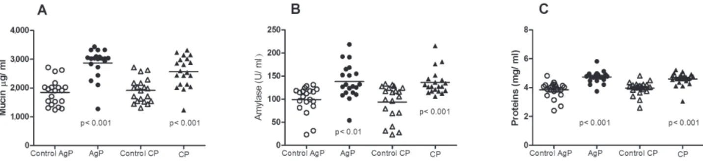
Figure 1- Salivary levels of mucin (A), amylase (B) and proteins (C) in unstimulated saliva from patients with aggressive and chronic periodontal disease and their matched controls
Figure 2- Pearson’s correlation analysis between mucin (A), amylase (B) and proteins (C) and clinical attachment level (CAL) in the entire subjects studied
Figure 4- Pearson’s correlation analysis between mucin (A), amylase (B) and proteins (C) and salivary unstimulated low
test. Patients with AgP had more periodontal destruction than patients with CP, as evidenced from the higher CAL and PPD observed.
The levels of mucin, amylase and proteins in unstimulated saliva differed widely in the patient and control groups. There was a significantly
higher mucin, amylase and protein concentration in saliva from patients with aggressive and chronic periodontitis than in the control groups (Figures 1A, B and C). On the other hand, there were no differences between the control groups or disease groups.
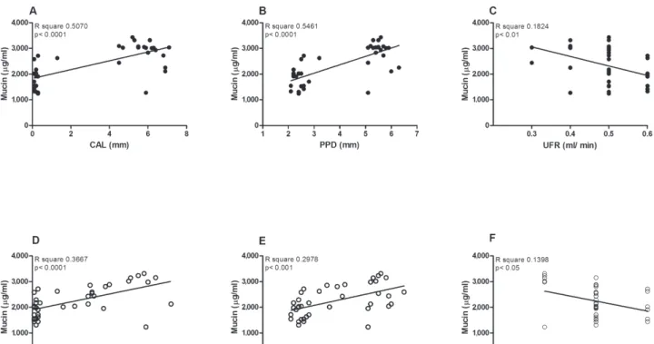
Figure 5- Pearson’s correlation analysis between mucin and clinical attachment level (CAL) (A and D), probing pocket depth (PPD) (B and E) and unstimulated low rate (UFR) (C and F) in aggressive periodontitis (AgP) (upper panel) and chronic periodontitis (CP) (lower panel) groups
The correlations, among the levels of mucin, amylase and proteins and the clinical parameters, determined in all studied subjects, are shown in Figures 2, 3 and 4. The salivary mucin, amylase and protein concentration was positively correlated with CAL (Figure 2) and PPD (Figure 3). Conversely,
a signiicant but negative correlation was observed
between the salivary levels of mucin, amylase
and proteins and the unstimulated low rate. The correlations, despite being signiicant, were weak.
On the other hand, the comparison by Fisher’s z transformation between those correlations showed
no signiicant differences.
In order to determine whether there were differences between the AgP and CP groups, based on the relationship among mucin, amylase and proteins and the clinical parameters, Pearson’s correlation analysis was carried out in each group separately (Figures 5 to 7). Fisher’s z transformation
showed that there was no signiicant difference
between both groups (p>0.05), the only exception being the correlation between UFR and proteins (p<0.01).
DISCUSSION
In this study, the relationship between salivary mucin, amylase and protein levels and clinical parameters in patients with AgP and CP were compared. Unstimulated saliva were used because there is a prevalent resting condition in the oral cavity
for most of the 24-hour period and, as the salivary
secretion is a relex response, it can be inluenced
by several stimuli such as the accumulation of plaque-derived substances or inflammatory products18. Thus, a positive relationship between
basal values of mucin, amylase and proteins with CAL and PPD would be expected17. As a matter of
fact, there is evidence that the salivary glands may respond to periodontitis by enhanced synthesis of some acinar proteins and thereby an increase in the protective potential of saliva9,11,16.
Both patient groups showed higher CAL and
PPD but la ower low rate than the control groups.
Patients with AgP showed higher CAL and PPD than patients with CP, without differences in unstimulated
low rate.
Levels of mucin, amylase and proteins in saliva from adults with or without chronic periodontal disease were evaluated in a previous study16.
A higher concentration of mucin, amylase and proteins were found in saliva from patients compared with healthy subjects. This study was undertaken to determine whether there were any differences between AgP and CP. The results showed that patients with AgP presented similar levels of mucin, amylase and proteins than patients with CP
and because unstimulated low rate was similar in
both groups, this result must not be attributed to a different volume of saliva.
As previously described17, a positive and
amylase and protein with CAL and PPD. This fact is
in accordance with the hypothesis that inlammatory
products may trigger salivary secretion via neural pathways18. When correlations were made in each
group separately, no differences between AgP and PC were found.
It is known that AgP differs from CP not only in bacterial aetiology, but also in host immune and
inlammatory factors implicated in the pathogenesis
and progression of AgP. The presence of neutrophil abnormalities16 and a reduced total IgA, described
in subjects with AgP8, relects a general
down-regulation of the humoral immune system. However, as derived from our results, it can be concluded that there are no differences between AgP and CP in relation to mucin, amylase and protein salivary levels. In both clinical manifestations of the disease the increase in mucin, amylase and protein
concentrations is inluenced by clinical parameters.
CONCLUSION
Comparison of the salivary levels of mucin, amylase and protein and their relationship with clinical parameters of AgP patients with that of CP patients revealed no differences between both groups.
ACKNOWLEDGMENTS
This work was supported by Grant UBACYT Nº
20020110100135. The authors have no conlict of
interest to disclose.
REFERENCES
1- Armitage GC. Development of a classiication system for
periodontal diseases and conditions. Ann Periodontol. 1999;4:1-6. 2- Baik JE, Hong SW, Choi S, Jeon JH, Park OJ, Cho K, et al. Alpha-amylase is a human salivary protein with affinity to lipopolysaccharide of Aggregatibacter actinomycemcomitans. Mol Oral Microbiol. 2013;28:142-53.
3- Bernfeld P. Enzymes of starch degradation and synthesis. Adv Enzymol Relat Subj Biochem. 1951;12:379-428.
4- Fábián TK, Fejérdy P, Csermely P. Saliva in health and disease, chemical biology of. In: Begley TP, ed. Wiley encyclopedia of chemical biology. Hoboken: John Wiley & Sons, Inc.; 2008. vol. 4, p. 1-9.
5- Fábián TK, Fejérdy P, Csermely P. Salivary genomics, transcriptomics and proteomics: the emerging concept of the oral ecosystem and their use in the early diagnosis of cancer and other diseases. Curr Genomics. 2008;9:11-21.
6- Fábián TK, Fejérdy P, Nguyen MT, Söti C, Csermely P. Potential immunological functions of salivary Hsp70 in mucosal and periodontal defense mechanisms. Arch Immunol Ther Exp (Warsz). 2007;55:91-8.
7- Fábián TK, Gótai L, Beck A, Fábián G, Fejérdy P. The role of molecular chaperones (HSPAs/HSP70s) in oral health and oral
inlammatory diseases: a review. Eur J Inlamm. 2009;7:53-61.
8- Fábián TK, Hermann P, Beck A, Frejérdy P, Fábián G. Salivary defense proteins: their network and role in innate and acquired oral immunity. Int J Mol Sci. 2012;13:4295-320.
9- Henskens YM, van den Keijbus PA, Veerman EC, Van der Weijden GA, Timmerman MF, Snoek CM, et al. Protein composition of whole and parotid saliva in healthy and periodontitis subjects. Determination of cystatins, albumin, amylase and IgA. J Periodontal Res. 1996;31:57-65.
10- Highield J. Diagnosis and classiication of periodontal disease.
Aust Dent J. 2009;54:S11-26.
11- Kejriwal S, Bhandary R, Thomas B, Kumari S. Estimation of levels of salivary mucin, amylase and total protein in gingivitis and chronic periodontitis patients. J Clin Diagn Res. 2014;8:ZC56-60. 12- Lowry OH, Rosebrough NJ, Farr AL, Randall R. Protein measurement with the Folin phenol reagent. J Biol Chem. 1951;193:267-75.
13- Madhwani T, McBain AJ. Compositional modiication of nascent
in vitro dental plaques by human host-defence peptides. FEMS Immunol Med Microbiol. 2012;64:374-81.
14- Ochiai A, Harada K. Hashimoto K, Shibata K, Ishiyama Y, Mitsui
T, et al. α-Amylase is a potential growth inhibitor of Porphyromonas
gingivalis, a periodontal pathogenic bacterium. J Periodont Res. 2014;49:62-8.
15- Rocha DM, Zenóbio EG, Van Dyke T, Silva KS, Costa FO, Soares RV. Differential expression of salivary glycoproteins in aggressive and chronic periodontitis. J Appl Oral Sci. 2012;20:180-5. 16- Sánchez GA, Miozza V, Delgado A, Busch L. Determination of salivary levels of mucin and amylase in chronic periodontitis patients. J Periodontal Res. 2011;46:221-7.
17- Sánchez GA, Miozza VA, Delgado A, Busch L. Relationship between salivary mucin or amylase and the periodontal status. Oral Dis. 2013;19:585-91.
18- Seeman R, Hägewald SJ, Sztankay V, Drews J, Bizhang M, Kage A. Levels of parotid and submandibular/sublingual salivary immunoglobulin A in response to experimental gingivitis in humans. Clin Oral Invest. 2004;8:233-7.
19- Wu Y, Shu R, Luo LJ, Ge LH, Xie YF. Initial comparison of
proteomic proiles of whole unstimulated saliva obtained from