*Correspondence: G. Bonfanti. Centro de Ciências da Saúde e Agrárias. Universidade de Cruz Alta - UNICRUZ. Campus Universitário Dr. Ulysses Guimarães. Rodovia Municipal Jacob Della Méa, Km 5.6 - Parada Benito - 98020-290 - Cruz Alta - RS, Brasil. E-mail: [email protected]
<http://dx.doi.org/10.1590/S1984-82502016000100006
Safety assessment and behavioral effects of Solanum guaraniticum
leaf extract in rats
Gabriela Bonfanti
1,*, Paula Eliete Rodrigues Bitencourt
3, Karine Santos De Bona
2, Luis Ricardo
Peroza
4, Lariane Oliveira Cargnelutti
3,
Raphaela Maleski Borges
3, Aline Grohe Schirmer Pigatto
5,
Roselei Fachinetto
2,4, Aline Augusti Boligon
2, Margareth Linde Athayde
3, Thissiane de Lima
Gonçalves
2, Maria Beatriz Moretto
2,31Centro de Ciências da Saúde e Agrárias, Universidade de Cruz Alta, UNICRUZ, Cruz Alta, RS, Brasil, 2Centro de Ciências da Saúde, Universidade Federal de Santa Maria, UFSM, Santa Maria, RS, Brasil, 3Centro de Ciências da Saúde, UFSM, 4Centro
de Ciências Naturais e Exatas, UFSM, 5Centro Universitário Franciscano, UNIFRA, Santa Maria, RS, Brasil
Solanum guaraniticum is a medicinal plant traditionally used to treat gastric and liver diseases. However, there is no documented evidence corroborating its safety. The present study evaluated the potential toxicity of S. guaraniticum leaf extract after acute administration in rats. Single doses of the extract (1.250, 2.500, and 5.000 mg/kg) were administered by gavage, and the rats were then monitored for
48 h and/or 14 days. Mortality, acute signs of toxicity, and general activity in the open ield test were assessed as well as hematological and biochemical parameters, enzymatic activity (δ-aminolevulinate
dehydratase and acetylcholinesterase), and oxidative stress parameters (lipid peroxidation level, non-protein thiol content, tissue catalase activity, and serum ferrous reducing power). Phytochemical analysis was also performed by HPLC. The results showed that extract administration produced no deaths (LD50 > 5,000 mg/kg), and no signiicant adverse efects regarding food consumption, body weight gain, gross pathology, or other parameters. However, the open ield tests showed a decrease in spontaneous
activity (crossing and rearing) mainly at 48 h after treatment. The results suggest that S. guaraniticum
extract is not acutely toxic, but causes alterations in central nervous system activity.
Uniterms:Solanum guaraniticum/leaf extract/toxicity. Solanum guaraniticum/phytochemistry. Medicinal plants. Solanaceae.
Solanum guaraniticum é uma planta medicinal tradicionalmente usada para tratar doenças gástricas e hepáticas. Porém, não há evidências documentadas sobre sua segurança. O presente estudo avaliou a toxicidade do extrato das folhas de S. guaraniticum após administração aguda em ratos. Doses únicas do extrato (1.250, 2.500 and 5.000 mg/kg) foram administradas por gavagem e os animais foram monitorados por 48 h ou 14 dias. Mortalidade, sinais de toxicidade aguda e atividade geral, através do teste de campo aberto, foram analisados, assim como parâmetros hematológicos e bioquímicos, atividades enzimáticas (δ-aminolevulinato desidratase e acetilcolinesterase) e parâmetros de estresse oxidativo (nível de peroxidação lipídica, conteúdo de tióis não protéicos, atividade da catalase em tecidos e poder
redutor em soro). A análise itoquímica também foi realizada por HPLC. Os resultados mostraram que a
administração do extrato não provoca mortes (LD50>5.000 mg/kg) ou efeitos adversos signiicativos com
relação ao consumo de comida, ganho de peso corporal, análise patológica, entre outros. Entretanto, o teste de campo aberto mostrou uma diminuição na atividade espontânea geral (cruzamentos e levantadas), principalmente em 48 h após o tratamento. Portanto, nossos resultados sugerem que o extrato de S. guaraniticum não é agudamente tóxico, mas causa alterações na atividade do sistema nervoso central.
Unitermos:Solanum guaraniticum/extrato de folhas/toxicidade. Solanum guaraniticum/itoquímica.
INTRODUCTION
The consumption of medicinal plants has increased in recent years and has received much attention. According to the World Health Organization, between 70% and 90% of the world´s population (primarily in developing countries), rely on plant-derived medicines for their healthcare (WHO, 2011). However, such traditional plants
used for the treatment of diseases need further scientiic
investigation of their toxic side effects (WHO, 2008). Moreover, systematic toxicity studies, and especially pharmacological activity studies of these plants must be conducted in order to make their use safe and appropriate (Andrade, et al., 2012).
The Solanum genus is widely distributed throughout the world with approximately 1,400 species; it has a special importance to local economies, agriculture, and to the pharmaceutical sciences (Bohs, 2005). Of particular importance is Solanum guaraniticum A. St.-Hil, a yearlong
lowering and fruiting shrub with broad and simple leaves, white lowers, and globose fruits that are yellow when
ripe (Rech et al., 2006). The species occurs in Paraguay, Argentina, and Brazil (Stehmann et al., 2012), and is popularly known as “false-jurubeba”. The tea from its leaves is used to treat anemias, fevers, spleen and liver diseases, and as a tonic and digestive stimulant (Costa, 1940; Simões et al., 1999).
Recent in vitro studies have demonstrated that
Solanum guaraniticum leaf extract displays radical
scavenger activity, and protective efects against oxidative
injury in cells and tissues (Bonfanti et al., 2013, 2014; Zadra et al., 2012). The Brazilian Pharmacopoeia describes only S. paniculatum L. as the true jurubeba (Corrêa, 1984), yet interchangeable use of these two species by folk medicine is common.
Certain members of Solanaceae, including tomatoes potatoes and eggplants are known to contain varied steroidal glycol-alkaloids that are toxic to both humans and
animals (Baker, Keeler, Gaield, 1991; Jadhav, Sharma,
Salunkhe, 1981). Yet there is paucity of pharmacological and toxicological data on such S. guaraniticum properties. Previous studies of a similar species, S. fastigiatum in high doses, have evidenced liver toxicity (Sabir, Rocha, 2008),
which was also related to bovine intoxication, afecting the
central nervous systems of the animals (Rech et al., 2006; Riet-Correa et al., 1983; Paulovich et al., 2002).
Toxicity in medicinal plants occurs at several levels in biochemical systems, and metabolic enzymes might well help for identification of these impairments. The enzyme acetylcholinesterase (AChE, EC 3.1.1.7) plays a major role in the regulation of several physiological
events, and the activity of the cholinergic system is vital to normal behavior and muscular function (Schetinger et al., 2000; Payne et al., 1996). As such, the crucial role of this enzyme in neural transmission makes it a primary target for a large number of cholinesterase-inhibiting drugs and toxins (Pohanka, 2011), and a valuable diagnostic tool for verifying exposure to chemical agents (Shenouda, Green, Sultatos, 2009). The heme pathway enzyme
delta-aminolevulinate dehydratase (δ-ALA-D, EC 4.2.1.24)
presents high sensitivity to pro-oxidant situations, and to impairment of metabolic processes, thus being used to evaluate toxicity as well (Nogueira et al., 2003; Souza et al., 2009). Previous studies by our group have already demonstrated the inhibitory in vitro efects of S. guaraniticum extract on erythrocyte and brain δ-ALA-D,
suggesting possible extract caused hematological and central nervous systems impairments (Bonfanti et al., 2013, 2014).
Despite the popular use of S. guaraniticum for the treatment of various disorders, little scientific data is available regarding its safety, and there are no documented toxicological studies which can be used to ascertain safe dosages for its herbal preparations. The present study aimed to carry out an extensive acute toxicological evaluation of S. guaraniticum leaf extract in rats, including biochemical, hematological, and oxidative parameters,
and also its efects on the general activity of the animals in an open-ield test.
MATERIAL AND METHODS
Plant material
Leaves of S. guaraniticum were collected in the city of Santa Maria (Rio Grande do Sul, State of Brazil). The material was authenticated by Prof. Aline S. Pigatto, and a voucher specimen was deposited at the herbarium of the Federal University of Santa Maria (registration number: 12980). The leaves were dried in a greenhouse, smashed in a knife mill, and submitted to extraction with ethanol 80% in a Soxhlet apparatus until exhaustion. The extract was prepared at the proportion of 5 g of dried leaves to 100 mL of solvent. After extraction, the solvent was completely evaporated in a rotavapor system (Eidi, Eidi, Esmaeili, 2006), and analyzed by high-performance liquid chromatography (HPLC). Reverse phase chromatographic analyses were carried out under gradient conditions using a C18 column (4.6 mm × 250 mm), packed with
5 μm diameter particles. The mobile phases were: (A)
until 10 min, and changing to obtain 20%, 40%, 60%, 70% and 100% A, at 20, 30, 40, 50 and 60 min, respectively. The flow rate was 0.7 mL/min, injection volume was
40 μL, and the wavelengths were 254 nm for gallic
acid, 280 nm for catechin and epicatechin, 327 nm for
chlorogenic, ellagic, and cafeic acids, and 365 nm for
rutin, isoquercitrin, quercitrin, kaempferol and quercetin.
The chromatographic peaks were conirmed by comparing
their retention times with those of reference standards, and by DAD spectra (200 to 500 nm).
Animals
Male adult albino Wistar rats (200-250 g) were maintained at 22 ± 2 °C and on a 12-h light/dark cycle allowing adaptation to laboratory conditions for one week prior to the start of the experiment. The animals were divided according to dosages, in groups of three rats for each treatment; the treatments were duplicated with
diferent animals. Tap water and a standard pellet diet were
provided ad libitum throughout the experiment, except for the short fasting period where the drinking water was still free to access but no food supply was provided for 12 h prior to treatment. The animals were used according to the guidelines of the Committee on Care and Use of Experimental Animal Resources of the Federal University
of Santa Maria, Brazil (Protocol 053/2012), and all eforts
were made to minimize the number of animals used and
their sufering.
Study design
Acute oral toxicity of the S. guaraniticum leaf extract was evaluated based on the procedures outlined by the Organization for Economic Co-operation and
Development with minor modiications (OECD, 2001,
2008; Rosidah et al., 2009; Hor et al., 2011). Following the 12 h fast, the extracts (suspended in water) were administered by oral gavage at single doses of 1.250, 2.500 and 5.000 mg/kg to the respective groups (n = 6). Six animals received distilled water and were regarded as the control group. All treatments were administered at 5 mL/kg body weight.
Food was provided again to the rats approximately one hour after treatment. Visual observations: for mortality, behavioral pattern changes such as aggressiveness, food or water refusal, diarrhea, salivation, discharge from the eyes and ears, noisy breathing, changes in locomotor activity, piloerection, tremors, convulsion, coma, injury, pain, or any signs of illness in each treated group were monitored
continuously for the irst 4 h after dosing, and periodically
until 48 h. At the end of the observation time, all the
surviving rats were sacriiced by decapitation.
In a second experiment, animals were treated with the same doses of extract as described before, and observed for 14 days for abnormal clinical signs and death. Rats were weighed on days 1, 7, and 14 after treatment (Rosidah et al., 2009), with food and water consumption
measured daily. Rats were also submitted to an open ield
test to analyze changes in spontaneous locomotor activity at 3 h, 48 h, and 14 days after the single dose treatments.
All the surviving rats were sacriiced by decapitation on
day 15.
Open field test
To analyze changes in spontaneous locomotor activity caused by the S. guaraniticum treatment, the
animals were placed individually in an open ield arena
(100 × 100 × 50 cm) with black plywood walls and a
white loor divided into 20 equal squares, as previously
described (Broadhurst, 1960). The number of line crossings and rearings were measured over 5 min as well as the defecation rate.
Collection of blood and organ samples
At the end of both experiments (48 h and 14 days after the extract administration), the fasted animals were anesthetized by intraperitoneal injection of ketamine-xylazine (90-13 mg/kg) and blood samples were collected by cardiac puncture into tubes respectively with or without EDTA for the hematological or biochemical analyses. Samples of the brain, liver, and kidney tissues were rapidly dissected, weighed, and placed on ice. The tissues were immediately homogenized in 10 mM Tris-HCl, ph 7.4 (1/10 w/v). The homogenates were centrifuged at 4,000 x g at 4°C for 10 min to yield a low-speed supernatant (S1)
that was used for the determination of δ-ALA-D, AChE,
and catalase (CAT) activity, thiobarbituric acid reactive substances (TBARS) levels, and thiol content (NPSH). The protein content of the S1 also was measured (Peterson, 1977).
Organ macroscopic examination and organ-to-body weight ratio
spleen were necropsied and examined macroscopically for any lesions or abnormalities and the appearance of the individual organs in the treated and control groups was compared. Body and organ weights were measured and recorded. The relative organ weight of each animal was then calculated as follows: (absolute organ weight ×
100%)/ body weight of rat on the day of sacriice (Malathi,
Gomaz, 2008).
Biochemical and hematological analysis
EDTA-blood samples were analyzed for white blood cell count (WBC), red blood cell count (RBC), hemoglobin (Hb) concentration, hematocrit (Ht), red blood cell distribution width (RDW), mean corpuscular volume (MCV), mean corpuscular hemoglobin (MCH), mean corpuscular hemoglobin concentration (MCHC), platelet
and diferential leukocyte counts. Hematological analyses
were performed using a fully automated hematological analyzer (KX-21N, Sysmex, Japan).
Blood samples for biochemical analyses were allowed to clot and centrifuged at 3.400 rpm for 10 min (CELM LS-3 Plus, Brazil). The serum was collected and analyzed for the following biochemistry parameters: alanine aminotransferase (ALT), aspartate aminotransferase (AST), alkaline phosphates (ALP), albumin, glucose, total cholesterol, triglycerides, urea, and creatinine. Measurements of biochemical parameters were performed using commercial kits and a semi-automated analyzer (BIO-2000, Bioplus, Brazil).
Enzymatic assays
The δ-ALA-D activity in the liver, kidney, and
cerebral cortex was assayed by Sassa method (1982). After a pre-incubation period, the enzymatic reaction was
initiated by adding the substrate δ-aminolevulinic acid and
incubation was carried out at 37° C for 1 h for the liver and kidney, and 3 h for the cortical homogenate. The incubation was stopped by adding a 10% trichloroacetic acid solution with 10 mM HgCl2. Porphobilinogen (PBG), which is
formed during the incubation period, was mixed with a
modiied Ehrlich’s reagent, and the color developed was
measured spectrophotometrically at 555 nm against a blank. The results were expressed as nmol PBG/mg protein/h.
The AChE enzymatic assay in the cerebral cortex was determined by a modification of the Ellman et al. spectrophotometric method (1961), as previously described (Rocha, Emanuelli, Pereira, 1993). The reaction was initiated by adding 0.8 mM of acetylthiocholine iodide (AcSCh) to the reaction mixture (2 mL final volume),
containing 100 mM TFK, pH 7.5, and 1 mM DTNB. The method is based on the formation of a yellow anion, 5,5´-dithio-bis-acid nitrobenzoic, measured by absorbance at 412 nm during 2 min of incubation at 25 °C. The enzyme activity was expressed in µmol AcSCh/h/mg protein.
Oxidative stress parameters
Lipid peroxidation levels in liver, kidney and cerebral cortex tissues were estimated colorimetrically by measuring TBARS levels at 532 nm, according to the method of Buege and Aust (1978), and presented in nmol of MDA/mg protein. NPSH content in tissues was determined according to Ellman (1959) based on the development of the color yellow with DTNB, which is measured at 412 nm and presented as mmol GSH/mg protein. CAT activity was assayed by the decomposition of hydrogen peroxide according to the method of Aebi (1984)and expressed as mmol H2O2 consumed/min/mg
protein. In addition, the total serum antioxidant power was determined using the ferric reducing (FRAP) assay, a colorimetric method based on the reduction of a ferric tripyridyltriazine complex to its ferrous form (Benzie, Strain, 1996). The results are presented as mM Fe2+.
Statistical analysis
The analyses were performed using Statistica for Windows, version 6.0 (StatSoft Inc. Tulsa, OK, USA). All data were analyzed using one way ANOVA followed by Duncan´s multiple range test and presented as mean ± standard error of mean (SEM). A value of p < 0.05
was considered statistically signiicant for all analyses.
RESULTS
Phytochemical screening of S. guaraniticum
extract
H P L C r e v e a l e d t h e p r e s e n c e o f g a l l i c (tR = 10.36 min), chlorogenic (tR = 19.98 min) and
ellagic acids (tR = 28.15 min), catechin (tR = 16.53 min), epicatechin (tR = 32.08 min), rutin (tR = 38.26 min),
quercitrin (tR = 42.31 min), isoquercitrin (tR = 45.09 min), quercetin (tR = 48.63 min) and kaempferol (tR = 53.27 min)
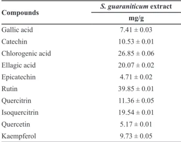
(Figure 1 and Table I).
General clinical observations
showed piloerection immediately after dosing, which lasted approximately 15 min. Thereafter, all animals
behaved normally and no signiicant changes in general
appearance or behavioral pattern were noted till the end of the observation times. Further, the consumption of food and water was similar between treated and control groups throughout the experiment (data not shown).
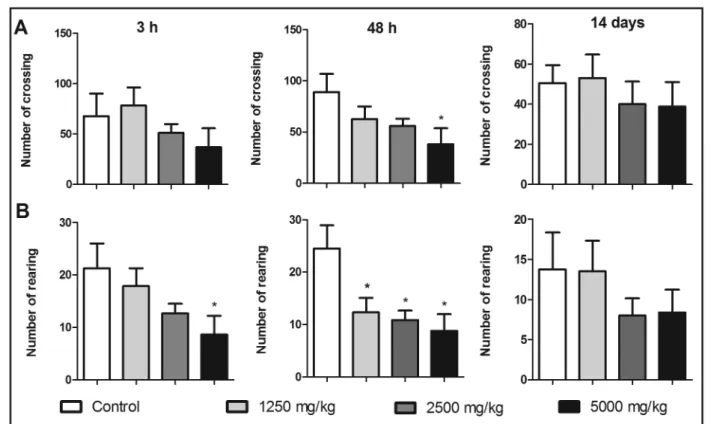
Open field test
In the open field test, the rats treated with 5,000 mg/kg of S. guaraniticum extract exhibited reduced total locomotor activity compared to the control group at
48 h after dosing. When this parameter was evaluated at
3 h and 14 days after treatment, no diferences between
treated and control animals were found (Figure 2). The group treated with 5,000 mg/kg of extract also showed reduced rearing frequencies in the vertical exploration test at 3 h after dosing. However, a dose-dependent reduction in the number of rearings was observed in all treated rats at 48 h after S. guaraniticum
administration. Interestingly, this efect was not observed at 14 days after treatment, when there was no diference
between treated and control groups (Figure 2).
In relation to defecation frequency, which may indicate anxiety levels, none of the treated animals
presented diferences from the control group for number
of fecal pellets (data not shown).
Effect of the extract on organ weight and body weight gain
Normal body weight gainswere observed in all groups after 14 days of treatment (control = 46.4 g ± 3.2; group 1,250 mg/kg = 45.2 g ± 6.5; group 2,500 mg/kg = 40.6 g ± 4.1; group 5,000 mg/kg = 41 g ± 10.4). No significant difference was observed in the relative organ weights of the treated rats when compared to the controls, at 48 h or 14 days after dosing (Table II). Organ examination did not reveal any abnormalities or gross lesions.
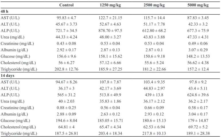
Effect of the extract on hematological and biochemical parameters
The hematological and biochemical proiles of the
treated and control groups are presented in Tables III and IV, respectively. S. guaraniticum did not signiicantly alter
either hematological or serum biochemical parameters as compared to the control animals.
Enzymatic activities and oxidative stress parameters
Acute treatment with S. guaraniticum extract did
not promote any efects on the liver, kidney, or cerebral cortex, δ-ALA-D activity, or on cerebral cortex AChE
activity (Table V).
In the kidney, there was a decrease in TBARS levels only at day 14 after treatment. Notwithstanding, the extract did not alter NPSH levels or CAT activity in the organs of
the treated animals. Moreover, no signiicant diference
was observed in FRAP levels between the treated and control groups (Table VI).
TABLE I - Quantiication of phenolic compounds found by HPLC
analyses of Solanum guaraniticum leaf extract
Compounds S. guaraniticum extract
mg/g
Gallic acid 7.41 ± 0.03
Catechin 10.53 ± 0.01
Chlorogenic acid 26.85 ± 0.06
Ellagic acid 20.07 ± 0.02
Epicatechin 4.71 ± 0.02
Rutin 39.85 ± 0.01
Quercitrin 11.36 ± 0.05
Isoquercitrin 19.54 ± 0.01
Quercetin 5.17 ± 0.01
Kaempferol 9.73 ± 0.05
Results are expressed as mean ± SEM (n = 3)
DISCUSSION
Though medicinal plants remain the first line of treatment for many ailments in developing countries where access to modern health facilities is limited, very little is generally known about their toxicity. Since there are just few published studies about its toxicological
profile, this paradigm also applies to S. guaraniticum. Despite the fact that efficacy is one of the overriding criterions in the selection of medicinal plants for use in health care systems, safety should never be overlooked and formal toxicological evaluation of medicinal plants should be performed as part of the validation process (Chan, 2003). In this sense, the present acute oral toxicity TABLE II - Body weight and organ body index of rats after 48 h and 14 days of S. guaraniticum treatment
Control 1250 mg/kg 2500 mg/kg 5000 mg/kg
48 h
Body weight (g) 216.8 ± 6.6 216.3 ± 11.6 214.5 ± 8.6 205.2 ± 7.1
Liver (%) 4.1 ± 0.3 4.23 ± 0.2 4.06 ± 0.3 3.87 ± 0.2
Kidney (%) 0.42 ± 0.01 0.41 ± 0.01 0.43 ± 0.02 0.40 ± 0.01
Spleen (%) 0.26 ± 0.02 0.27 ± 0.01 0.28 ± 0.01 0.26 ± 0.01
14 days
Body weight (g) 277.2 ± 7.3 278.8 ± 9.9 281.8 ± 11.6 249.3 ± 3
Liver (%) 3.66 ± 0.1 3.49 ± 0.1 3.54 ± 0.1 3.67 ± 0.2
Kidney (%) 0.36 ± 0.01 0.34 ± 0.01 0.36 ± 0.01 0.38 ± 0.03
Spleen (%) 0.21 ± 0.01 0.20 ± 0.01 0.21 ± 0.01 0.21 ± 0.03
Data are expressed as mean ± SEM of six rats per group.
study has revealed the safety proile of S. guaraniticum
leaf extract. On the basis of the presented results and under the conditions of this study, the lethal dose (LD50) of S.
guaraniticum extract after a single oral administration in male Wistar rats is expected to be more than 5.000 mg/kg. Therefore, the extract may be characterized as nontoxic
according to the classiication proposed by Loomis and
Hayes (Loomis, Hayes, 1996).
The reduction in the number of rearings and crossings may suggest that S. guaraniticum caused alterations in the activity of the central nervous system
(Morais, Barbosa-Filho, Almeida, 1998; Mora et al., 2005; Yasar et al., 2012). Further, it is interesting to note
that the behavioral efects were more pronounced 48 h
after treatment, and the behavior of the rats was similar to the controls at day 14 after treatment. In line with this, it
is possible that the efect of S. guaraniticum extract may be delayed and short-lasting, since the animals showed normal behavior at the end of the second experiment.
General activity is an index for evaluating behavioral changes induced in animals, by not only physiological and genetic manipulation, but also by toxicological TABLE III - Hematological proile of rats after 48 h and 14 days of S. guaraniticum treatment
Control 1250 mg/kg 2500 mg/kg 5000 mg/kg
48 h
WBC (x103/mm3) 8.63 ± 1.28 9.05 ± 0.35 7.90 ± 1.42 7.21 ± 0.76
RBC (x106/mm3) 7.21 ± 0.14 7.08 ± 0.08 7.13 ± 0.19 7.35 ± 0.16
Hb (g/dL) 13.18 ± 0.18 13.33 ± 0.11 13.36 ± 0.19 13.42 ± 0.25
HCT (%) 42.03 ± 0.67 40.93 ± 0.45 41.76 ± 1.08 42.02 ± 0.65
MCV (fL) 58.35 ± 0.62 57.82 ± 0.47 58.58 ± 0.74 57.15 ± 0.58
MCH (pg) 18.28 ± 0.29 18.83 ± 0.18 18.68 ± 0.52 18.27 ± 0.38
MCHC (g/dL) 31.37 ± 0.2 32.58 ± 0.14 32.04 ± 0.56 31.93 ± 0.41 Platelets (x103/mm3) 1232 ± 66.2 1133 ± 66.5 1140 ± 83 1173 ± 58.76
RDW (%) 13.02 ± 0.24 13.33 ± 0.34 13 ± 0.41 13.88 ± 0.64
Eosinophils (%) 0.66 ± 0.21 0.83 ± 0.3 0.8 ± 0.2 1.5 ± 0.71
Monocytes (%) 2.66 ± 0.33 2.33 ± 0.33 2.6 ± 0.4 2.83 ± 0.47
Lymphocytes (%) 81.67 ± 2.34 72.17 ± 6.97 79.2 ± 2.78 76.83 ± 2.6 Neutrophils (%) 16.33 ± 2.49 23 ± 7.12 14.5 ± 3.5 19.17 ± 2.28 14 days
WBC (x103/mm3) 10.05 ± 1.05 9.93 ± 0.73 8.53 ± 1.05 8.91 ± 0.82
RBC (x106/mm3) 7.2 ± 0.06 7.4 ± 0.27 7.53 ± 0.11 7.85 ± 0.14
Hb (g/dL) 13.58 ± 0.2 13.43 ± 0.33 13.82 ± 0.19 14.17 ± 0.26
HCT (%) 41.65 ± 0.56 41.42 ± 1.04 42.85 ± 0.38 44.38 ± 0.58*
MCV (fL) 57.82 ± 0.47 56.07 ± 0.76 56.92 ± 0.74 56.57 ± 0.72
MCH (pg) 18.83 ± 0.21 18.2 ± 0.34 18.37 ± 0.44 18.03 ± 0.25
MCHC (g/dL) 32.62 ± 0.22 32.45 ± 0.18 32.27 ± 0.42 31.92 ± 0.35 Platelets (x103/mm3) 1045 ± 28 1210 ± 64.2 955.7 ± 104.6 937.3 ± 177
RDW (%) 11.87 ± 0.3 13.03 ± 0.4 12.05 ± 0.44 12.05 ± 0.21
Eosinophils (%) 0.83 ± 0.4 1 ± 0.51 0.33 ± 0.21 0.5 ± 0.34
Monocytes (%) 1.5 ± 0.42 2 ± 0.51 2.16 ± 0.47 0.83 ± 0.3
interference (Karl, Pabst, Von Hörsten, 2003). Among the techniques used to assess general activity, the open field enables various behavioral parameters related to emotional, exploratory and motor behavior to be measured (Ribeiro de Assis et al., 2006). The decrease in locomotor
and rearing frequencies caused by S. guaraniticum
extract administration suggests depressant and/or motor
disturbance. This efect is similar to symptoms of cattle
intoxication caused by Solanum species (Rech et al., 2006; Riet-Correa et al., 1983; Paulovich et al., 2002), TABLE IV - Serum biochemical parameters of rats after 48 h and 14 days of S. guaraniticum treatment
Control 1250 mg/kg 2500 mg/kg 5000 mg/kg
48 h
AST (U/L) 95.83 ± 4.7 122.7 ± 21.15 115.7 ± 14.4 87.83 ± 3.45
ALT (U/L) 45.67 ± 3.73 52.67 ± 4.63 51.17 ± 7.78 42.33 ± 3.2
ALP (U/L) 721.7 ± 34.5 878.70 ± 97.5 612.80 ± 68.2 677.3 ± 75.9 Urea (mg/dL) 44.33 ± 4.24 48.00 ± 3.27 43.83 ± 3.88 47.33 ± 4.31 Creatinine (mg/dL) 0.43 ± 0.08 0.53 ± 0.04 0.53 ± 0.04 0.49 ± 0.06
Albumin (g/dL) 2.92 ± 0.17 2.87 ± 0.13 2.87 ± 0.1 3.07 ± 0.29
Glucose (mg/dL) 156.6 ± 9.6 158.1 ± 15.62 150.6 ± 9.18 148.2 ± 13.53 Cholesterol (mg/dL) 56 ± 6.27 57.12 ± 6.66 55.6 ± 5.24 56.62 ± 4.38 Triglyceride (mg/dL) 202.8 ± 12.76 185.9 ± 27.25 181.2 ± 22.66 157.2 ± 12.4 14 days
AST (U/L) 94.67 ± 8.26 107.8 ± 7.87 103.4 ± 9.35 97.8 ± 9.2
ALT (U/L) 36.17 ± 3 42.17 ± 3.69 44.83 ± 2.97 43.4 ± 5.11
ALP (U/L) 565 ± 31.2 513.8 ± 49.9 439 ± 13.8 624.8 ± 39.6
Urea (mg/dL) 40 ± 2.03 35.83 ± 1.86 36.17 ± 2.12 36.2 ± 2.17
Creatinine (mg/dL) 0.88 ± 0.25 0.56 ± 0.04 0.66 ± 0.09 0.58 ± 0.17 Albumin (g/dL) 2.88 ± 0.09 2.63 ± 0.12 2.93 ± 0.12 3.04 ± 0.17 Glucose (mg/dL) 194.6 ± 8.04 185.05 ± 15.71 180.6 ± 15.13 179 ± 14.87 Cholesterol (mg/dL) 64.81 ± 4 65.47 ± 4.34 62.53 ± 6.94 69.72 ± 5.2 Triglyceride (mg/dL) 187.5 ± 26.81 203.4 ± 18.34 217.8 ± 10.13 189.1 ± 28.38 Aspartate transaminase (AST), alanine transaminase (ALT), alkaline phosphatase (ALP). Data are expressed as mean ± SEM.
TABLE V - Tissue enzyme activity of rats after 48 h and 14 days of S. guaraniticum treatment
Control 1250 mg/kg 2500 mg/kg 5000 mg/kg
48 h
Liver δ-ALA-D 8.61 ± 0.57 9.82 ± 0.48 9.67 ± 0.91 8.55 ± 0.41
Kidney δ-ALA-D 3.66 ± 0.12 3.9 ± 0.33 3.96 ± 0.32 3.74 ± 0.23
Cortex δ-ALA-D 1.1 ± 0.08 1.17 ± 0.13 1.32 ± 0.13 1.09 ± 0.05
Cortex AChE 0.68 ± 0.06 0.9 ± 0.03 0.81 ± 0.1 0.82 ± 0.05
14 days
Liver δ-ALA-D 11.01 ± 0.32 11.64 ± 0.32 11.04 ± 0.28 10.32 ± 0.42
Kidney δ-ALA-D 4.58 ± 0.34 5.28 ± 0.27 4.24 ± 0.31 4.25 ± 0.29
Cortex δ-ALA-D 1.85 ± 0.44 1.82 ± 0.33 1.65 ± 0.33 1.78 ± 0.33
Cortex AChE 0.62 ±0.14 1.05 ± 0.08 0.93 ± 0.12 0.88 ± 0.2
which includes locomotive incoordination and loss of equilibrium. Although this study does not present microscopic evaluation of brain tissues, these results should be further investigated prior to indication of this plant as a phytotherapeutic agent. Further, it is quite possible that the extract´s compounds are related to this activity, since it is known that a wide range of phenolic compounds interact directly with neurotransmitter systems
and exert depressant efects (Kennedy, Wightman, 2011).
The similarity between the treated and control groups for food and water consumption and weight gain during the study period, as well as the compared hematological
and biochemical proiles indicate that S. guaraniticum
treatment did not cause significant adverse effects.
Moreover, the treatment did not modify signiicantly the
antioxidant status in animal tissues or serum, as assessed by the TBARS, NPSH and FRAP levels, and CAT activity, this excepting the kidneys where TBARS levels were
decreased. The results suggest that the extract could have renal protective action.
Similarly, previous results in our laboratory have demonstrated the in vitro inhibitory efect of the extract
on erythrocyte and brain δ-ALA-D activity, suggesting a hematologic and neurotoxic efect (Bonfanti el al., 2013,
2014). However, the efect was not observed after in vivo
acute treatment. This might be explained by the complex pharmacokinetic and pharmacodynamic characteristics of plant extracts (Rabbani, Sajjadi, Mohammadi, 2008; Akindele, Adeyemi, 2010). Plant extracts are composed of various components, with mostly unknown pharmacological and dose-response data (Ahmed, Azmat, Azeem, 2004). The numerous bioactivities found in in vitro
studies are not always meaningful to human health because the bioavailability of the natural product is often limited and only low plasma concentrations are detected in vivo
(Villegas, Sánchez-Fidalgo, Alarcón de la Lastra, 2008). TABLE VI - Oxidative status of rats after 48 h and 14 days of S. guaraniticum treatment
Control 1250 mg/kg 2500 mg/kg 5000 mg/kg
48 h
Liver TBARS 0.54 ± 0.11 0.65 ± 0.19 0.46 ± 0.07 0.63 ± 0.13
Kidney TBARS 0.83 ± 0.14 0.76 ± 0.11 0.67 ± 0.12 0.54 ± 0.07
Cortex TBARS 2.66 ± 0.36 2.6 ± 0.31 2.62 ± 0.13 2.49 ± 0.12
Liver NPSH 2.79 ± 1.18 1.66 ± 0.09 1.57 ± 0.27 2.19 ± 0.22
Kidney NPSH 2.51 ± 0.46 3.38 ± 0.33 3.23 ± 0.68 3.22 ± 0.4
Cortex NPSH 9.11 ± 2.91 8.09 ± 2.19 7.6 ± 1.11 8.9 ± 2.94
Liver CAT 10.55 ± 0.52 10.98 ± 0.74 8.78 ± 0.25 9.87 ± 0.29
Kidney CAT 5.97 ± 0.2 5.92 ± 0.2 5.70 ± 0.51 5.59 ± 0.41
Cortex CAT 0.93 ± 0.21 0.89 ± 0.25 0.61 ± 0.09 0.55 ± 0.08
Serum FRAP 0.46 ± 0.03 0.53 ± 0.07 0.55 ± 0.08 0.51 ± 0.09
14 days
Liver TBARS 0.45 ± 0.12 0.69 ± 0.2 0.47 ± 0.08 0.59 ± 0.23
Kidney TBARS 1.50 ± 0.29 0.83 ± 0.08* 0.69 ± 0.1** 0.89 ± 0.08*
Cortex TBARS 4.04 ± 0.89 3.03 ± 0.89 2.39 ± 0.5 2.75 ± 0.32
Liver NPSH 3.24 ± 0.94 2.38 ± 0.7 2.60 ± 0.4 2.96 ± 0.75
Kidney NPSH 3.41 ± 0.47 2.73 ± 0.35 2.69 ± 0.32 3.73 ± 0.41
Cortex NPSH 5. 18 ± 0.33 6.53 ± 0.72 7.35 ± 1.03 7.44 ± 0.94
Liver CAT 8.52 ±0.3 8.46 ± 0.23 8.20 ± 0.14 8.28 ± 0.41
Kidney CAT 4.87 ± 0.12 4.58 ± 0.2 4.72 ± 0.15 4.79 ± 0.18
Cortex CAT 1.57 ± 0.6 1.59 ± 0.49 1.17 ± 0.39 1.03 ± 0.3
Serum FRAP 0.47 ± 0.04 0.56 ± 0.04 0.56 ± 0.02 0.48 ± 0.02
Thiobarbituric acid reactive substances (TBARS; nmol MDA (malondialdehyde)/mg protein). Non-protein thiol groups (NPSH; mmol GSH/mg protein). Catalase (CAT; mmol H2O2 consumed/min/mg protein). Ferric reducing power (FRAP; mM Fe
2+). Data
In this context, in vivo studies are helpful in clarifying the
efective biological properties of a plant’s constituents thru
considering metabolism and pharmacokinetics.
Toxicity studies in appropriate animal models are commonly used to assess potential health risks to humans. This is a key stage in ensuring the safety of drugs and an acute toxicity study is just one of the many types of toxicity tests that are used for such purposes (Asare et al., 2011).
The fact that no lethal toxic efects were observed in the
S. guaraniticum acute toxicity study indicates that plant preparations are safe for short-term treatments; and this low toxicity may have been responsible for its widespread use in varied ethno-therapeutic interventions. However, this assertion of safety may not be applicable to herbal preparation taken for a long period. To add further safety information a chronic toxicity study is recommended.
CONCLUSION
In conclusion, this study provides valuable data on
the acute oral toxicity proile of S. guaraniticum. Based on the toxicological parameters evaluated, it can be concluded that this medicinal plant is not acutely toxic yet appears to possess some action on the general behavior of rats, a biological property that deserves greater attention. The results presented may be helpful for therapeutic decisions with respect to the toxicology of S. guaraniticum extract, and should be useful for any future in vivo or clinical study of this medicinal plant.
ACKNOWLEDGMENTS
The authors acknowledge the doctoral fellowship to Gabriela Bonfanti given by the Fundação Coordenação de Aperfeiçoamento de Pessoal de Nível Superior (CAPES), as well as the Federal University of Santa Maria (UFSM),
RS, Brazil, for inancial support.
CONFLICT OF INTEREST
The authors declare that they have no conlicts of
interest to disclose.
REFERENCES
AEBI, H. Catalase in vitro. Methods Enzymol., v.105, p.121-126, 1984.
AHMED, M.; AZMAT, A.; AZEEM, M.A. Dose-response curve of Somina (Herbal preparation): a study on frog heart. Pak. J. Pharmacol., v.21, n.2, p.19-22, 2004.
AKINDELE, A.; ADEYEMI, O. Anxiolytic and sedative effects of Byrsocarpus coccineus Schum. and Thonn. (Connaraceae) extract. Int. J. Appl. Res. Nat. Prod., v.3, n.1, p.28-36, 2010.
ANDRADE, F.; ALBUQUERQUE, C.A.C.; MARASCHIN, M.; SILVA, E.L. Safety assessment of yerba mate (Ilex paraguariensis) dried extract: results of acute and 90 days subchronic toxicity studies in rats and rabbits. Food Chem. Toxicol., v.50, n.2, p.328-334, 2012.
ASARE, G.A.; ADDO, P.; BUGYEI, K.; GYAN, B.; ADJEI, S.; OTU-NYARKO, L.S.; WIREDU, E.K.; NYARKO, A. Acute toxicity studies of aqueous leaf extract of Phyllanthus niruri. Interdiscipl. Toxicol., v.4, n.4, p.206-210, 2011.
BAKER, D.C.; KEELER, R.F.; GAFFIELD, W. Toxicosis from steroidal alkaloids of Solanum species: toxicology of plant and fungal compounds. In: KEELER, R.F.; TU, A.T., eds. Handbook of natural toxins. New York: Marcel Dekker, 1991. p.589-610.
BENZIE, I.F.; STRAIN, J.J. The ferric reducing ability of plasma (FRAP) as a measure of antioxidant power: the FRAP assay. Anal. Biochem., v.239, n.1, p.70-76, 1996.
BOHS, L. Major clades in Solanum based on ndhF sequences. In: KEATING, R.C.; HOLLOWELL, V.C.; CROAT, T.B., eds. A festschrift for William G. D’Arcy: the legacy of a taxonomist. Saint Louis: Missouri Botanical Garden Press, 2005. p.27-49. (Monographs in systematic botany from the Missouri Botanical Garden, 104).
BONFANTI, G.; BITENCOURT, P.R.; DE BONA, K.S.; SILVA, P.S.; JANTSCH, L.B.; PIGATTO, A.S.; BOLIGON, A.; ATHAYDE, M.L.; GONÇALVES, T.L.; MORETTO, M.B. Syzygium jambos and Solanum guaraniticum show similar antioxidant properties but induce diferent enzymatic activities in the brain of rats. Molecules, v.18, n.8, p.9179-9194, 2013.
BROADHURST, P.L. Experiments in psychogenetics, application on biometrical genetics to the inheritance of behavior. In: EYSENCK, H.J., ed. Experiments in personality. London: Routledge & Kegan Paul, 1960. v.1, p.1-120.
BUEGE, J.A.; AUST, S.D. Microsomal lipid peroxidation. Methods Enzymol., v.52, p.302-310, 1978.
CHAN, K. Some aspects of toxic contaminants in herbal medicines. Chemosphere, v.52, n.9, p.1361-1371, 2003.
CORRÊA, M.P. Dicionário das plantas úteis do Brasil e das exóticas cultivadas. [S.l.]: Ministério da Agricultura, Instituto Brasileiro de Desenvolvimento Florestal, 1984. v.3.
COSTA, O.A. Jurubeba. Rev. Bras. Farm., v.21, p.404-416, 1940.
EIDI, A.; EIDI, M.; ESMAEILI, E. Antidiabetic efect of garlic (Allium sativum L.) in normal and streptozotocin-induced diabetic rats. Phytomedicine, v.13, n.9/10, p.624-629, 2006.
ELLMAN, G.L. Tissue sulfhydryl groups. Arch. Biochem. Biophys., v.82, n.1, p.70-77, 1959.
ELLMAN, G.L.; COURTNEY, K.D.; ANDRES Jr., V.; FEATHERSTONE, R.M. A new and rapid colorimetric determination of acetylcholinesterase activity. Biochem. Pharmacol., v.7, n.2, p.88-95, 1961.
HOR, S.Y.; AHMAD, M.; FARSI, E.; LIM, C.P.; ASMAWI, M.Z.; YAM, M.F. Acute and subchronic oral toxicity of Coriolus versicolor standardized water extract in Sprague-Dawley rats. J. Ethnopharmacol., v.137, n.3, p.1067-1076, 2011.
JADHAV, S.J.; SHARMA, R.P.; SALUNKHE, D.K. Naturally occurring toxic alkaloids in foods. Crit. Rev. Toxicol., v.9, n.1, p.21-104, 1981.
KARL, T.; PABST, R.; VON HÖRSTEN, S. Behavioral phenotyping of mice in pharmacological and toxicological research. Exp. Toxicol. Pathol., v.55, n.1, p.69-83, 2003.
KENNEDY, D.O.; WIGHTMAN, E.L. Herbal extracts and phytochemicals: pant secondary metabolites and the enhancement of human brain function. Adv. Nutr., v.2, n.1, p.32–50, 2011.
LOOMIS, T.A.; HAYES, A.W. Loomis’s essentials of toxicology. 4.ed. San Diego: Academic Press, 1996. 282 p.
MALATHI, R.; GOMAZ, P. Evaluation of preliminary toxicity studies on the methanolic leave extract of Tylophora asthmatica in experimental rats. J. Pharmacol. Toxicol., v.3, n.1, p.34-40, 2008.
MORA, S.; DÍAZ-VÉLIZ, G.; MILLÁN, R.; LUNGENSTRASS, H.; QUIRÓS, S.; COTO-MORALES, T.; HELLIÓN-IBARROLA, M.C. Anxiolytic and antidepressant-like effects of the hydroalcoholic extract from Aloysia polystachya in rats. Pharmacol., Biochem. Behav., v.82, n.2, p.373-378, 2005.
MORAIS, L.C.S.L.; BARBOSA-FILHO, J.M.; ALMEIDA, R.N. Central depressant efects of reticuline extracted from Ocotea duckei in rats and mice. J. Ethnopharmacol., v.62, n.1, p.57-61, 1998.
NOGUEIRA, C.W.; BORGES, V.C.; ZENI, G.; ROCHA, J.B.T. Organochalcogens efects on δ-aminolevulinate dehydratase activity from human erythrocytic cells in vitro. Toxicology, v.191, n.2/3, p.169-178, 2003.
OECD. Organization for Economic Cooperation and Development. OECD Guidelines for the testing of chemicals: acute oral toxicity – acute toxic class method. Paris: Organization for Economic Cooperation and Development, 2001. 14p. (OECD/OCDE Test n.423). Available at: https://ntp.niehs.nih.gov/iccvam/suppdocs/ feddocs/oecd/oecd_gl423.pdf>. Accessed on: April 2015.
OECD. Organization for Economic Cooperation and Development. OECD Guidelines for the testing of chemicals: repeated dose 28-day oral toxicity study in rodents. Paris: Organization for Economic Cooperation and Development, 2008. 13p. (OECD/OCDE Test n.407). Available at: <http://www.oecd-ilibrary.org/docserver/ download/9740701e.pdf?expires=1462907307&id=id&a ccname=guest&checksum=4C051DAF7247577C6E48A 97B48885D63>. Accessed on: April 2015.
PAYNE, J.F.; MATHIEW, A.; MELVIN, W.; FANCEY, L.L. Acetylcholinesterase, an old biomarker with a new future? Field trials in association with two urban rivers and a paper mill in Newfoundland. Mar. Pollut. Bull., v.32, n.2, p.225-231, 1996.
PETERSON, G.L. A simpliication of the protein assay method of Lowry et al. which is more generally applicable. Anal. Biochem., v.83, n.2, p.346-356, 1977.
POHANKA, M. Cholinesterases, a target of pharmacology and toxicology. Biomed. Pap. Med. Fac. Univ. Palacký, Olomouc, Czech. Repub., v.155, n.3, p.219-229, 2011.
RABBANI, M.; SAJJADI, S.; MOHAMMADI, A. Evaluation of the anxiolytic efect of Nepeta persica Boiss in mice. Evid. Based Complement. Alternat. Med., v.5, n.2, p.181-186, 2008.
RECH, R.R.; RISSI, D.R.; RODRIGUES, A.; PIEREZAN, F.; PIAZER, J.V.M.; KOMMERS, G.D.; BARROS, C.S.L. Intoxicação por Solanum fastigiatum (Solanaceae) em bovinos: epidemiologia, sinais clínicos e morfometria das lesões cerebelares. Pesq. Vet. Bras., v.26, n.3, p.183-189, 2006.
RIBEIRO DE ASSIS, J.C.; SUFFREDINI, I.B.; MORENO, P.R.; YOUNG, M.C.; VARELLA, A.D.; YOUNES, R.N.; BERNARDI, M.M. Analysis of the toxic potential of Palicourea corymbifera (Mull Arg.) Standl. in laboratory animals. Res. Vet. Sci., v.80, n.2, p.209-217, 2006.
RIET-CORREA, F.; MÉNDEZ, M.C.; SCHILD, A.L.; SUMMERS, B.A.; OLIVEIRA, J.A. Intoxication by Solanum fastigiatum var. fastigiatum as a cause of cerebellar degeneration of cattle. Cornell Vet., v.73, n.3, p.240-256, 1983.
ROCHA, J.B.; EMANUELLI, T.; PEREIRA, M.E. Effects of early undernutrition on kinetic parameters of brain acetylcholinesterase from adult rats. Acta Neurobiol. Exp., v.53, n.3, p.431-437, 1993.
ROSIDAH; YAM, M.F.; SADIKUN, A.; AHMAD, M.; AKOWUAH, G.A.; ASMAWI, M.Z. Toxicology evaluation of standardized methanol extract of Gynura procumbens. J. Ethnopharmacol., v.123, n.2, p.244-249, 2009.
SABIR, S.M.; ROCHA, J.B.T. Antioxidant and hepatoprotective activity of aqueous extract of Solanum fastigiatum (false “Jurubeba”) against paracetamol-induced liver damage in mice. J. Ethnopharmacol., v.120, n.2, p.226-232, 2008.
SASSA, S. Delta-aminolevulinic acid dehydratase assay. Enzyme, v.28, n.2/3, p.133-145, 1982.
SCHETINGER, M.R.C.; PORTO, N.M.; MORETTO, M.B.; MORSCH, V.M.; ROCHA, J.B.T.; VIEIRA, V.; MORO, F.; NEIS, R.T.; BITTENCOURT, S.; BONACORSO, H.G.; ZANATTA, N. New benzodiazepines alter acetylcholinesterase and ATPase activities. Neurochem. Res., v.25, n.7, p.949-955, 2000.
SHENOUDA, J.; GREEN, P.; SULTATOS, L. An evaluation of the inhibition of human butyrylcholinesterase and acetylcholinesterase by the organophosphate chlorpyrifos oxon. Toxicol. Appl. Pharmacol., v.241, n.2, p.135-142, 2009.
SIMÕES, C.M.O.; FALKENBERG, M.; MENTZ, L.A.; SCHENKEL, E.P.; AMOROS, M.; GIRRE, L. Antiviral activity of south Brazilian medicinal plant extracts. Phytomedicine, v.6, n.3, p.205-214, 1999.
SOUZA, A.C.G.; LUCHESE, C.; SANTOS NETO, J.S.; NOGUEIRA, C.W. Antioxidant efect of a novel class of telluroacetilene compounds: studies in vitro and in vivo. Life Sci., v.84, n.11/12, p.351-357, 2009.
STEHMANN, J.R.; MENTZ, L.A.; AGRA, M.F.; VIGNOLI-SILVA, M.; GIACOMIN, L. Solanaceae. In: Lista de espécies da Flora do Brasil. Rio de Janeiro: Jardim Botânico do Rio de Janeiro, 2012. Available at: <http://loradobrasil. jbrj.gov.br/jabot/listaBrasil/PrincipalUC/PrincipalUC.do ;jsessionid=881E70462293A552A0EC8BF5902633B7>. Accessed on: 08 Oct. 2013.
VILLEGAS, I.; SÁNCHEZ-FIDALGO, S.; ALARCÓN DE LA LASTRA, C. New mechanisms and therapeutic potential of curcumin for colorectal cancer. Mol. Nutr. Food Res., v.52, n.9, p.1040-1061, 2008.
WORLD HEALTH ORGANIZATION. WHO. The world medicines situation 2011: traditional medicines: global situation issues and challenges. Geneva: World Health Organization, 2011.
YASAR, S.N.; CAN, O.D.; OZTURK, N.; SAGRATINI, G.; RICCIUTELLI, M.; VITTORI, S.; MAGGI, F. Central nervous system activities of Hypericum origanifolium extract via gabaergic and opioidergic mechanisms. Phytother. Res., v.27, n.6, p.877-884, 2012.
ZADRA, M.; PIANA, M.; BRUM, T.F.; BOLIGON, A.A.; FREITAS, R.B.; MACHADO, M.M.; STEFANELLO, S.T.; SOARES, F.A.A.; ATHAYDE, M.L. Antioxidant activity and phytochemical composition of the leaves of Solanum guaraniticum A. St.-Hil. Molecules, v.17, n.11, p.12560-12574, 2012.
Received for publication on 20th April 2015