*Correspondence: P. C. P. Rosa. Faculty of Pharmaceutical Sciences. State University of Campinas. Rua Alexandre Fleming, s/n - Distrito Barão Geraldo - 13083-887 - Campinas-SP, Brazil. E-mail: paulo.rosa@fcf.unicamp.br
<http://dx.doi.org/10.1590/S1984-82502016000100008
Performance evaluation of Cryo Laser Phoresis technique as
biophysical method to promote diclofenac sodium cutaneous
perfusion
Marcio Adriano Andréo
1, Iasmin Mimoto Ruino
1, Daniela Cecilia Ortiz de Orue Ubaldo
1, Estela
Boaro Herbst
1, Heron Dominguez Torres da Silva
1, Pedro Gonçalves de Oliveira
2, Marcio Ferrari
3,
Paulo Cesar Pires Rosa
4,*, Patrícia Santos Lopes
1, Vânia Rodrigues Leite-Silva
11Institute of Environment, Chemistry and Pharmaceutical Sciences, Federal University of São Paulo Diadema, SP, Brazil, 2Faculty of Pharmacy, Anhembi Morumbi, University, São Paulo, SP, Brazil, 3Department of Pharmacy, Federal University of Rio
Grande do Norte, Natal, RN, Brazil, 4Faculty of Pharmaceutical Sciences State University of Campinas, Campinas, SP, Brazil
Aiming to alter and/or improve permeation of active compounds in the skin, many strategies have been developed, including biophysical methods. One of the physical absorption techniques, currently known as Cryo Laser Phoresis (CLP), consists of an apparatus that emits radiation on polar or nonpolar molecules of the active substance, resulting in faster penetration when in comparison to the standard topical application. The goal of this work was to evaluate the eicacy of a method that proposes to increase cutaneous permeation of diclofenac sodium by using CLP technique. The inluence on permeation was evaluated ex vivo, using Franz cell and human skin obtained from cosmetic surgery. The results were evaluated using statistical methods and data exploratory analysis: clusters, k-means and Principal Component Analysis. The results showed a larger increase in the concentration of diclofenac sodium in the dermis with the use of laser. In all samples (with or without laser application) it was observed that skin surface showed an amount of diclofenac sodium and that there was no active passage to the receptor liquid, suggesting that diclofenac sodium was not absorbed. These results indicate that CLP, when used under the conditions described in this study, is able to increase diclofenac sodium penetration and its retention into deeper layers.
Uniterms: Cryo Laser Phoresis/dematologic use. Cutaneous permeation. Diclofenac sodium/cutaneous permeation. Franz cell.
No sentido de alterar e/ou melhorar a penetração de substâncias na pele, diversas estratégias têm sido desenvolvidas, variando desde a aplicação de novos veículos e ativos encapsulados, até equipamentos que atuam por métodos biofísicos. Uma das técnicas de absorção física, atualmente conhecida como Crio Laser Forese (CLF), consiste em um aparato que emite radiação sobre moléculas polares ou apolares da substância ativa, tornando sua penetração mais rápida, se comparada à administração tópica comum. O objetivo deste trabalho foi avaliar a eicácia de um método que propõe aumentar a permeação cutânea do diclofenaco de sódio incorporado a um gel, por meio do uso da CLF. A inluência sobre a permeação foi avaliada ex vivo, utilizando célula de Franz e pele humana obtida de cirurgia plástica. Os resultados foram balizados mediante aplicação de métodos estatísticos e análise exploratória de dados: clusters, k-means e Análise por Componentes Principais. Os resultados demonstraram aumento na concentração do diclofenaco de sódio na derme com o uso do laser. Em todas as amostras (com ou sem aplicação de laser), observou-se, uma quantidade de diclofenaco de sódio na superfície da pele e que não houve passagem de ativo para o líquido do receptor, sugerindo que o diclofenaco de sódio não foi absorvido. Estes resultados indicam que CLF usada sob as condições descritas neste estudo é capaz de aumentar a penetração do diclofenaco de sódio e sua retenção em camadas mais profundas da pele
INTRODUCTION
The skin has many functions and amongst them it is possible to emphasize its role as a barrier against exogenous agents, its utility to keep the homeostasis of the organism and to prevent internal water loss. Beyond that, it has been shown that it is an appropriate route for treatment systems, through transdermal administration of drugs (Leite-Silva et al., 2012). Several methods have been developed to increase the penetration of substances through skin. This includes the use of substances that act as permeation enhancers (chemical enhancers) in formulations or the development of new methods that modify the lipid and protein organization, inter and intracellular of the stratum corneum (biophysical enhancers) and can reversibly decrease the skin’s barrier property (Lopes et al., 2008; Silva et al., 2010).
Throughout the years there was a signiicant growth
of knowledge in biochemical and metabolic processes
involving substance low through skin (Fluhr, Feingold,
Elias, 2006). There are many factors that influence substance permeation through skin, such as formulation, particle size, component concentration, skin thickness, pH and sebum production, among others (Gallagher et al., 2003; Benson, 2005; Trommer, Neubert, 2006; Baroli, 2010; Leite-Silva et al., 2012).
Several methods have been formulated to promote augmentation of transdermic transportation, having obtained varying levels of success. The promoting agents of chemical absorption can improve the skin’s permeability to small molecules, but also can lead to irritation of the surface of the skin or other factors that may limit their
use. Iontophoresis uses electric ields to permeate ionized
molecules through the skin using electrophoresis and electroosmosis for non-ionized molecules. This technique can be useful for applications that contain peptides and small proteins. Physical methods that temporarily improve skin permeability such as electroporation and ultrasound are used, representing perspectives in the application of drugs of low or high molecular weight (Patil, Saraogi, 2014). However, not much studying has been done about the use of CryoLaser as a method of biophysical permeation.
In spite of the use of chemical permeation enhancers being widely studied, remarkable attention has been given to the benefits of biophysical permeation promoting techniques. One of them is known as Cryo Laser Phoresis (CLP), also named Cryopass Therapy, developed by C.I.R.C.E srl – Italy (Bonizzoni, 2007). It is applied with a device called Lasericemed, which emits a laser radiation on polar or nonpolar active substances and claims to make
their absorption faster and more eicient when compared
to the standard topical administration. The laser emission releases photons that collide with electrons of active substance molecules. This impact leads to photon energy transfer to electrons from more external orbitals, as kinetic energy form. This increases gravitational acceleration, allowing molecules to pass through skin and be delivered to the desired site (Terraneo et al., 2011).
The aim of this work was to evaluate the Cryo Laser Phoresis efficiency as biophysical method to promote diclofenac sodium cutaneous permeation.
MATERIAL AND METHODS
Material
H y d r o x y e t h y l c e l l u l o s e ( C A S 9 0 0 4 - 6 2 - 0 , Mapric Produtos Farmacosmeticos).The diclofenac s o d i u m w o r k i n g s t a n d a r d ( m o n o s o d i u m a c i d 2-[(2,6-dichlorophenyl) amino] benzene acetic salt) was donated by Medley Pharmaceutical Industry and methylchloroisothiazolinone and methylisothiazolinone were donated by Pro-ServQuimica LTDA. Acetonitrile (J. T. Baxter™) and methanol (J. T. Baxter™) were from HPLC gradeand phosphate buffer (PBS) components (NaCl - MW: 58,44; KCl - MW: 74,55; Na2HPO4 - MW: 141,96 and KH2PO4 - MW: 136,09) were P. A. grade from Synth®. The tape stripping tapes (Standard S-Squame Disc
CuDe 15109) were from CuDerm. The syringe iltersMilex
were from Milipore®. The C18 chromatography column
within 25 cm x 4.6 mm dimensions, 5 μm particle size
was from ACE™.
Formulation
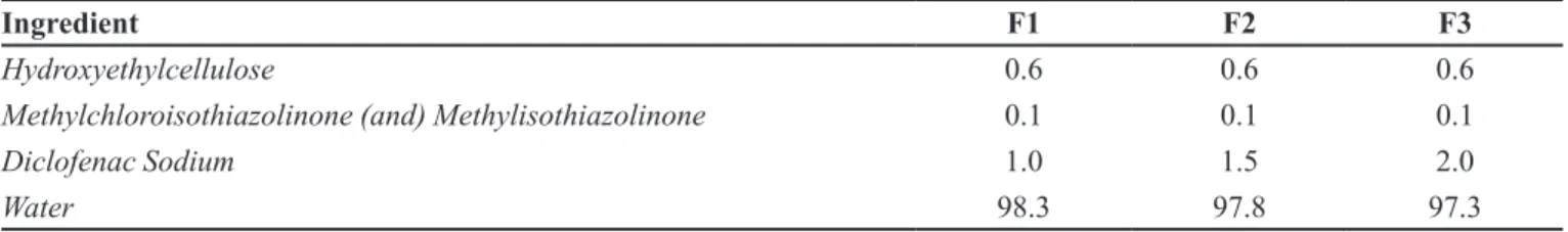
A gel was formulated using low irritant power and low cost raw materials (Table I). The gel was frozen until -20 ºC in suitable a package to attach to the device.
Permeation test
UNIFESP’s Ethics Committee previously approved the use of skin obtained by excision in cosmetic surgery, with protocol number 631972. After preparation, the skin was stored at a temperature of approximately -20 ºC.
The experiment was performed using dual compartment vertical diffusion cells (Franz cells) that
were speciically modiied and adapted exclusively for
phosphate buffer (PBS) pH 7.4. PBS was added to the receptor compartment and the system was immersed in a bath at 37 ºC, and stirred continuously for half an hour to stabilize the system.
The bottle containing the active frozen gel was attached to the device and placed in contact with the skin for 10 min, with or without laser contact, in this case, employing the device Lasericemed turned on (Figure 1B). It is just an illustration, without the human skin and the receptor liquid only to demonstrate how the laser is used. This study was performed in triplicate.
The receptor liquid was collected at intervals of 10 and 20 min. After 20 min, the gel applied to skin surface was removed with swabs™. Next, stratum corneum (SC) extraction was performed by “tape stripping” technique and the detachment of epidermis from dermis was performed (water at 60 ºC for 90 s). The samples were stored after being soaked in methanol for approximately 15 min.
Afterwards, an aliquot from the supernatant liquid from each sample was removed and submitted to HPLC
analysis. The inal samples were: 10 min receptor liquid,
20 min receptor liquid, swab, tapes 1-5, tapes 6-10, epidermis and dermis, with and without laser application.
Determination of diclofenac sodium in the samples
The methodology of diclofenac sodium analysis was developed based on conditions described on USP 35 (2013) monograph and also on the methodology described by Klimes et al. (2001).
Considering these aspects, the chromatographic
quantiication of diclofenac sodium was performed by
using HPLC degree solvents (acetonitrile and methanol) and C18 chromatography column within 25 cm x 4.6 mm dimensions, 5 μm particle size. Diclofenac sodium (working standard), with 99% purity was used as secondary standard. The analyses were performed in a liquid chromatograph model ACELLA 600, from Thermo Scientific™, equipped with quaternary pump system, column oven, automatic injector and diode array detector (DAD). Data was processed by ChromQuest™ software.
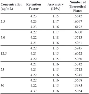
In addition, the following chromatography system parameters were also evaluated: retention factor, asymmetry (10%) and number of theoretical plates, according to (USP 35, 2013) recommended parameters.
In connection with the sample preparation, with
0.5 mL ixed volume, all samples were previously iltered through 0.45 μm syringe filters and stored in 1.5 mL
vials. The samples were diluted, when necessary, so active expected concentrations were coherent with the ones determined at calibration curve. Chromatography conditions for diclofenac sodium analysis were based on
the US Pharmacopoeia (USP 35), with some modiications
for optimizing the method, according to gel matrix and biological matrix content, assessing parameters as: stationary phase and chromatographic column dimensions,
content, mobile phase low, temperature, injection volume,
sample solution and standard solution concentration related to HPLC equipment response.
After optimization, an isocratic method was established with mobile phase composed of 30% phosphate
bufer (pH 2.5) and 70% acetonitrile, with 1.0 mL.min-1
flow, using C18 column with 25 cm x 4.6 mm, 5 μm
particle size and DAD detector monitoring 254 nm TABLE I - Composition of formulations (%, w/w) evaluated
Ingredient F1 F2 F3
Hydroxyethylcellulose 0.6 0.6 0.6
Methylchloroisothiazolinone (and) Methylisothiazolinone 0.1 0.1 0.1
Diclofenac Sodium 1.0 1.5 2.0
Water 98.3 97.8 97.3
wavelength. The injected volume of each sample was
25 μL and 9 min run time.
A stock solution was prepared in a volumetric lask,
transferring 12.5 mg of diclofenac sodium in 25 mL of
methanol, obtaining a 500 μg.mL-1 solution. From the
stock solution and using 10 mL volumetric lasks, dilutions
were prepared leading to the concentrations: 2.5; 5.0; 12.5; 25; 50; 100 and 150 µg.mL-1 in methanol. The standard concentrations for the calibration curve were prepared in triplicate and analyzed in the previously described chromatography conditions.
Statistical analysis
At the data exploratory phase, t Student test was
applied, which allowed verifying signiicant diferences
between “With Laser” and “Without Laser” situations, to the Dermis (D). Given the hypothesis absence, a priori, to all other results, cluster analysis method application was chosen (p-value < 0.05 was considered to be statistically
signiicant).
RESULTS AND DISCUSSION
Diclofenac sodium was chosen as a marker due to
its hydrophilic characteristics that hinder its low through
the skin’s lipid layer (Lopes et al., 2008) and because it can be a good alternative for challenging the biophysical enhancer. Gaur et al. (2014) had studied the transdermal formulations containing permeation enhancers that cause skin damage and diclofenac was used as a model drug. The presence of ceramid specifically promotes drug permeation through SC and dermis and also contributes to stability and non-irritancy.
Escribano et al. (2003) evaluated the transdermal permeation of sodium diclofenac. Permeation studies were carried out in vitro using human skin from plastic surgery as a membrane – as we did in our research. However, they used chemical enhancers instead of biophysical methods to improved diclofenac permeation.
The influence of an erbium:YAG laser on the transdermal delivery of drugs across skin was studied
in vitro by Lee et al. (2001). The researchers concluded that the use of an erbium:YAG laser is a good method for enhancing transdermal absorption of both lipophilic and hydrophilic drugs, because it allows precise control of SC removal, and this ablation of SC can be reversible to its original normal status. Nevertheless, the laser used in this work does not have ablation potential.
The Figure 2 shows the comparative performance of samples submitted to the laser treatment and not submitted
to the laser treatment; highlighted are the Swab and dermis samples, in gray are the other samples with inexpressive graphic representation.
The statistical analysis reinforces the validity of these data. The Figure 3 shows a hierarchical grouping analysis dendrogram (K-means) that allowed verifying the evident dissimilarity of results obtained from the swab (with and without laser and dermis (with laser)), when compared to the other experimental data.
Complementarily to cluster analysis, data was treated by Principal Component Analyses (PCA) method application, permitting the gathering of similar behavior data groups (Figure 4). It basically consists of the diagonalization of a symmetric data matrix, resulting in a new group of variables (principal components), which are linear combinations of the original variables and non-correlated. In this way, a new space is generated in which
the variables can be projected and classiied into categories
or groups. The application of this method allowed the
clear diferentiation (grouping) between the variables to be veriied, highlighting the swab and the dermis (Figure 4A), conirming that the laser treatment promotes results
to the other result groups. Figure 4B highlights the
diferentiation of dermis with laser (DCL) and Swab with
and without laser (SCL and SSL), compared to the results set considering all tested concentrations.
According Yilmaz and Ciltas (2015), diverse methodologies are described in the literature to diclofenac qualitative and quantitative analysis, using many analytical techniques. However, most of them use high performance liquid chromatography with ultraviolet detection (HPLC-UV) (Arcelloni et al., 2001; Bhattacharya et al., 2013). Therefore, this work methodology was developed based on conditions of analysis described on USP 35 (2013) monograph and on the methodology described by Klimes
et al. (2001).
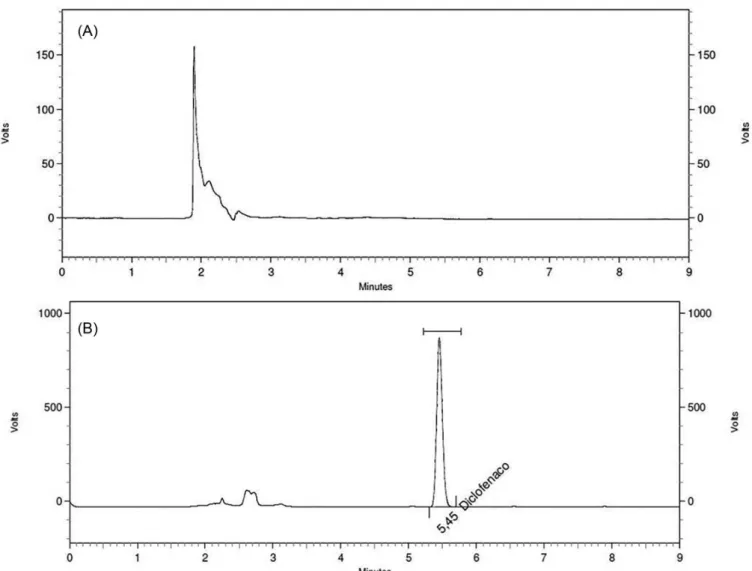
The method presented suitable specificity as it considered the excipients used in the gel vehicle. Representative chromatograms from samples with or without diclofenac sodium are shown on Figure 5.
The analytical curve, based on the expected active concentrations on the different skin layers, presented
results and Coeicient of Variation (CV) suitable with
the aim of the analysis. These analyses were performed in triplicate and their results are shown in Table II.
The obtained coefficient of determination was 0.9966, which shows good explaining ability and linearity
coeicient of variation of 4.7% from Detection Limit (DL), determined in 3.3 μg.mL-1 until maximum concentration
established in calibration (50 μg.mL-1).
The chromatographic system evaluation parameter values to analyze the standards were: retention factor = 4.2, asymmetry (10%) = 1.16, number of theoretical plates = 15582, indicating chromatographic system adequacy FIGURE 3 - Cluster Analysis about diferentiation of dermis with
laser and Swab with and without laser when compared to the other experimental data. Notes: SCL = Swab with laser; SSL= Swab without laser; E1CL = tape strip 1 to 5 with laser; E1SL = tape strip 1 to 5 without laser; E2CL = tape strip 6 to 10 with laser; E2SL = tape strip 6 to 10 without laser; E3CL = tape strip 11 to 15 with laser; E3SL = tape strip 11 to 15 without laser; E4CL = tape strip 16 to 20 with laser; E4SL = tape strip 16 to 20 without laser; EPCL= epidermis with laser; EPSL= epidermis without laser; DCL= Dermis with laser; DSL= Dermis without laser; L10CL= receptor liquid 10 min with laser; L10SL= receptor liquid 10 min without laser, L20CL= receptor liquid 20 min with laser; L20SL= receptor liquid 20 min without laser.
FIGURE 4 - Principal Component Analysis method application, allowing the gathering of similar behavior data groups. (A)
Diferentiation of variables swab and dermis when submitted to laser treatment,(B)diferentiation of dermis with laser (DCL) and
and compatibility with the USP 35 (2013) recommended parameters. The values of all evaluation parameters to the standards are on Table III.
These results of performance parameters, including
speciicity, range, linearity, precision (CV), LOD (limit of
detection) and LOQ (limit of quantiication), presented by
the HPLC method used for the determination of diclofenac sodium in the samples obtained from the permeation tests,
assure the sensibility and conidence necessary for the
assessment of the data connected with the potential of the TABLE II - Results obtained from diclofenac sodium solution analysis to build an analytical curve
Concentration (mg.mL-1) Area 1 Area 2 Area 3 Mean ± SD CV (%)
0.0025 564670 564970 558828 562823 ± 3463 0.6
0.005 734929 742080 737941 738317 ± 2831 0.4
0.0125 2042415 2043344 2053681 2046480 ± 5106 0.25
0.025 3781948 3794774 3786593 3787772 ± 5302 0.14
0.05 7237005 7245341 7855082 7445809 ± 289419 0.4
Notes: CV = Coeicient of Variation. SD = Standard Deviations
FIGURE 5 - Chromatogram analysis obtained from samples without diclofenac sodium addition (A) and samples with diclofenac sodium (B). Chromatographic conditions: chromatographic column ACE® C18 (25 cm x 4.6 mm, 5 μm particle size), mobile phase
containing 30% phosphate bufer (pH 2,5) and 70% acetonitrile, 1.0 mL.min-1 low, 254 nm detection, 25 μL injected volume and
CLP to increase the cutaneous permeation of diclofenac sodium.
The performed analysis, in all three investigated concentrations (1.0%, 1.5%, 2.0%) presented the same behavior, as shown on Figure 2. All showed a higher increase of the substance in the dermis with the laser use, suggesting that laser employment in these conditions increases diclofenac sodium penetration and its retention into deeper layers. In all samples (with or without laser application), it is observed that skin surface (swab) showed, as expected, a much higher diclofenac sodium quantity (out of method work range) and that there is no active passage to the receptor liquid, suggesting that diclofenac sodium is not absorbed.
Using an intermediate concentration as a numeric example, it is possible to verify that without laser
employment, the analysis representing the irst 5 layers
from stratum corneum (0.049 mg.mL-1), layers 6 to 10 (0.014 mg.mL-1), layers 11 to 15 (0.008 mg.mL-1) and layers 16 to 20 (0.004 mg.mL-1) show that the found diclofenac sodium quantity decreases according to the analyzed stratum corneum depth, remaining low in dermis (0.011 mg.mL-1). As in the samples that underwent laser treatment, the samples that represent the first 5 stratum corneum layers (0.024 mg.mL-1), layers 6 to 10 (0.011 mg.mL-1), layers 11 to 15 (0.006 mg.mL-1) and layers 16 to 20 (0.004 mg.mL-1) also presented as a result
a decrease in concentrations according to the analyzed
stratum corneum depth and a signiicant increase in the
dermis (0.117 mg.mL-1).
New studies must be conducted with distinct physicochemical properties molecules, aiming to measure
the permeation enhancer capacity on diferent situations.
Finally it must be highlighted that, because of operational limitations, this experiment was adapted to evaluate only
the efects of laser application on a frozen active solution
in a hydrophilic gel, when applied over the skin. According to Bonizzoni (2007), after applying the gel with the laser, skin surface should be exposed to a light beam again by a scanning device, which could have contributed to the permeation process if it had been used in this experiment.
CONCLUSION
The results indicate that the Cryo Laser Phoresis technique, when used under the conditions described in this study, is able to enhance skin permeation of diclofenac sodium and potentially of other molecules with similar physical-chemical proprieties, thus constituting an interesting alternative to drug administration, especially in situations in which penetration in deeper skin layer is necessary and where the systemic use may be unfeasible or may be capable to promote unpleasant reactions.
REFERENCES
ARCELLONI, C.; LANZI, R.; PEDERCINI, S.; MOLTENI, G.; FERMO, I.; PONTIROLIC, A.; PARONI, R. High-performance liquid chromatographic determination of diclofenac in human plasma after solid-phase extraction.
J. Chromatogr. B: Biomed. Sci. Appl., v.763, n.1/2, p.195-200, 2001.
BAROLI, B. Penetration of nanoparticles and nanomaterials
in the skin: iction or reality? J. Pharm. Sci., v.99, n.1/2,
p.21-50, 2010.
BENSON, H.A. Transdermal drug delivery: penetration enhancement techniques. Curr. Drug Deliv., v.2, n.1, p.23-33, 2005.
BHATTACHARYA, S.S.; BANERJEE, S.; GHOSH, A.K.; CHATTOPADHYAY, P.; VERMA, A.; GHOSH, A. A RP-HPLC method for quantiication of diclofenac sodium released from biological macromolecules. Int. J. Biol. Macromol., v.58, p.354-359, 2013.
TABLE III - Evaluation parameters values to all standard solution analysis Concentration (µg/mL) Retention Factor Assymetry (10%) Number of Theoretical Plates 2.5
4.23 1.15 15842
4.23 1.17 16097
4.23 1.16 16192
5.0
4.22 1.17 16000
4.22 1.18 15713
4.21 1.16 15961
12.5
4.22 1.15 15945
4.21 1.15 16022
4.22 1.15 15980
25
4.21 1.16 15742
4.21 1.15 15712
4.22 1.16 15745
50
4.22 1.16 15658
4.22 1.15 15685
B O N I Z Z O N I , E. M e d i c a l a p p a r a t u s f o r c u t a n e o u s administration of medicaments. European Patent Application EP 1752190A1, 14 fev. 2007.
ESCRIBANO, E.; CALPENA, A.C.; QUERALT, J.; OBACH, R.; DOMÉNECH, J. Assessment of diclofenac permeation with diferent formulations: anti-inlammatory study of a selected formula. Eur. J. Pharm. Sci., v.19, n.4, p.203-210, 2003.
FLUHR, J.W.; FEINGOLD, K.R.; ELIAS, P.M. Transepidermal water loss relects permeability barrier status: validation in human and rodent in vivo and ex vivo models. Exp. Dermatol., v.15, n.7, p.483–492, 2006.
GALLAGHER, S.J.; TROTTET, L.; CARTER, T.P. HEARD, C.M. Effects of membrane type and liquid/liquid phase boundary on in vitro release of ketoprofen from gel formulations. J. Drug Target., v.11, n.6, p.373-379, 2003.
GAUR, P.K.; PUROHIT, S.; KUMAR, Y.; MISHRA, S.; BHANDARI, A. Preparation, characterization and permeation studies of a nanovesicular system containing diclofenac for transdermal delivery. Pharm. Dev. Technol., v.19, n.1, p.48-54, 2014.
KLIMES, J.; SOCHOR, J.; DOLEZAL, P.; KÖRNER, J. HPLC evaluation of diclofenac in transdermal therapeutic preparations. Int. J. Pharm., v.217, n.1/2, p.153-160, 2001.
LEE, W.-R.; SHEN, S.-C.; LAI, H.-H.; FANG, J.-Y. Transdermal drug delivery enhanced and controlled by erbium: YAG laser: a comparative study of lipophilic and hydrophilic drugs. J. Control. Release, v.75, n.1/2, p.155-156, 2011.
LEITE-SILVA, V.R.; ALMEIDA, M.M.; FRADIN, A.; GRICE, J.E.; ROBERTS, M.S. Delivery of drugs applied topically to the skin. Expert Rev. Dermatol., v.7, n.4, p.383-397, 2012.
LOPES, P.S.; PINTO, C.A.S.O.; BABY, A.R.; VELASCO, M.V.R.; TAQUEDA, M.E.; KANEKO, T.M. Evaluation of in vitro percutaneous enhancement efect of papain and
pequi oil on diclofenac sodium permeation through human skin. Rev. Bras. Ciênc. Farm., v.44, n.2, p.225-231, 2008.
PATIL, U.K.; SARAOGI, R. Natural products as potential drug permeation enhancer in transdermal drug delivery system.
Arch. Dermatol. Res., v.306, n.5, p.419-426, 2014.
SILVA, J.A.; APOLINÁRIO, A.C.; SOUZA, M.S.R.; D A M A S C E N O , B . P. G . L . ; M E D E I R O S , A . C . D . Administração cutânea de fármacos: desaios e estratégias para o desenvolvimento de formulações transdérmicas.
Rev. Ciênc. Farm. Básica Apl., v.31, n.3, p.125-131, 2010.
TERRANEO, L.; FINATI, E.; VIRGILI, E.; DEMARTINI, G.; DE ANGELIS, L.; DALL’AGLIO, R.; FRASCHINI, F.; SAMAJA, M.; PARONI, R. LNCaP prostate cancer growth
in vivo: oncostatic effects of melatonin as compared to hypoxia and reoxygenation. In: SPIESS, P.E., ed. Prostate cancer:original scientiic reports and case studies. Rijeka: In Tech Europe, 2011. cap.5, p.77-90.
TROMMER, H.; NEUBERT, R.H. Overcoming the stratum corneum: the modulation of skin penetration: a review. Skin Pharmacol. Physiol., v.19, n.2, p.106-121, 2006.
UNITED States Pharmacopeia: USP 35; National Formulary: NF 30. Rockville: United States Pharmacopeial Convention, 2013.
YILMAZ, B.; CILTAS, U. Determination of diclofenac in pharmaceutical preparations by voltammetry and gas chromatography methods. J. Pharm. Anal., v.5, n.3, p.153-160, 2015.