*Correspondence: Raju Senthil Kumar. Natural Products Laboratory. Swamy Vivekanandha College of Pharmacy, Elayampalayam Post, Tiruchengodu - 637 205, Tamilnadu, India. E-mail: [email protected].
<http://dx.doi.org/10.1590/S1984-82502016000100013
Beneficial effects of methanolic extract of
Indigofera linnaei
Ali. on
the inflammatory and nociceptive responses in rodent models
Raju Senthil Kumar
1,*, Balasubramanian Rajkapoor
2, Perumal Perumal
3, Sekar Vinoth Kumar
1,
Arunachalam Suba Geetha
11Natural Products Laboratory, Swamy Vivekanandha College of Pharmacy, Tiruchengodu, Tamilnadu, India, 2Department
of Pharmacology, Faulty of Medicine, Sebha University, Sebha, Libya, 3Department of Pharmaceutical Chemistry, Nirmala College of Pharmacy, Kizhakkekara, Muvattupuzha, Kerala, India
Indigofera linnaei Ali. (Tamil Name: Cheppu Nerinjil) belongs to the family Fabaceae, used for the treatment of various ailments in the traditional system of medicine. In the present study, the beneicial efects of methanol extract of whole plant of I. linnaei (MEIL) were evaluated on inlammation and nociception responses in rodent models. In vitro nitric oxide (NO), lipoxygenase (LOX) and cyclooxygense (COX) inhibitory activities were also performed to understand the mode of action. MEIL at the dose of 200 & 400 mg/kg, p.o. signiicantly inhibited carrageenan induced rat paw volume and reduced the weight of granuloma in cotton pellet granuloma model. The results obtained were comparable with the standard drug aceclofenac. The anti-nociceptive efect of MEIL in mice was evaluated in hot plate and acetic acid induced writhing model. The plant extract signiicantly reduced the number of writhes and the analgesic efect was higher than that of the standard drug aspirin. However, the extract fails to increase the latency period in hot plate method suggesting that the extract produce nociception by peripheral activity. The extract produced inhibitory efect on NO, LOX and COX in concentration dependent manner. The extract exhibited pronounced and selective COX-2 inhibition. Altogether, these results suggested that the methanol extract of Indigofera linnaei could be considered as a potential anti-inlammatory and analgesic agent.
Uniterms: IIndigofera linnaei/pharmacognosy. Indigofera linnaei/anti-inflammatory activity. Indigofera linnaei/analgesic activity. Indigofera linnaei/anti-nociceptive efect. Nitric oxide/scavengers. Cyclooxygenase/inhibitors. Lipoxygenase/inhibitors. Medicinal plants.
Indigofera linnaei Ali pertence à família Leguminosae e é utilizada para o tratamento de várias doenças na medicina tradicional. No presente estudo, os efeitos benéicos do extrato metanólico da planta inteira de I. linnaei (MEIL) foram avaliados em respostas inlamatórias e nocicepção em modelos de roedores. Testes in vitro de atividade inibitória do óxido nítrico (NO), lipoxigenase (LOX) e ciclooxigenase (COX) também foram realizados para compreender o modo de ação. MEIL nas doses de 200 e 400 mg/kg, p.o. inibiu signiicativamente o volume da pata de rato induzido por carragenana e reduziu o peso do granuloma no modelo de pélete de algodão. Os resultados obtidos foram comparáveis ao do fármaco padrão, aceclofenaco. O efeito anti-nociceptivo de MEIL foi avaliado em camundongos no modelo de placa quente e de contorção induzida por ácido acético. O extrato da planta reduziu signiicativamente o número de contorções e o efeito analgésico foi maior do que o do fármaco padrão, ácido acetilsalicílico. Porém, o extrato não conseguiu aumentar o período de latência no método da placa quente, sugerindo que este produz nocicepção por atividade periférica. O extrato produziu efeito inibitório sobre o NO, LOX e COX dependente da concentração. O extrato exibiu inibição acentuada e seletiva da COX-2. No seu conjunto, estes resultados sugerem que o extrato metanólico de Indigofera linnaei poderia ser considerado como agente anti-inlamatório e analgésico potencial.
INTRODUCTION
Inlammation is a protective response of the body
towards various injurious stimuli like infections and trauma (Vijayalakshmi et al., 2011; Yonathan et al.,
2006). At the same time it is accompanied with pain,
redness, swelling and malfunctioning of the afected part
of the body (Amira et al., 2012). Inlammatory process
involves the release of various chemical mediators that are responsible for signs and symptoms associated with such conditions. Prostaglandins are ubiquitous substances that indicate and modulate cell and tissue responses involved in
inlammation. Their biosynthesis has also been implicated
in the pathophysiology of cardiovascular diseases, cancer, colonic adenomas and Alzheimer’s disease. To alleviate the pain and other associated symptoms various anti-inflammatory agents are used, most of which are
synthetic drugs, associated with various side efects such
as peptic ulcer, bleeding etc. (Dharmasiri et al., 2003; Bepary et al., 2008). Based on ethnopharmacological uses, many medicinal plants have attracted considerable interest, particularly in the treatment of various disease conditions including chronic inflammatory diseases (Moro et al., 2012). The research into medicinal plants
with alleged folkloric use as analgesics, anti-inlammatory
agents, should therefore be viewed as a fruitful and logical research strategy in the search for new analgesic and
anti-inlammatory drugs.
Indigofera linnaei Ali (Syn: Indigofera enneaphylla
L.) belongs to the family Fabaceae and is a reputed indigenous medicine. It is a small trailing, much branched annual or biennial herb, distributed throughout India. The juice of the plant is used as antiscorbutic and diuretic, for burns and epilepsy. It has long been used by the ethnic people and native medical practitioners to treat rheumatism, arthritis, inflammation, tumor and liver diseases (Wealth…, 1992). Literature review reveal that three nitropropanoyl esters of glucose namely 1,2,6-tri-O-(3-nitropropanoyl)-β-D-glucopyranose,
2,3,4,6-tetra-O-(3-nitropropanoyl)-α-D-glucopyranose and 3,4,6-tri-O-(3-nitropropanoyl)-α-D-glucopyranose were isolated from the aerial parts of Indigofera linnaei
(Majak et al., 1992). The plant exhibit wound healing
activity in rats (Hemalatha et al., 2001). A new isolavone
namely 7,8-methylenedioxy-4’-methoxyisoflavone
has been isolated from the entire plant of Indigofera
linnaei (Rajendraprasad,Chakradhar, 2004). In our
earlier studies, the plant extract exhibited a potent free radical scavenging and antioxidant activity (Kumar et al., 2011a) and anticancer activity (Kumar et al., 2011b). In continuation of our previous works on this plant,
the present study was aimed to evaluate the beneicial efects of methanol extract of Indigofera linnaei (MEIL)
on inlammatory and nociceptive responses in rodent
models.
MATERIAL AND METHODS
Chemicals
Kappa carrageenan type III, acetylsalicylic acid,
linoleic acid, lipoxygenase (lipoxidase from soyabean),
Dulbecco’s Modified Eagle’s Medium (DMEM), fetal bovine serum (FBS), penicillin, streptomycin, naphthylethylenediamine dihydrochloride, LPS were purchased from Sigma Chemicals Co., (St. Louis, USA).
Pentazocin was obtained from Ranbaxy Laboratories
Ltd. (New Delhi, India). Aceclofenac was obtained from
Micro Labs (Hosur, India). All other chemical used were
of analytical grade. Murine monocytic macrophage cell line RAW 264.7 was procured from National Centre of Cell Science (Pune, India).
Plant material and Extraction
Entire plants of I. linnaei were collected from the foothills of Yercaud in the month of November 2014. The plant was authenticated by Dr. G.V.S. Murthy, Joint Director, Botanical Survey of India, Coimbatore, Tamilnadu, India. A voucher specimen is preserved in our laboratory for future reference (Voucher No. P. Ch. IL 002). The plant material was shade dried, pulverized
and extracted (500 g) by cold maceration process with 80% methanol at room temperature for 72 h. The extract was iltered and concentrated to dryness under reduced
pressure and controlled temperature (40 ºC to 50 ºC) in a
rotary evaporator. The extract was a dark yellowish brown
solid weighing 50.2 g (yield, 10.4 %) and was preserved in a vacuum desiccator until further use.
Animals
Male Wistar albino rats (150-200 g) and Swiss albino mice (20-25 g) were procured from Venkatershwara Enterprises, Bangalore, Karnataka, India, and used throughout the study. The animals were housed in microlon
boxes in a controlled environment (temperature 25 ± 2 ºC
In vitro anti-inflammatory activities of MEIL
Cell culture and stimulation
The murine monocytic macrophage cells RAW
264.7 was grown in a plastic culture lask in Dulbecco’s Modiied Eagle’s Medium (DMEM) with L-glutamine
supplemented with 10% fetal bovine serum (FBS) and
1% penicillin/streptomycin solution under 5% CO2 at 37
°C. After 4-5 days, cells were removed from the culture
lask and centrifuged for 10 min at 1500 rpm. The medium
was then removed and the cells re-suspended with fresh DMEM. The cell concentration was adjusted to 1.106
cells/mL in the same medium. 100 μL of the above concentration were cultured in a 96-well plate for one day
to become nearly conluent. Concentrations ranging from
10-100 µg/mL of the extract or standard (Aceclofenac) were prepared in DMEM to give a volume of 100 µL in each well of a microtitre plate. Then wells were cultured
with the vehicle, test extract and standard in the presence
of 1 µg/mL of LPS for 24 h.
Nitric oxide inhibitory assay
The nitrite concentration in the culture medium
was measured as an indicator of NO production based on the Griess reaction (Hu et al., 2008). 100 µL of each
supernatant was mixed with the same volume of Griess
reagent (1% sulphanilamide in 5% phosphoric acid and 0.1% naphthylethylenediamine dihydrochlorie in
water) and the remaining mixture was then incubated at
room temperature for 10 min. The absorbance at 540 nm was measured using a mictrotitre plate reader. Nitrite concentration in the supernatant was determined by comparison with a sodium nitrite standard curve.
5-Lipoxygenase Inhibitory assay
The 5-lipoxygenase inhibitory activity was used as an indicator of the anti-inlammatory activity. The assay was done using linoleic acid as substrate and 5-lipoxygenase as enzyme. A total volume of 200 µL assay mixture containing 160 µL sodium phosphate bufer (100 mM, pH 8.0), 10 µL test extract (10-100 µg in 100 mM Tris bufer, pH 7.4) and 20 µL of lipoxygenase enzyme was used. The contents
were incubated at 25 °C for 10 min. The reaction was then initiated by the addition of 10 µL linoleic acid solution. The change in absorbance was observed after 6 min at 234 nm (Komatou et al., 2010). All reactions were performed in triplicates in 96-well microplates. Aceclofenac was used as reference standard. The percentage inhibition was calculated by the following formula:
Inhibition (%) = [AControl – ATest/AControl] × 100
Cyclooxygenase inhibitory assay
In vitro COX-2 inhibitory activities of MEIL have
been evaluated using ‘COX (Ovine) inhibitor screening
kit’ (Cayman Chemical Company, Ann Arbor, MI) with 96-well plates. All the reagents added were prepared just before use. This screening assay directly measures PGF2α produced by SnCl2 reduction of COX-derived
PGH2. COX-1 and COX-2 initial activity tubes were
prepared taking 950 µL of reaction buffer, 10 µL of
heme and 10 µL of COX-1 and COX-2 enzymes in respective tubes. Similarly, COX-1 and COX-2 inhibitor tubes were prepared by adding 20 µL of the extract at
various concentrations (10-100 µg/mL) in each tube in addition to the above ingredients. The background tubes
corresponding to inactivated COX-1 and COX-2 enzymes
obtained after keeping the tubes containing enzymes in boiling water for 3 min along with vehicle control. Reactions were initiated by adding 10 µL of arachidonic acid in each tube and quenched with 50 µL of 1 M HCl.
PGH2 thus formed was reduced to PGF2α by adding
100 µL of SnCl2. The prostaglandin produced in each well
was quantiied using prostaglandin speciic antiserum that
binds with major prostaglandins and reading the plate at 405 nm.
In vivo anti-inflammatory activities of MEIL
Carrageenan-induced rat paw edema
The rats were divided into four groups (n = 6) and were treated orally with MEIL (200 & 400 mg/kg), aceclofenac (10 mg/kg), and vehicle control (0.3% CMC,
1 mL/kg/p.o.). The administration of extract and standard
drug was 1 h prior to injection of 0.1 ml of 1% freshly prepared suspension of carrageenan in normal saline in the right hind paw sub plantar of each rat. The paw volume was measured initially and then at 1, 3 and 5 h after the carrageenan injection by using plethysmometer (Sulaiman et al., 2010). The anti-inlammatory efect of
MEIL was calculated by the following formula:
Anti-inlammatory activity (%) = (1-Vt/Vc) × 100
where Vt represents the paw volume in drug treated animals and Vc represents the paw volume of control groups animals.
Cotton pellet granuloma method
The animals were divided into four groups of six
animals in each group. The rats were anaesthetized and
sterile cotton pellets weighing 10 ± 1 mg were implanted
each rat. Group I served as control and received the vehicle (0.3% CMC, 1 mL/kg/p.o.). Group II animals received aceclofenac at a dose of 10 mg/kg for the same period. MEIL at the concentration of 200 & 400 mg/kg was administered orally to groups III and IV animals for seven consecutive days from the day of cotton pellet
implantation. On 8th day the animals were killed by
giving excess anesthesia and the pellets together with the
granuloma tissues were carefully removed and made free
from extraneous tissues. The wet pellets were weighed
initially and then dried in an oven at 60 °C for 24 h to constant weight and were weighed again. Increment in the dry weight of the pellets was taken as a measure of
granuloma formation. The antiproliferative efect of MEIL
was compared with control (Jothimanivannan et al., 2010).
In vivo antinociceptive activity of MEIL
Acetic acid induced writhing test
The writhes were induced by intraperitoneal injection of 0.6 %(v/v) acetic acid (80 mg/kg). Group I served as control (0.3% CMC, 1 mL/kg/p.o.) and group II animals received aspirin at a dose of 300 mg/kg.
Two diferent doses of MEIL (200 & 400 mg/kg) were
administered orally to the group III and group IV animals.
The extract and standard drug were administered 30
min before chemical stimulus. The number of muscular contractions was counted over a period of 20 min and is
expressed as writhing numbers (Purnima et al., 2009).
Hot plate method
the hot plate method in rats was performed by using Eddy’s hot plate method. The evaluated parameters were the latency time for paw licking and jumping responses
on exposure to the hot plate surface, kept at 55 ± 1 °C. The
animal was kept in the hot plate until it lifted one of its hind paws. For this method, the animals were divided into
four groups of six animals each. Group I served as control
(0.3% CMC, 1 mL/kg/p.o.). Group II received pentazocin at a dose of 5 mg/kg. Group III and IV received MEIL at a dose of 200 & 400 mg/kg/p.o. respectively. All the treatments were given 30 min before the thermal stimulus and the response was determined at 60, 120 and 180 min (Nikajoo, 2009).
Statistical analysis
All values were expressed as mean ± SEM.
Statistical analysis was performed with one way analysis
of variance (ANOVA) followed by Tukey Kramer Multiple
Comparison test. P values <0.05 were considered to be statistically significant when compared to control.
Determination of IC50 values and statistical analysis were
done by using GraphPad Prism v5.01 software.
RESULTS AND DISCUSSION
Inflammation is a reaction occurring in several types of tissue injuries, infections or immunological stimulation as a defense against foreign or altered endogenous substances. The process of inflammation comprises of a series of changes of the terminal tissues, which tend to eliminate the injurious agents and to
repair the damaged tissue. Nitric oxide is a short-lived free radical produced from L-arginine by nitric oxide synthase (NOS) that mediates diverse functions by
activating on various cells through interactions with
diferent molecular targets. Although nitric oxide acts as
an essential multifunctional mediator in various biological
systems, excessive production of nitric oxide by inducible nitric oxide synthase (iNOS) is involved in various types of inlammation and multistage carcinogenesis at inlammatory sites (Lee et al., 2009; Wang et al., 2013). In the present study, a dose dependent and gradual increase in the percentage inhibition was observed with MEIL.
Higher percentage inhibition (95.7) was observed at
100 μg/mL concentration of MEIL. The IC50 value was
found to be 60.67 μg/mL for MEIL. Aceclofenac at a concentration range of 10-100 μg/mL was employed and served as reference standard. The lowest dose (10 μg/mL) produced an inhibitory percentage of 20.1 and the highest concentration produced 97.03% inhibition. Like MEIL, a dose dependent increase in the percentage of inhibition was observed in all the concentrations tested. The IC50
value of aceclofenac against nitric oxide inhibitory assay
was found to be 79.43 μg/mL.The results reveal that the
extract inhibits the production of nitric oxide as stimulated
by LPS in mouse macrophage cell line RAW 264.7. The results obtained are presented in Table I.
Lipoxygenases (LOX) are members of a class of non-heme iron-containing dioxygenases that catalyze the addition of molecular oxygen to fatty acids containing a
cis, cis-1,4-pentadiene system to give an unsaturated fatty
acid hydroperoxides. It has been proved that these LOX
products play a key role in variety of disorders such as
bronchial asthma, inlammation and tumor angiogenesis. In human tissues, LOX is expressed in platelet, eosinophils,
neutrophils, colonic tissues, lung tissues and bone marrow
cells. Neutrophils contain 5-LOX which converts the arachidonic acid to 5-hydroxy-6,8,11,14-eicosatetraenoic acid (5-HPETE). This 5-HPETE is converted into a series of leukotrienes and the nature of the inal product varies
allergic response and inlammation. Inhibitors of 5-LOX
are used in the treatment of inflammation (Shah et al.,
2013). Various concentrations (10-100 μg/mL) of MEIL
were analyzed for 5-lipoxygenase inhibitory activity. A proportionate increase in the percentage of lipoxygenase
was observed for MEIL. The IC50 value of MEIL was found to be 59.99 μg/mL. Maximum inhibition (97.6%) by MEIL was observed at the concentration of 100 μg/mL. Aceclofenac at the concentration of 100 μg/mL exhibits a percentage inhibition of 97.8 and the IC50 value was found to be 45.97 μg/mL. A gradual increase in percentage of inhibition was observed for increasing concentration of
aceclofenac. However the concentration of MEIL required for inhibiting lipoxygenase enzyme was higher than that of the standard drug aceclofenac. The methanolic extract
of I. linnaei inhibits 5-LOX in concentration dependent
manner and this inhibitory activity might be attributed to
the anti-inlammatory potential of the extract. The results
are presented in Table I.
The enzyme cyclooxygenase (COX) has been played as a key role in inlammatory process. It is a membrane
bound glycoprotein found in the endoplasmic reticulum of prostanoid forming cells. It occurs catalytically in
two active forms i.e. COX-1 and COX-2. These two isoforms of COX are almost identical in structure but have important diferences in substrate and inhibitor selectivity
and in their intracellular locations (Gacche et al.,
2011). Among these two isoforms, COX-2 involved in mediating the inlammatory process produces PGE2 from
endogenous arachidonic acid. COX-2 is inducible and
dramatically up-regulated by a variety of stimuli such as cytokines, mitogens, oncogenes, growth factor and tumor promoters and is detectable in only certain type of
tissues. Non-steroidal anti-inlammatory drugs (NSAIDs) are COX inhibitors and they also inhibit COX-1. This is problematic because COX-1 is essential for the repair and
maintenance of stomach linings, which result in varying degrees of gastric ulcerations, perforation or obstructions (Gately, 2000). So there is a need for agents which inhibit
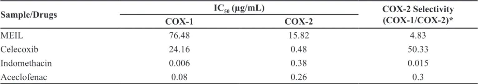
COX-2 selectively without afecting COX-1. In the present study, the methanolic extract of I. linnaei was screened for
its inhibitory eicacy against COX-1 and COX-2.
The in vitro cyclooxygenase inhibitory study
demonstrated that the plant extract had good and selective inhibitory activity against 2 as compared to
COX-1. The results obtained revealed that MEIL selectively
inhibited COX-2 catalysed prostaglandin biosynthesis
with an IC50 value of 15.82 μg/mL, compared with COX-1 derived prostaglandin synthesis which showed an IC50
value of 76.48 μg/mL. The resulting COX-2 selectivity was 4.83 (Table II). A comparison was also made with
COX-1/COX-2 selectivity ratio of some traditional NSAIDs (Indomethacin and aceclofenac) and Celecoxib, a synthetic selective COX-2 inhibitor, obtained in the same assay
system. Indomethacin and aceclofenac non-selectively
inhibited COX-1/2 ratio. The percentage inhibition produced by MEIL against COX-1 ranged between 12.6 and 65.4, whereas it was 30.17 to 94.47 for COX-2. Maximum enzyme inhibitory efect was observed at 100 μg/mL for
both COX-1 and COX-2.The extract inhibits both isoforms
TABLE I - Nitric oxide and 5-lipoxygenase Inhibitory activity of MEIL
Sample Concentration (µg/mL)
NO Inhibitory Activity 5-lipoxygenase Inhibitory Activity Enzyme Inhibition
(%)
IC50 (µg/mL)
Enzyme Inhibition (%)
IC50 (µg/mL)
MEIL
10 20.73 ± 0.75
60.67
19.9 ± 0.96
59.99
20 26.3 ± 0.9 24.8 ± 1.16
40 36.83 ± 1.57 39.27 ± 1.7
60 54.87 ± 3.48 61.87 ± 1.36
80 77.7 ± 2.14 91.87 ± 2.62
100 95.7 ± 1.55 97.6 ± 1.76
Aceclofenac
10 20.1 ± 1.38
79.43
21 ± 1.13
45.97
20 24.56 ± 4.15 32.4 ± 1.53
40 37.9 ± 2.23 49.83 ± 1.45
60 60.93 ± 2.04 73.4 ± 1.69
80 84.15 ± 1.49 92.36± 2.75
100 97.03 ± 1.43 97.8 ± 1.6
of cyclooxygenase in concentration dependent manner. At the same time, COX-2 selectivity ratio is more than the
standard NSAIDs such as indomethacin and aceclofenac.
The results obtained indicate that the extract is having selective COX-2 inhibition.
In the present study, the anti-inflammatory and
anti-nociceptive activities of the methanolic extract of
Indigofera linnaei (MEIL) has been established in both acute and chronic inflammation models as employed. Carrageenan induced rat paw edema is a suitable test for
evaluating anti-inlammatory drugs which has been used
to assess the anti-edematous effect of natural products (Panthong et al., 2003). The development of edema in the paw of the rat after injection of carrageenan is believed to be biphasic event. The initial phase observed during the first hour is attributed to the release of histamine and serotonin; the second phase is due to the release of prostaglandin, protease and lysosome (Paviaya et al., 2013). Based on this, it could be hypothesized that the
suppression of the irst phase may be due to inhibition
of the release of early mediators, such as histamine and serotonin, and the action in the second phase may be
explained by an inhibition of cyclooxygenase (Gobianand
et al., 2010). Previous studies suggest that the injection of carrageenan into the rat paw induces the liberation of bradykinin, which later induces the biosynthesis of prostaglandins and other autocoids, which are responsible
for the formation of the inlammatory exudates. Besides,
in the carrageenan induced rat paw edema model, the production of prostanoids has been through the serum
expression of COX-2 by a positive feedback mechanism
(Ueno et al., 2000).
Sub-plantar injection of carrageenan in rats showed to a time-dependent increase in paw volume (Table III),
which was observed at 1 h and maximum swelling at 5 h
after administration of carrageenan injection in the control
group. However, carrageenan induced inflammation
was significantly (p < 0.001) reduced in all phases of
the experiment by the treatment with MEIL 200 and 400 mg/kg, as well as aceclofenac. No signiicant diference
in reduction was observed by MEIL when compared to the standard drug aceclofenac, which indicates the equipotent
nature of the extract. Maximum inhibition was observed at
the dose of 400 mg/kg. The results reveal that the methanol
extract of I. linnaei had a significant anti-inflammatory
effect on the tested dose levels. The extract at a dose of 400 mg/kg, had higher anti-inlammatory potential than aceclofenac (NSAID), a cyclooxygenase inhibitor, while
at the dose of 200 mg/kg the activity was similar to that of aceclofenac. Therefore, the results obtained suggest that the mechanism of action of MEIL may be related to the inhibition of prostaglandin biosynthesis.
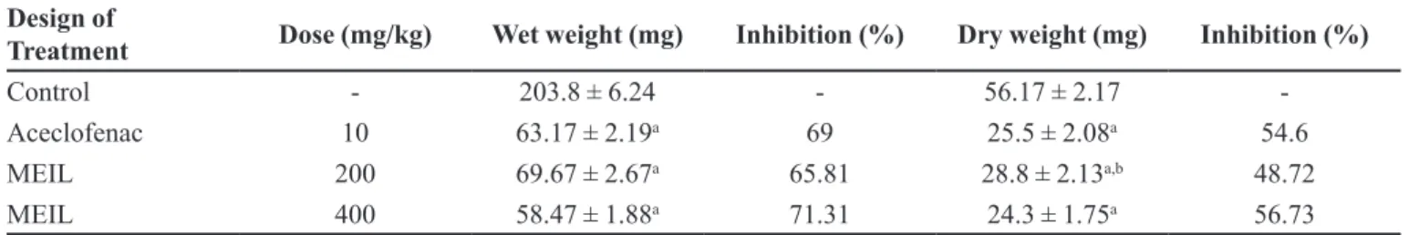
The cotton pellet granuloma method has been
widely employed to assess the transductive, exudative
TABLE II - Comparison of IC50 values of various COX inhibitors with MEIL
Sample/Drugs IC50 (µg/mL) COX-2 Selectivity (COX-1/COX-2)*
COX-1 COX-2
MEIL 76.48 15.82 4.83
Celecoxib 24.16 0.48 50.33
Indomethacin 0.006 0.38 0.015
Aceclofenac 0.08 0.26 0.3
*Ratio of the IC50 values for COX-1 and COX-2 can be used as an indication of the COX-2 selectivity of inhibitors. A COX-1/
COX-2 ratio of more than 1 indicates preferential COX-2 selectivity.
TABLE III - Efect of MEIL on Carrageenan Induced Paw Edema in Rats
Design of Treatment Dose (mg/kg) Paw Volume (mL)
1 h 3 h 5 h
Control - 0.37 ± 0.01 0.52 ± 0.03 0.95 ± 0.03
Aceclofenac 10 0.24±0.01 (35.13) 0.30 ± 0.02a (42.3) 0.28 ± 0.01a (70.52)
MEIL 200 0.27 ± 0.01 (27.02) 0.35 ± 0.01a (32.69) 0.31 ± 0.02a (67.37)
MEIL 400 0.26± 0.012 (29.73) 0.31 ± 0.02a (40.38) 0.25 ± 0.06a (73.68)
n=6; Data were expressed as Mean ± SEM; ap < 0.001 vs Control; Data were analyzed by One way ANOVA followed by Tukey
and proliferative components of chronic inlammation is a typical feature of established chronic inlammatory reaction. The luid absorbed by the pellet greatly inluences
the wet weight of the granuloma and dry weight correlates well with the granuloma of the granulomatous tissue formed (Joseph et al., 2010). In the present investigation,
the methanol extract of I. linnaei registered profound
anti-inlammatory activity against cotton pellet granuloma in rats (Table IV). The extract exhibited a significant (p < 0.001) anti-inlammatory activity and the results were
comparable with that of standard drug aceclofenac. The
extract at the dose of 400 mg/kg showed the maximum
granuloma inhibition, which is equal to aceclofenac. In the writhing test, high levels of prostaglandins PGE2α and PGF2α was observed during the irst 30 min after acetic acid injection. Nevertheless, it was found that the intraperitoneal administration of acetic acid induces not only the liberation of prostaglandins, but also of the release of pro-inflammatory cytokines, such as tumor necrosis factor-a (TNF-α), interleukine-1b (IL-1b) and IL-8 from resident peritoneal macrophages and mast cells (Mothana, 2011). The oral administration of MEIL
induced the analgesic activity signiicantly and the results
are presented in Table V. The treatment of animals with MEIL at the dose of 200 and 400 mg/kg, produced a
signiicant (p < 0.001) and dose dependent inhibition in
abdominal writhes produced by acetic acid. The inhibition by MEIL at the dose of 400 mg/kg was nearly similar
to that produced by the standard drug aspirin. Thus, the results obtained for the writhing test using acetic acid are similar to those obtained for the edematogenic test
using carrageenan, since MEIL efectively inhibited the
writhings in mice in dose dependent manner. The results were comparable with the group treated with standard
drug aspirin, which indicate that the antinociceptive efect
of I. linnaei might be attributed to the inhibition of the
synthesis of some pro-inlammatory mediators such as
prostaglandins and cytokines.
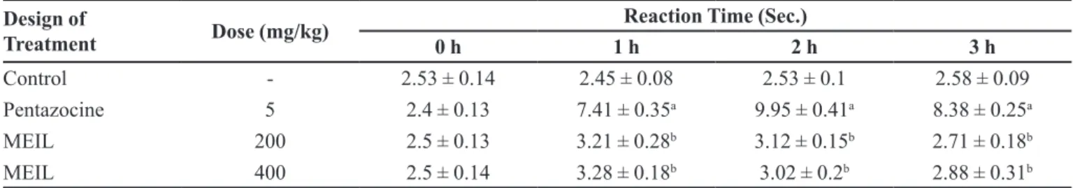
The hot plate test is a classical approach widely applied in the analgesic investigations for several decades. The hot plate test when associated with the writhing test, can usually distinguish central from peripheral effects (Srinivasan et al., 2003). Some
analgesic drugs such as aspirin usually have few efects
in the hot plate test indicating their peripheral analgesic activities. But some other analgesic drugs such as ibuprofen and morphine can decrease prostaglandin
synthesis via central inhibition of cyclooxygenase (Zhu
et al., 2011) or bind to speciic opioid receptors in the
central nervous system, exhibiting both their peripheral and central analgesic activities. However, the hot plate
test was undertaken to verify if MEIL would have any
central analgesic efect and the results are presented in
Table VI. In this analgesic testing model, pentazocine at
a dose of 5 mg/kg, signiicantly prolonged the reaction time of animals with relatively extended duration of
TABLE IV - Efect of MEIL on Cotton Pellet Granuloma in Rats
Design of
Treatment Dose (mg/kg) Wet weight (mg) Inhibition (%) Dry weight (mg) Inhibition (%)
Control - 203.8 ± 6.24 - 56.17 ± 2.17
-Aceclofenac 10 63.17 ± 2.19a 69 25.5 ± 2.08a 54.6
MEIL 200 69.67 ± 2.67a 65.81 28.8 ± 2.13a,b 48.72
MEIL 400 58.47 ± 1.88a 71.31 24.3 ± 1.75a 56.73
n=6; Data were expressed as Mean ± SEM; ap < 0.001vs Control; bp < 0.01 vs Aceclofenac; Data were analyzed by One way ANOVA
followed by Tukey Kramer Multiple Comparison test.
TABLE V - Efect of MEILon Chemical Stimulus Induced Pain in Mice
Design of Treatment Dose (mg/kg) No. ofwrithings Inhibition (%)
Control - 105.66 ± 3.20
-Aspirin 300 49.67 ± 2.99a 53
MEIL 200 66.83 ± 2.23a,b 36.75
MEIL 400 50 ± 2.15a 52.68
n=6; Data were expressed as Mean ± SEM; ap < 0.001vs Control; bp < 0.001 vs Aspirin; Data were analyzed by One way ANOVA
stimulation, conirming centrally mediated activity. The results obtained from the extract treated groups revealed no signiicant activity when compared to control group and a highly signiicant diference was observed with the group treated with pentazocin (5 mg/kg). Hence, it is assumed that MEIL has no signiicant analgesic efect
on the central nervous system.
Recently our biological studies on methanol
extract of I.linnaei reported potent antioxidant and free
radical scavenging activities as well as a good anticancer activity (Kumar et al., 2011a; Kumar et al., 2011b). Many
Indigofera species such as I. colutea, I. macrocalyx, I. nigritiana, I. puchra, I. tinctoria, I. trita and I. cassioides
exhibited potent anti-inflammatory, analgesic and lipoxygenase inhibitory activities (Bakaso et al., 2013; Kumar et al., 2009; Kumar et al., 2013). Reports of the previous studies and the traditional use of this medicinal
plant encouraged us to extend our evaluation using
several in vitro and in vivo models of inlammation and
nociception in rodent models. It is important to point
out that this work represents the irst report on the anti-inlammatory and antinociceptive activities of I. linnaei.
Reactive Oxygen Species (ROS) and Reactive
Nitrogen Species (RNS) participate in normal cell function as well as in pathological disorders such as inflammation. Several studies have demonstrated the
participation of ROS and RNS in inlammation models.
Preliminary phytochemical and quantitative analysis of
MEIL indicated the presence of rich amount of lavonoids
and phenolic compounds in MEIL (Kumar et al., 2011a). Anti-inflammatory and antinociceptive activities as
well as antioxidant potential of many plants have been attributed to their lavonoids (Garcia-Mediavilla et al.,
2007; Gonzalez et al., 2011; Shariifar et al., 2009) or to their high triterpene (Ammon, 2006; Mothana et al.,
2011). Flavonoids are known as important secondary plant products in many medicinal plants. They have attracted much attention in relation to their wide range
of activities in the prevention of cancer, inlammation
and coronary heart disorders (Garcia-Mediavilla et al.,
2007). Previous reports suggest that these compounds may inhibit several enzymes, which are activated in certain inflammatory conditions. Prostaglandins
and nitric oxide (NO) biosynthesis is involved in inlammation. Moreover, inducible nitric oxide synthase (iNOS), cyclooxygenase (COX-2) and lipooxygenase
are responsible for the production of large amounts of
these pro-inlammatory mediators (Garcia-Mediavilla
et al., 2007). Diferent studies conirm that lavonoids
like quercetin and kaempferol significantly decrease
the levels of iNOS, COX-2, lipoxygenase and reactive
C-protein (RCP) in a concentration dependent manner (Morales et al., 2006; Tunon et al., 2009). Furthermore, some investigated flavonoids such as kaempferol, quercetin and daidzein also inhibit the activation of the signal transducer and activator of transcription 1 (STAT-1), another important transcription factor for
iNOS. Luteolin 7-glucoside, kaempferol and quercetin
effectively inhibit lipopolysaccharide (LPS)-induced PGE2 production. Four lavonoids signiicantly inhibit
LPS-induced COX-2 expression, while mPGES-1 expression was downregulated by kaempferol and isorhamnetin (Hamalainen et al., 2011). The observed
beneficial effects of methanol extract of I. linnaei on
inlammatory and nociceptive responses in rodents may
be due to the presence of these phytochemicals.
CONCLUSION
All the data obtained in the present work demonstrate
the possible development of methanol extract of Indigofera linnaei as a novel and potential drug in the management
of inlammation and pain, which are probably mediated
via inhibition of various autacoids formation and release. Further detailed investigation is underway to determine
the exact phytoconstituents that are responsible for these
activities.
TABLE VI - Efect of methanolic extract of Indigofera linnae on Thermal Stimulus Induced Pain in Mice
Design of
Treatment Dose (mg/kg)
Reaction Time (Sec.)
0 h 1 h 2 h 3 h
Control - 2.53 ± 0.14 2.45 ± 0.08 2.53 ± 0.1 2.58 ± 0.09
Pentazocine 5 2.4 ± 0.13 7.41 ± 0.35a 9.95 ± 0.41a 8.38 ± 0.25a
MEIL 200 2.5 ± 0.13 3.21 ± 0.28b 3.12 ± 0.15b 2.71 ± 0.18b
MEIL 400 2.5 ± 0.14 3.28 ± 0.18b 3.02 ± 0.2b 2.88 ± 0.31b
n=6; Data were expressed as Mean ± SEM; ap < 0.001 vs Control; bp < 0.001 vs Pentazocine; Data were analyzed by One way
ACKNOWLEDGEMENT
We thank the Management and Principal of Swamy Vivekanandha College of Pharmacy, for rendering support
to carry out this work. We have no inancial or personal conlictsof interest related to this work.
REFERENCES
AMIRA, S.; DADE, M.; SCHINELLA, G.; JOSE-LUIS, R. Anti-inlammatory, anti-oxidant and apoptotic activities of
four plant species used in folk medicine in the Mediterranean basin. Pak. J. Pharm. Sci., v.25, n.1, p.65-72, 2012.
AMMON, H.P. Boswellic acids in chronic inflammatory
diseases. Planta. Med., v.72, n.12, p.1100-1116, 2006.
BAKASSO, S.; LAMIEN-MEDA, A.; LAMIEN, C.E.; KIENDERBEOGO, M.; COULIBALY, A.Y.; COMPAORE, M.; MEDA, N.R.; NACOULMA, O.G. In vitro inhibition
of acetylcholinesterase, lipoxygenase, xanthine oxidase and antibacterial activities of ive Indigofera (Fabaceae)
aqueous acetone extracts from Burkina Faso. Curr. Res. J. Biol. Sci., v.5, n.3, p.115-122, 2013.
BEPARY, S.; BIPLAB, K.D.; SITESH, C.B.; JOYDEV, K.K.; ABU, S.S.R.; BIDYUT, K.D. Anti-inlammatory activity
of indanyltetrazole derivatives. Pak. J. Pharm. Sci., v.21, n.3, p.295-298, 2008.
DHARMASIRI, M.G.; JAYAKODY, J.R.A.C.; GALHENA, G.; LIYANAGE, S.S.P.; RATNASOORIYA, W.D.
Anti-inflammatory and analgesic activities of mature fresh leaves of Vitex negundo. J. Ethnopharmacol., v.87, n.2/3, p.199-206, 2003.
GACCHE, R.; SHAIKH, R.; PUND, M.; DESHMUKH, R. Cyclooxygenase inhibitory, cytotoxicity and free radical
scavenging activities of selected medicinal plants used in Indian traditional medicine. Phcog. J., v.3, n.19, p.57-64, 2011.
GARCIA-MEDIAVILLA, V.; CRESPO, I.; COLLADO, P.S.; ESTELLER, A.; SANCHEZ-CAMPOS, S.; TUNON, M.J.; GONZALEZ-GALLEGO, J. The anti-inlammatory
flavones quercetin and kaempferol cause inhibition of
inducible nitric oxide synthase, cyclooxygenase-2 and
reactive C-protein, and down regulation of the nuclear factor
κB pathway in Chang Liver cells. Eur. J. Pharmacol., v.557, n.2/3, p.221-229, 2007.
GATELY, S. The contributions of cyclooxygenase-2 to tumor
angiogenesis. Cancer Metastasis Rev., v.19, n.1/2, p.19-27, 2000.
GOBIANAND, K.; VIVEKANANDAN, P.; PRADEEP, K.; MOHAN, C.V.R.; KARTHIKEYAN, S. Anti-inlammatory
and antipyretic activities of Indian medicinal plant Cassia
istula Linn. (Golden Shower) in Wistar albino rats. Int. J. Pharmacol., v.6, n.6, p.719-725, 2010.
GONZALEZ, R.; BALLESTER, I.; LOPEZ-POSADAS, R.; SUAREZ, M.D.; ZARZUELO, A.; MARTINEZ-AUGUSTIN, O.; DE MEDINA, F. Efects of lavonoids and other polyphenols on inlammation. Crit. Rev. Food Sci. Nutr., v.51, n.4, p.331-362, 2011.
HAMALAINEN, M.; NIEMINEN, R.; ASMAWI, M.Z.; VUORELA, P.; VAPAATALO, H.; MOILANEN, E. Efects of lavonoids on prostaglandin E2 production and on COX-2 and mPGES-1 expressions in activated macrophages. Planta Med., v.77, n.13, p.1504-1511, 2011.
HEMALATHA, S.; SUBRAMANIAN, N.; RAVICHANDRAN, V.; CHINNASWAMY, K. Wound healing activity of Indigofera enneaphylla Linn. Indian J. Pharm. Sci., v.63, n.4, p.331-333, 2001.
HU, X.; JIN, H.; XU, W.; ZHANG, W.; LIU, X.; YAN, S.; CHEN, M.; LI, J.; ZHANG, W. Anti-inlammatory and analgesic efect of Daphne retusa Hemsl. J. Ethnopharmacol., v.120, n.1, p.118-122, 2008.
JOSEPH, J.M.; SOWNDHARARAJAN, K.; MANIAN, S. Evaluation of analgesic and anti-inlammatory potential of Hedyotis puberula (G. Don) R. Br. ex Arn. in experimental animal models. Food Chem. Toxicol., v.48, n.7, p.1876-1880, 2010.
JOTHIMANIVANNAN, C.; KUMAR, R.S.; SUBRAMANIAN, N. Anti-inlammatory and analgesic activities of ethanol extract of aerial parts of Justicia gendarussa Burm. Int. J. Pharmacol., v.6, n.3, p.278-283, 2010.
KOMATOU, G.P.P.; VILJOEN, A.M.; STEENKAMP, P. Antioxidant, anti-inflammatory activities and HPLC
analysis of South African Salvia species. Food Chem., v.119, n.2, p.684-688, 2010.
KUMAR, R.S.; RAJKAPOOR, B.; PERUMAL, P. Anti-inlammatory and analgesic activities of ethanol extract of Indigofera trita Linn. PharmacologyOnline, v.1, p.278-289, 2009.
KUMAR, R.S.; RAJKAPOOR, B.; PERUMAL, P. Anti-inlammatory and anti-nociceptive activities of methanolic leaf extract of Indigofera cassioides Rottl. Ex. DC. J. Acute Dis., v.2, n.4, p.322-326, 2013.
KUMAR, R.S.; RAJKAPOOR, B.; PERUMAL, P. Antioxidant
potential of Indigofera linnaei Ali: an in vitro study. PharmacologyOnline, v.1, p.710-720, 2011a.
LEE, J.; KIM, K.A.; JEONG, S.; LEE, S.G.; PARK, H.J.; KIM, N.J.; LIM, S. Anti-inlammatory, nociceptive, and anti-psychiatric efects by the rhizomes of Alpinia oicinarum
on complete Freund’s adjuvant induced arthritis in rats. J. Ethnopharmacol., v.126, n.2, p.258-264, 2009.
MAJAK, W.; BENN, M.; MCEWAN, D.; PASS, M.A. Three nitropropanoyl esters of glucose from Indigofera linnaei. Phytochemistry, v.31, n.7, p.2393-2395, 1992.
MORALES, A.I.; VICENTE-SANCHEZ, C.; JERKIC, M.; SANTIAGO, J.M.; SANCHEZ-GONZALEZ, P.D.; PEREZ-BARRIOCANAL, F.; LOPEZ-NOVOA, J.M. Efect of quercetin on metallothionein, nitric oxide synthase and cyclooxygenase-2 expression on experimental chronic cadmium nephrotoxicity in rats. Toxicol. Appl. Pharmacol., v.210, n.1/2, p.128–135, 2006.
MORO, C.; IRENE, P.; MIGUEL, L.; MATILDE, D.A.; EVA, G.; ANA, V.; JOSE, A.M.; GARCIA-LAFUENTE, A. Antiinflammatory activity of methanolic extracts
from edible mushrooms in LPS activated RAW 264.7 macrophages. Food Chem., v.130, n.2, p.350-355, 2012.
MOTHANA, R.A. Anti-inflammatory, antinociceptive and antioxidant actitivities of the endemic Soqotraen Boswellia elongata Balf. f. and Jatropha unicostata Balf. f. in diferent experimental models. Food Chem. Toxicol., v.49, n.10, p.2594-2599, 2011.
MOTHANA, R.A.A.; HASSON, S.S.; SCHULTZE, W.; MOWITZ, A.; LINDEQUIST, U. Phytochemical
composition and in vitro antimicrobial and antioxidant activities of essential oils of three endemic Soqotraen Boswellia species. Food Chem., v.126, n.3, p.1149-1154, 2011.
NIKAJOO, L.T. Analgesic activity of aqueous and alcohol root extracts of Pergularia daemia (forsk.) chiov. Int. J. Pharm. Pharm. Sci., v.1, suppl.1, p.33-37, 2009.
PANTHONG, A.; KANJANAPOTHI, D.; TAESOTIKUL, T.; WONGCOME, T.; REUTRAKUL, V. Anti-inlammatory
and antipyretic properties of Clerodendrum petasites S. Moore. J. Ethnopharmacol., v.85, n.1, p.151-156, 2003.
PAV I AYA , U . S . ; K U M A R , P. ; WA N J A R I , M . M . ;
THENMOZHI, S.; BALAKRISHNAN, B.R. Analgesic and anti-inlammatory activity of root bark of Grewia asiatica Linn. in rodents. Ancient Sci. Life., v.32, n.3, p.150-155, 2013.
P U R N I M A , A . ; K O T I , B . C . ; T I K A R E , V. P. ; VISWANATHASWAMY, A.; THIPPESWAMY, A.H.;
DABADI, P. Evaluation of analgesic and antipyretic activities of Centratherum anthelminticum(L) kuntze seed. Indian J. Pharm. Sci., v.71, n.4, p.461-464, 2009.
RAJENDRAPRASAD, Y.; CHAKRADHAR, V. A new isolavone from Indigofera linnaei. Indian J. Chem., v.43B, n.8, p.1807-1808, 2004.
SHAH, S.M.A.; ASHRAF, M.; AHMED, I.; ARSHAND, S.; YAR, M.; LATIF, A. Anti-lipoxygenase activity of some
indigenous medicinal plants. J. Med. Plants Res., v.7, n.6, p.219-222, 2013.
SHARIFIFAR, F.; DEHGHN-NUDEH, G.; MIRTAJALDINI, M. Major flavonoids with antioxidant activity from Teucrium polium L. Food Chem., v.112, n.4, p.885–888, 2009.
SRINIVASAN, K.; MURUGANANDAN, S.; LAL, J.;
CHANDRA, S.; TANDAN, S.K.; RAVIPRAKASH, V.;
KUMAR, D. Antinoniceptive and antipyretic activities of Pongamia pinnata leaves. Phytother. Res., v.17, n.3, p.259–264, 2003.
TUNON, M.J.; GARCIA-MEDIAVILLA, M.V.; SANCHEZ-CAMPOS, S.; GONZALEZ-GALLEGO, J. Potential of
flavonoids as anti-inflammatory agents: modulation of
pro-inlammatory gene expression and signal transduction
pathways. Curr. Drug Metab., v.10, n.3, p.256-271, 2009.
UENO, A.; NARABA, H.; IKEDA, Y.; USHIKUBI, F.; MURATA, T.; NARUMIYA, S.; OH-ISHI, S. Intrinsic prostacyclin contributes to exudation induced by bradykinin
or Carrageenan: a study on the paw edema-induced in
IP-receptor- deicient mice. Life Sci., v.66, n.12, p.155-160, 2000.
VIJAYALAKSHMI, A.; RAVICHANDIRAN, V.; VELRAJ, M.; HEMALATHA, S.; SUDHARANI, G.; JAYAKUMARI, S. Anti-anaphylactic and anti-inlammatory activities of a
bioactive alkaloid from the root bark of Plumeria acutifolia Poir. Asian Pac. J. Trop. Biomed., v.1, n.5, p.401-405, 2011.
WANG, B.S.; HUANG, G.J.; LU, Y.H.; CHANG, L.W. Anti-inlammatory efects of an aqueous extract of Welsh onion
green leaves in mice. Food Chem., v.138, n.2/3, p.751-756, 2013.
WEALTH of India: a dictionary of Indian raw materials and
industrial products. New Delhi: CSIR, 1992. v.5, p.178-179.
YONATHAN, M.; ASRES, K.; ASSEFA, A.; BUCAR, F. In vivo anti-inflammatory and anti-nociceptive activities of Cheilanthes farinose. J. Ethnopharmacol., v.108, n.3, p.462-470, 2006.
ZHU, Z.Z.; MA, K.J.; RAN, X.; ZHANG, H.; ZHENG, C.J.; HAN, T.; ZHANG, Q.Y.; QIN, L.P. Analgesic, anti-inlammatory and antipyretic activities of the petroleum ether fraction from the ethanol extract of Desmodium podocarpum. J. Ethnopharmacol., v.133, n.3, p.1126-1131, 2011.
Received for publication on 8th August 2015