Licence Creative Commom
CC
RBCDH
1 Universidade Estadual Paulista “Júlio de Mesquita Filho”. Rio Claro, SP. Brasil.
2 Faculdades de Dracena. De-partamento de Educação Física. Dracena, SP, Brasil.
3 Universidade Estadual Paulista. Departamento de Fisioterapia, Presidente Prudente, SP. Brasil.
4 Universidade de Marília. Labora-tório de Avaliação Física e Prática Esportiva. Marília, SP. Brasil.
5 Universidade Cruzeiro do Sul. Instituto de Ciências da Atividade Física e Esporte. Departamento de Educação Física. São Paulo, SP. Brasil.
Received: 10 October 2013 Accepted: 9 January 2014
Gait stability in diabetic peripheral
neuropathy
Estabilidade da marcha na neuropatia diabética
periférica
Ana Claudia de Souza Fortaleza1,2
Eliane Ferrari Chagas3
Dalva Minonroze Albuquerque Ferreira3
Alessandra Madia Mantovani1
Eduardo Federighi Baisi Chagas4
José Ângelo Barela5
Cristina Elena Prado Teles Fregonesi3
Abstract– he aim of this study was to evaluate gait stability in diabetic patients with peripheral neuropathy in three conditions: habitual walking with eyes open, walking with eyes closed, and walking with eyes open and narrow base of support. he study included 41 subjects, 18 with neuropathy (NG) and 23 controls. Gait stability was evaluated on a baropodometer using the Footwalk Pro sotware. he following data were obtained: gait speed and percentage of time spent in double stance and single stance. Signiicant dif-ferences were observed between groups in all three conditions for gait speed and single stance time, which were reduced in NG (p<0.05), and for double stance time, which was increased in NG (p<0.05). For gait speed, double stance time and single stance time, the eyes open condition difered from the eyes closed (p<0.001) and narrow base of support (p<0.001) conditions. In the three conditions studied, patients of NG presented a deicit in gait stability and this performance was even more compromised in the two conditions that required greater postural control. hese gait changes resulting from the complex-ity imposed by the diferent conditions suggest the inclusion of these conditions in the evaluation and treatment of this population.
Key words: Balance; Diabetic neuropathies; Gait; Physiotherapy.
Gait stability in diabetic neuropathy Fortaleza et al.
INTRODUCTION
he number of people with diabetes has increased as a result of factors such as population growth and aging, urbanization, obesity, and physical inactivity1. In Brazil, the number of people with diabetes is estimated to
increase from 7,633,000 in 2010 to 12,708,000 in 2030, with Brazil becom-ing the country with the ith largest population of diabetics in the world2.
One of the complications caused by diabetes mellitus is diabetic pe-ripheral neuropathy (DPN)3, a condition characterized by sensory4 and
motor5 alterations that can culminate in gait impairment6.
During walking, patients with a certain disease may develop changes in neuromotor control that can impair the maintenance of gait stability7.
he sensory component, particularly the proprioceptive system7, is
important for the maintenance of gait stability. his component conducts aferents to the central nervous system so that this system, together with other information for postural control, can make the necessary adjustments to maintain gait stability8. hese adjustments also depend on visual and
vestibular information, which permits to obtain information from the body and environment and relationship between these two9.
In this respect, gait performance in diabetic neuropathic patients has been a matter of concern and studies have evaluated diferent variables such as speed10, variation in gait cycle time10, step time variability11, and
double and single stance time12.
In addition, falls are common among diabetic neuropathic patients and generally occur during walking13. It is therefore important to study
gait characteristics and stability under diferent conditions.
Few studies have investigated gait behavior during more complex activities, such as walking on irregular surfaces, in an attempt to detect clinically relevant deiciencies11,14-16. However, there are still gaps that involve
visual or base of support variations, demonstrating greater requirement of the neuromuscular control system, particularly the proprioceptive system. It is therefore necessary to evaluate gait stability in patients with neuropa-thy in diferent conditions; for example, walking with the eyes closed in order to determine the capacity of these individuals to compensate the absence of vision through more efective participation of the proprioceptive system, and a narrow base of support which requires greater neuromuscular control.
he objective of the present study was to evaluate gait stability in sub-jects with DPN in three diferent conditions: habitual walking with eyes open, walking with eyes closed, and walking with eyes open and narrow base of support.
METHODOLOGICAL PROCEDURES
by the local Ethics Committee (Protocol No. 30/2010). he participants received detailed information about the procedures and objectives of the study and agreed to participate by signing a free informed consent form.
Sample
Forty-one subjects of both genders were divided into two groups: neuropa-thy group consisting of patients with type 2 diabetes mellitus and DPN (n=18), and a control group consisting of healthy non-diabetic subjects (n=23). Patients of the neuropathy group were recruited from the University Extension Project “Diabetic Foot Program” (Projeto de Extensão Univer-sitária “Programa Pé Diabético”) of FCT/UNESP, Presidente Prudente.
Procedures
Anthropometric data (body weight, height, and body mass index) were collected from all participants. Postprandial blood glucose was measured to conirm the diagnosis of diabetes in the neuropathy group and to exclude possible asymptomatic diabetic patients in the control group.
he diagnosis of DPN was made by somatosensory evaluation using Semmes-Weinstein monoilaments (SorriBauru®, Bauru, Brazil). he mono-ilaments were applied to the plantar and dorsal surfaces of the feet, which correspond to the sensitive dermatomes of the anterior tibial and common ibular nerves, bilaterally. he test was performed with the subject in dorsal decubitus and wearing a blindfold. he examiner exerted pressure of the monoilament on the skin until it bended, permitting standardization of the pressure exerted. he subject was asked to always report when he/she felt the touch. he test is deined as positive in the presence of insensitivity to the 10-g monoilament17.
he Michigan Neuropathy Screening Instrument was used for conir-mation of the diagnosis of DPN. he instrument consists of a questionnaire and physical assessment of the feet. he score ranges from 0 to 23, with a score ≥ 8 indicating the presence of neuropathy18.
Criteria for exclusion from the two groups were osteoarticular deformi-ties; plantar ulcers; amputation of regions of the foot; assisted walking; claudication; neurological disease of central origin or other peripheral diseases; inability to understand the tests; uncorrected visual impairment, and presence of some symptom detected by a dizziness questionnaire.
Evaluation of gait stability
Gait stability was analyzed with a baropodometer (FootWalk Pro, AM
CUBE, France; sampling rate of 200 Hz) consisting of a 2-m pressure
Gait stability in diabetic neuropathy Fortaleza et al.
he subjects walked on the walkway at a comfortable and self-selected speed in three conditions: habitual walking with eyes open (EO); walk-ing with eyes closed (EC), and walkwalk-ing with eyes open and narrow base of support (NB). For assessment of the EC condition, the subjects walked only in the working area of the baropodometer and were asked to close the eyes, walk the 2-m distance, open the eyes, turn, and walk back the same distance again with the eyes closed. For evaluation of the NB condition, the subjects also walked only in the working area of the baropodometer within two parallel lines marked on the surface of the walkway separated at a distance of 21 cm. hus, the maximum width of the base of support was reduced to this value.
For all conditions, the subjects walked once before the recording was started: in the irst condition, to minimize alterations due to the lack of adaptation to the equipment, and in the second and third conditions so that the subjects would understand the test. Six gait cycles were recorded automatically by the platform for each condition.
he following variables were calculated for the two lower limbs: gait speed, double stance time and single stance time corresponding to three gait cycles.
Double stance and single stance
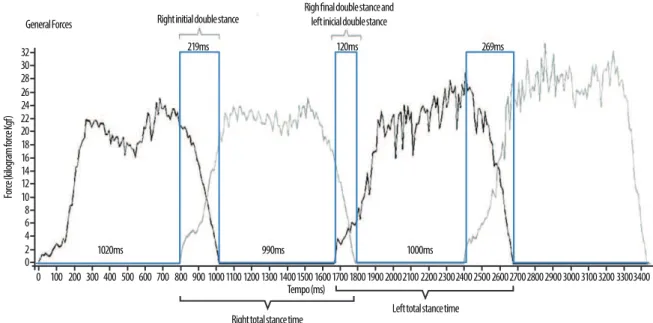
he stance time variables (in milliseconds) were extracted from the graph generated by the sotware, separately for the right and let foot (Figure 1).
Figure 1. Schematic representation of the image generated by the FootWorkPro software, version 3.2.0.1, used for the analysis of stance time.
Total stance time and initial and inal double stance time were col-lected. Single stance time, which was not provided by the sotware, was calculated by subtracting the double stance times from total stance time using the following equation (Equation 1):
Gait speed
Gait speed was calculated by dividing the step length by the cycle time, separately for each limb, and the mean of this value was then calculated (Equation 2):
Speed (m/s) = Step length
Equation 2
Cycle time
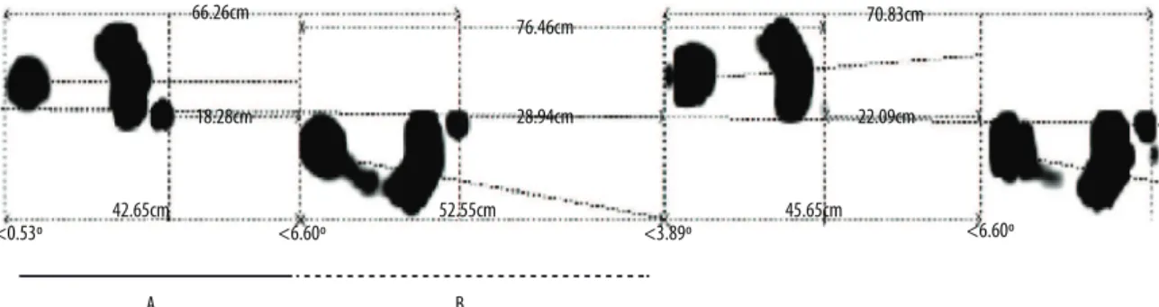
Step length was calculated as the sum of the length of two consecutive steps, expressed in centimeters, and these values were transformed into meters (Figure 2; Equation 3):
Figure 2. Schematic representation of the image generated by the FootWork Pro software, version 3.2.0.1, used for the calculation of step length.
Length of the left step = A+B Equation 3
Gait cycle time was obtained by the sum of total stance times of the right and let foot and subtracting double stance times, expressed in mil-liseconds, and was then transformed into seconds.
Statistical analysis
Descriptive statistics (mean and standard deviation) was used for charac-terization of the sample. he variables met the assumptions for normality (Kolmogorov-Smirnov) and sphericity (Mauchly’s). he Student t-test for independent samples was applied to the separate analysis of diferences in the quantitative variables between groups. Repeated-measures ANOVA was used to determine the relationship between group and gait condition for the dependent variables (gait speed, percentage of time spent in double and single stance), followed by the post hoc Fisher least signiicant difer-ence (LSD) test to localize diferdifer-ences.
he data obtained for the two lower limbs were compared and no sig-niicant diference was observed. herefore, since DPN is a symmetrical disease19, the data obtained for all variables were analyzed together by
calculating the mean of the right and let limb.
Gait stability in diabetic neuropathy Fortaleza et al.
RESULTS
Table 1 shows the characteristics of the sample. Diabetic peripheral neu-ropathy was diagnosed based on insensitivity to a 10-g monoilament and on a score ≥ 8 in the Michigan Neuropathy Screening Instrument.
Table 2 shows the gait variables obtained for the two groups in the diferent conditions.
Table 1. Characteristics of the sample (n=41).
Variable CG NG p-value
Age (years) 62.96 ± 5.97 64.33 ± 6.45 0.483
Body weight (kg) 67.73 ± 11.47 76.71 ± 16.37 0.046*
Height (m) 1.59 ± 0.10 1.61 ± 0.08 0.603
BMI (kg/m2) 26.63 ± 3.25 31.51 ± 6.92 0.005*
Glycemia (mg/dL) 124.09 ± 24.99 164.56 ± 43.34 0.001*
Values are the mean ± standard deviation. CG: control group (n=23); NG: neuropathy group (n=18); BMI: body mass index. *p<0.05.
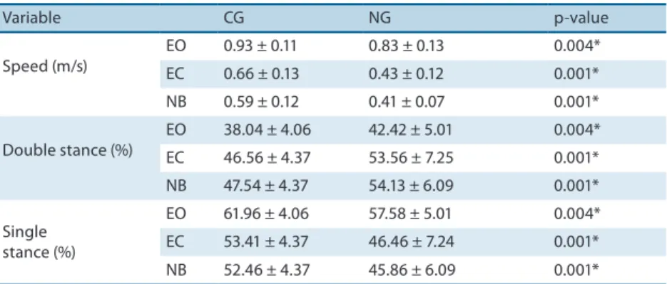
Table 2. Gait variables of speed, percentage of time spent in double and single stance obtained for patients with neuropathy and control subjects in the conditions of eyes open (EO), eyes closed (EC), and narrow base of support (NB).
Variable CG NG p-value
Speed (m/s)
EO 0.93 ± 0.11 0.83 ± 0.13 0.004*
EC 0.66 ± 0.13 0.43 ± 0.12 0.001*
NB 0.59 ± 0.12 0.41 ± 0.07 0.001*
Double stance (%)
EO 38.04 ± 4.06 42.42 ± 5.01 0.004*
EC 46.56 ± 4.37 53.56 ± 7.25 0.001*
NB 47.54 ± 4.37 54.13 ± 6.09 0.001*
Single stance (%)
EO 61.96 ± 4.06 57.58 ± 5.01 0.004*
EC 53.41 ± 4.37 46.46 ± 7.24 0.001*
NB 52.46 ± 4.37 45.86 ± 6.09 0.001*
Values are the mean ± standard deviation. CG: control group (n=23); NG: neuropathy group (n=18). *p<0.05.
Since the gait speed of patients with DPN was lower than that of con-trol subjects, the absolute values of double and single stance times were transformed into percentages, taking total stance as 100%
he Student t-test for independent measures also detected a diference in the percentage of time spent in double stance and single stance between groups (Table 2). Patients with DPN presented a higher percentage of time in double stance and a lower percentage of time in single stance in all conditions.
(p=0.001). he post hoc test indicated an increase in the percentage of time spent in double stance in the EC (p=0.001) and NB (p=0.001) conditions when compared to the EO condition. here was no diference between EC and NB conditions (p=0.899). he opposite was observed for the percentage of time spent in single stance, with a reduction in the EC (p=0.001) and NB (p=0.001) conditions compared to the EO condition. No diference was found between the EC and NB conditions (p=0.898). here was no group x condition interaction for the percentage of time spent in double stance (p=0.291) or single stance (p=0.295).
DISCUSSION
Characterization of the sample showed that neuropathic subjects had a higher BMI as a result of the higher body weight seen in this population12,15.
his inding can probably be explained by the reduced functionality and consequent decrease in mobility of the diabetic population.
Subjects with DPN presented poor gait stability in the three conditions tested. his performance was even more compromised in the two conditions that required greater contribution of the postural control system (EC and NB). Gait speed was reduced in subjects with DPN and in the two groups for the conditions requiring greater postural control. However, an interaction was observed between group and condition, with subjects with DPN being more susceptible to a reduction in gait speed in the EC and NB conditions.
In agreement with the present study, some authors reported a reduc-tion in gait speed in the diabetic populareduc-tion10,21. his reduction is more
pronounced as the degree of diiculty increases, such as when walking on irregular surfaces 10,14, a conditions associated with an increased risk
of falls21. his reduced gait speed may indicate an attempt to promote safe
walking in order to avoid instabilities22. However, there are no studies
in the literature that evaluate gait in other functional situations such as walking with eyes closed and with a narrow base of support as done in the present study. Although some studies have reported a reduction in gait speed in neuropathic patients with a history of falls when walking on an irregular surface under low light compared to those walking on a regular surface under good light conditions16, the authors did not diferentiate
whether the greater diiculty observed was due to the irregular surface, low light, or both.
Gait stability in diabetic neuropathy Fortaleza et al.
and deceleration as used in the EO condition to guarantee the safety of the subjects; however, this does not invalidate the diferences observed since the data were obtained in a common area for the three conditions.
Sacco et al.12 evaluated temporal and dynamic parameters of
self-selected gait and also found shorter single stance time and longer double stance time in patients with DPN. Costa et al.23 attributed this alteration
in stance times to a compensation in order to improve gait stability. he integrity of the postural control, sensory (visual, vestibular and somatosensory) and motor systems is necessary to maintain stability during static and dynamic tasks24. he diferences between the EO and EC
condi-tions may therefore be due to the importance of visual information for the control of stability, providing the spatial information necessary for body readjustment during locomotion. In this respect, both groups presented poorer performance during walking with the eyes closed.
Although both groups performed worse in the conditions that required greater postural control, subjects with DPN presented greater gait instabil-ity in the EC condition, characterized by lower gait speed, longer double stance time, and shorter single stance time. his result can be explained by the reduced sensory feedback25 in neuropathic patients, increasing gait
instability26.
According to Menz et al.15, the alterations in gait stability seen in
neuro-pathic patients are due to the importance of peripheral sensory information for the control of stability during locomotion, with this information exert-ing a predominant efect on vision and muscle strength. As a consequence, in the case of loss of proprioceptive and tactile input, the visual component becomes more necessary for the adjustment of postural control9. his fact
may explain the present inding that gait speed was more compromised in neuropathic subjects in the EC condition. he poor performance of subjects with DPN in the NB condition might be related to the reduction in sensitivity, muscle strength27 and range of motion28 generally observed
in this population.
he greater gait instability in the NB condition observed in the present study might be explained by a possible reduction in neuromuscular control of distal joints. Gomes et al.28, studying peak plantarlexor activity and
ankle range of motion at diferent gait cadences in diabetic neuropathic pa-tients, suggested a reduction in neuromuscular control around distal joints.
treatment and rehabilitation. We also emphasize the importance of inter-vention studies consisting of gait trainings under the conditions used in this study in order to determine the eicacy of this treatment to maintain gait stability in subjects with DPN.
CONCLUSIONS
Subjects with DPN presented greater gait instability than control subjects in the three conditions tested: habitual walking with eyes open; walking with eyes closed, and walking with eyes open and narrow base of support. Greater gait instability was also observed in neuropathic and control sub-jects in the EC and NB conditions when compared to the EO condition. his instability was characterized by a lower gait speed, longer double stance time, and shorter single stance time. On the basis of stance times, the response to the diiculties was similar in the two groups. However, neu-ropathic subjects presented poorer performance in terms of gait speed, i.e., DPN increased the diiculty during walking in the EC and NB conditions. he changes observed in the gait pattern resulting from instability in the diferent gait conditions suggest the use of these conditions for both the evaluation and elaboration of strategies that would detect alterations and support activities designed to improve stability and functionality in this population.
REFERENCES
1. Wild S, Roglic G, Green A, Sicree R, King H. Global prevalence of diabetes. esti-mates for the year 2000 and projections for 2030. Diabetes Care 2004;27 (5):1047-53.
2. Shaw JE, Sicree RA, Zimmet PZ. Global estimates of the prevalence of diabetes for 2010 and 2030. Diabetes Res Clin Pract 2010;87(1):4-14.
3. Bacarin TA, Sacco ICN, Hennig EM. Plantar pressure distribution patterns during gait in diabetic neuropathy patients with a history of foot ulcers. Clinics 2009;64(2):113-20.
4. Rao N, Aruin AS. Auxiliary sensory cues improve automatic postural responses in individuals with diabetic neuropathy. Neurorehabil Neural Repair 2011;25(2):110-117.
5. Van Schie CH. Neuropathy: mobility and quality of life. Diabetes Metab Res Rev 2008;24(1):45-51.
6. Allet L, Armand S, Aminian K, Pataky Z, Golay A, Bie RA et al. An exercise intervention to improve diabetic patients’ gait in a real-life environment. Gait Posture 2010;32(2):185-90.
7. Kuo AD, Donelan JM. Dynamic principles of gait and their clinical implications. Phys her 2010;90(2):157-76.
8. Vaugoyeau M, Viel S, Amblard B, Azulay JP, Assaiante C. Proprioceptive contribu-tion of postural control as assessed from very slow oscillacontribu-tions of the support in healthy humans. Gait Posture 2008;27(2):294-302.
9. Kleiner AFR, Schlittler DXC, Sánches-Ariaz MDR. O papel dos sistemas visual, vestibular, somatosensorial e auditivo para o controle postural. Rev Neurocienc 2011;19(2): 349-57
Gait stability in diabetic neuropathy Fortaleza et al.
Corresponding author
Ana Claudia de Souza Fortaleza Rua Barão do Rio Branco, 2194 Vila santa Helena
CEP: 19015-011, Presidente Pudente, SP. Brasil
E-mail: [email protected]
11. Richardson J, hies S, Ashton-Miller J. An exploration of step time variability on smooth and irregular surfaces in older persons with neuropathy. Clin Biomech 2008;23(3):349-56.
12. Sacco ICN, Amadio AC. A study of biomechanical parameters in gait analysis and sen-sitive chronaxie of diabetic neuropathic patients. Clin Biomech 2000;15(3):196-302.
13. Richardson JK, hies SB, Demott TK, Ashton-Miller JA. Gait analysis in a chal-lenging environment diferentiates between fallers and nonfallers among older patients with peripheral neuropathy. Arch Phys Med Rehabil 2005; 86(8):1539-44.
14. Allet L, Armand S, Bie RA, Pataky Z, Aminian K, Herrmann FR, et al. Gait alterations of diabetic patients while walking on diferent surfaces. Gait Posture 2009;29(3):488-93.
15. Menz HB, Lord SR, St George R, Fitzpatrick RC. Walking stability and sensorimo-tor function in older people with diabetic peripheral neuropathy. Arch Phys Med Rehabil 2004;85(2):245-52.
16. Richardson JK, hies SB, DeMott TK, Ashton Miller JA. Interventions improve gait regularity in patients with peripheral neuropathy while walking on an irregular surface under low light. J Am Geriatr Soc 2004;52(4):510-5.
17. Nather A, Neo SH, Chionh SB, Liew CFS, Sim EY, Chew JLL. Assessment of sensory neuropathy in diabetic patients without diabetic foot problems. J Diabetes Complications 2008;22(2):126-31.
18. Moghtaderi A, Bakhshipour A, Rashidi H. Validation of Michigan neuropathy screening instrument for diabetic peripheral neuropathy. Clin Neurol Neurosurg 2006;108(5):477-81.
19. Boulton A J, Malik R A, Arezzo, JC, Sosenko J M. Diabetic somatic neuropathies. Diabetes Care 2004;27(6):1458-86.
20. Petrofsky J, Lee ES, Bweir ES. Gait characteristics in people with type 2 diabetes mellitus. Eur J Appl Physiol 2005;93(5-6):640-7.
21. Allet L, Armand S, Golay A, Monnin D, Bie RA, Bruin ED. Gait characteristics of diabetic patients: a systematic review. Diabetes Metab Res Rev 2008;24(3):173-91.
22. Yavuzer G, Yetkin I, Toruner FB, Koca N, Bolukbas N. Gait deviations of patients with diabetes mellitus: Looking beyond peripheral neuropathy. Eura Medicophys 2006;42(2):127-33.
23. Lopes KT, Costa DF, Santos LF, Castro DP, Bastone AC. Prevalência do medo de cair em uma população de idosos da comunidade e sua correlação com mobilidade, equilíbrio dinâmico, risco e histórico de quedas. Rev Bras Fisioter 2009;13(3):223-9.
24. Barela JA, Júnior PF. Alterações no funcionamento do sistema de controle postural de idosos. Uso da informação visual. Rev Port Cien Desp 2006;6(1): 94-105.
25. Dingwel JB, Gu KH, Marin LC. he efects of sensory loss and walking speed on the orbital dynamic stability of human walking. J Biomech 2007; 40(8):1723-30.
26. Camargo MR, Fregonesi CEPT. Parâmetros da marcha em portadores de diabetes mellitus. Rev Bras Cineantropom Desempenho Hum 2010;12(2):155-63.
27. Ijzerman TH, Schaper NC, Melai T, Blijham P, Meijer K, Willems PJB, Hans HCM, Savelberg HH. Motor nerve decline does not underlie muscle weakness in type 2 diabetic neuropathy. Muscle Nerve 2011;44(2):241-5.
28. Turner D. E., Helliwell P. S,. Burton A. K, Woodburn J. he relationship between passive range of motion and range of motion during gait and plantar pressure measurements. Diabet Med 2007;24(11):1240-6.