268 268
Reliability of transverse plane pelvic alignment
measurement during the bridge test with
unilateral knee extension
Confiabilidade da mensuração do alinhamento pélvico no plano transverso durante
o teste da ponte com extensão unilateral do joelho
Juliana A. Andrade, Luisa C. Figueiredo, Thiago R. T. Santos, Ana C. V. Paula, Natália F. N. Bittencourt, Sérgio T. Fonseca
Abstract
Background: The bridge test with unilateral knee extension evaluates the stability of the trunk and pelvis. The evaluation of this stability can contribute to the understanding of the occurrence of musculoskeletal injuries. Objectives: To investigate the intra- and inter-rater reliability of a qualitative analysis and intra-test reliability of a quantitative analysis of transverse plane pelvic alignment during the bridge test with unilateral knee extension. Method: Thirty participants (24.73±4.24 years old) were tested. The qualitative analysis was conducted by asking two raters to judge the transverse plane pelvic alignment and its reliability was assessed with the weighted kappa coefficient (kw). The quantitative analysis was conducted by measuring the greatest pelvic tilt angle in transverse plane and
its reliability was assessed by use of the intraclass correlation coefficient (ICC); the mean change, which was evaluated using 95% confidence interval of the mean difference (95%CI d) and Bland-Altman plot; and the quantification of measurement variability, which was assessed using standard error of measurement (SEM) and the coefficient of variation of the typical error (CVTE). In addition, the
minimal detectable change (MDC95) was determined. Results: The intra-rater reliability ranged from fair to moderate (kw=0.32 to 0.58)
and the inter-rater reliability was substantial (kw=0.80). The intra-test reliability was excellent (ICC=0.82), the 95%CI d ranged from
-0.51º to 1.99º, the SEM was 2.38° and the CVTE was 28.75%. The MDC95 was 6.59°. Conclusions: The inter-rater reliability was greater
than the intra-rater reliability; the intra-test reliability was excellent and showed no systematic or random error.
Keywords: pelvis; bridge test; core stability; reliability; physical therapy.
Resumo
Contextualização: O teste da ponte com extensão unilateral do joelho avalia a estabilidade de tronco e pelve. A avaliação dessa estabilidade pode contribuir para o entendimento da ocorrência de lesões musculoesqueléticas. Objetivos: Investigar a confiabilidade intra e interexaminador de uma análise qualitativa e a confiabilidade intrateste de uma análise quantitativa do alinhamento pélvico no plano transverso durante o teste da ponte com extensão unilateral do joelho. Método: Foram avaliados 30 participantes (24,73±4,24 anos). A análise qualitativa foi realizada pelo julgamento do alinhamento pélvico no plano transverso por dois examinadores, e sua confiabilidade determinada pelo Coeficiente Kappa Ponderado (kw). A análise quantitativa foi realizada pela medida do maior ângulo de desalinhamento pélvico no plano transverso e
a confiabilidade determinada pelo Coeficiente de Correlação Intraclasse (CCI); pela análise da mudança na média dos dados, utilizando-se o intervalo de confiança de 95% da média da diferença (IC95% d) e método de Bland-Altman; pelo dimensionamento da variabilidade entre medidas, considerando-se o erro-padrão da medida combinado (EPM) e coeficiente de variação do erro típico (CVET). Além disso,
verificou-se a mudança mínima detectável (MMD95). Resultados: A confiabilidade intraexaminador variou de razoável a moderada (kw=0,32–0,58) e
a confiabilidade interexaminador foi substancial (kw=0,80). A confiabilidade intrateste foi excelente (CCI=0,82) e apresentou o IC95% d de
-0,51º a 1,99º, EPM de 2,38º e o CVET de 28,75%. O MMD95 foi de 6,59º. Conclusões: O índice de confiabilidade interexaminador foi superior ao
intraexaminador, a confiabilidade intrateste foi excelente e não apresentou erro sistemático e aleatório.
Palavras-chave: pelve; ponte; estabilização central; confiabilidade; fisioterapia.
Received: 09/03/2011 – Revised: 12/07/2011 – Accepted: 03/13/2012
Department of Physical Therapy, School of Physical Education, Physical Therapy and Occupational Therapy, Universidade Federal de Minas Gerais (UFMG), Belo Horizonte, MG, Brazil
Correspondence to: Sérgio Teixeira da Fonseca, Laboratório de Prevenção e Reabilitação de Lesões Esportivas, Centro de Excelência Esportiva, Escola de Educação Física, Fisioterapia e
269
Introduction
he presence of an adequate core stability maximizes body function by integrating proximal and distal segments in strength generation, balance and movement1-3. his stability is related to
the control of trunk over pelvic movements in response to inter-nal and exterinter-nal pertubations1. Studies have demonstrated the
inluence of trunk and pelvic characteristics in the occurrence of low back pain4,5, knee4, 6-8 and ankle4,8 injuries. Clinical
assess-ment of trunk and pelvic stability during tests that challenge the musculoskeletal system can be useful to identify patients who require rehabilitation and to monitor treatment progress9-11. To
be practical and useful, these tests need to be simple, valid and reliable11. herefore, clinical tests with appropriate clinimetric
properties are necessary in the assessment of trunk and pelvis because of the importance of these structures in integrating the proximal and distal parts of the body as well as in preventing musculoskeletal injuries.
Several tests can be used in the evaluation of trunk and pelvic stability. Tests that simulate tasks of higher demand can better represent the patient’s muscle performance in usual ac-tivities12. Hip bridge is described as a clinical test used to
evalu-ate lumbo-pelvic stability in patients with low back pain13. his
test, when progressed to an associated knee unilateral exten-sion, is used to evaluate muscle resistance11. Moreover, the
bridge with unilateral knee extension is also used as an exercise for treating patients with low back pain14-16 and for preventing
injuries in athletes15,17. hus, the bridge test with unilateral
knee extension evaluates trunk and pelvic stability in a task of high demand that can relect the patient’s pelvic control.
In the bridge test with unilateral knee extension, it is pos-sible to identify imbalances, asymmetries and compensations performed by the individual for the maintenance of trunk, pel-vic and lower limbs alignment13. Transverse plane pelvic
evalu-ations during this test can identify the capacities of the trunk and pelvis to withstand the demands of rotational torques gen-erated by knee extension11. During the execution of this test,
studies have identiied an increased electromyographic activity of the hip and spine extensors, in addition to the contralateral external oblique and ipsilateral internal oblique to the lower limb in elevation11,18. he identiication of a pelvic tilt on the
transverse plane might suggest low passive and active resis-tance torque of the abdominal obliques. his evaluation might contribute to the understanding of musculoskeletal injuries in the lower limbs that are commonly associated to excessive movements in the transverse plane such as the excessive hip internal rotation observed in patients with patelofemoral pain syndrome19.
In spite of the clinical relevance, there is no documenta-tion on the clinimetric properties of the bridge test with uni-lateral knee extension. he reproducibility of a test informs about its consistency and, thus, allows the safe use of the data collected both in clinical practice and research20. Several
statistical approaches are indicated in the literature for the assessment of the reproducibility of a test. he use of each test depends on, among other factors, the characteristic of the variable being measured21. In this view, reliability of
ordi-nal variables is commonly assessed using the weighted kappa coeicient21,22. On the other hand, there is less consensus in
literature on the use of reproducibility tests for continuous variables21. However, there is an indication for the use of tests
that address the relative reliability, in other words, the level in which the participants measurements maintain their position within the sample among repeated measures, as well as ab-solute reliability, which indicates the level to which repeated measures vary for the participants21,23. Among the tests that
measure relative reliability, intraclass correlation coeicient (ICC) is cited as the most indicated21,24. Absolute reliability
can be analyzed through indexes that verify the changes in data mean such as 95% conidence interval of the mean dif-ference between measures (95%CI d) and the Bland-Altman plots23,24. Absolute realiability can also be analyzed by indexes
that verify the measure variability, such as the standard error of measurement (SEM) and the coeicient of variation of the typical error (CVTE)
23,24. Another attribute of a measure is the
clinically signiicant diference that allows a better interpre-tation of the results of an instrument in relation to what they may clinically represent25. Among these indexes, the minimal
detectable change (MDC) indicates the minimal amount of change that is not probably due to the random variation of a measure25. herefore, prior to the use of the bridge test with
unilateral knee extension in the assessment of transverse plane pelvic alignment, it is necessary to verify its reliability through statistical approaches relevant to the characteristics of the data collected.
270
Method
Participants
hirty two participants (22 men and 10 women) were re-cruited by convenience at the university community. he inclu-sion criteria were: age between 18-35 years, absence of low back pain or musculoskeletal injuries in lower limbs. he exclusion criteria was the presence of cramps or pain that prevented test continuity, as well as the examiner’s impossibility to visualize the relexive markers, placed on the participant’s anterior su-perior iliac spines during testing. Participants’ characteristics are shown on Table 1. Two participants were excluded from this study, one due to the presence of hamstrings’ cramps and the other due to the diiculty in visualizing the relexive markers during test performance. An additional participant dropped out of the study and therefore, the intra-rater reli-ability of the qualitative analysis and the intra-test relireli-ability of the quantitative analysis were performed on 29 participants. he inter-rater reliability of the qualitative analysis was per-formed with the data collected on the irst day and included 30 participants. Among those who participated in the study, 17 (56.7%) performed physical activity regularly, and 13 (43.3%) were sedentary.
his study was approved by the Ethics in Research Com-mittee of the Universidade Federal de Minas Gerais (UFMG), Belo Horizonte, MG, Brazil (Protocol n° ETIC 280/09).
Procedures
After signing the free informed consent, participants an-swered to a demographic characteristics questionnaire and if
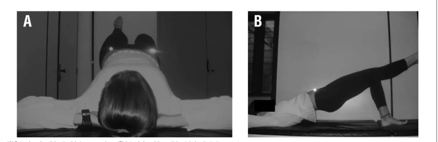
they practiced physical activity regularly. Two data collections were performed with each participant within an one week in-terval. In each data collection, a physical therapist, previously trained in a pilot study, placed a relexive marker of 10 mm on each participant’s anterior superior iliac spine to aid in the iden-tiication of these structures during analysis. Each participant was positioned in supine position, with hands placed under the head, with hips and knees lexed in a self-selected range of mo-tion and with the feet soles close together and supported on the assessment bed. he self-selected knee lexion range of motion was used to guarantee that participants were comfortable dur-ing testdur-ing and that the position selected was the most adequate to each individual’s anthropometric characteristics. he degree of knee lexion adopted on the irst day was measured for each participant to guarantee that the same joint position was kept in the second collection day. Participants were oriented to raise the pelvis from the assessment bed and perform the extension of one knee, maintaining the trunk, hip and lower limb on a straight line at the same level as the thigh of the opposite side (Figure 1). Before beginning data collection, participants per-formed the test once with the purpose of familiarization. During data collection, the test position was held for 10 seconds and then the test was repeated with the other lower limb. Each par-ticipant determined the order of the lower limbs to be tested. Instructions for the execution of the test was standardized and delivered by a physical therapist with experience in conducting these test in clinical evaluations.
Characteristics (n=30)
Age (years) 24.7 (4.2)
Weight (Kg) 66.9 (10.0)
Height (m) 1.70 (0.09)
BMI (Kg/m2) 23.0 (1.9)
Table 1. Participants’ characteristics presented as mean and standard deviation.
BMI=Body Mass Index.
Figure 1. Participants position during the assessment of pelvic alignment.
(A) Rater view of participant pelvic transverse plane; (B) Lateral view of the participant during the test.
271
he qualitative analysis was performed by two physical therapist raters, with distinct clinical practice experience (rater 1: two years; rater 2: six years). Both raters were experienced in conducting this test in clinical practice but not in using the proposed classiication. hus, before beginning data collection, raters performed training sessions on ten volunteers. During the test, the examiners were positioned behind the partici-pants’ head, with the eyes leveled with the participant’s pelvis. hese raters only judged the test; they did not provide the instructions for the test or the positioning of the volunteers. he evaluation consisted in judging the maintenance of an ad-equate alignment of the pelvis through the observation of the position of the anterior superior iliac spines in a line parallel to the assessment bed on a transverse plane. When a misalign-ment was observed, the rater classiied the amount that the anterior superior iliac spine dropped in relation to the opposite side. In order to assess this displacement, the rater judged the angle formed between the line that ran from each relective marker and the horizontal line parallel to the assessment bed. his angle was judged qualitatively in relation to the maximal possible excursion of the anterior superior iliac spine of the raised lower limb, from an horizontal line parallel to the assess-ment bed to the point at which the participant touched the assessment bed (Figure 2). he greater misalignment observed during the 10 seconds in which the participant remained with the knee extended was classiied as no pelvic tilt, a slight pel-vic tilt (0-25% of the possible excursion of the tilt), a moderate pelvic tilt (25-75% of the possible excursion of the tilt) or an accentuated pelvic tilt (>75% of the possible excursion of the tilt). Blinding between raters was performed by not allowing one rater to have access the other rater’s data22. Additionally,
within rater’s blinding was performed as they did not have ac-cess to their previous judgment and the participants’ order was randomized to reduce memory efect22.
he test performed during the qualitative assessment was registered with a digital camera (SC-D385, Samsung®, China) placed on a tripod at a distance of 80 cm of the extremity of the assessment bed. he camera on the tripod was aligned with the aid of an inclinometer (Mundo Sat, Brazil), to be parallel with the loor and with height determined by each participant an-thropometric characteristics in a way that the plane for image
capture would stay orthogonal to pelvic transverse plane during the test. his position allowed the pelvis to be centralized in the image captured during the test. Following imaging capture, a two-dimensional (2D) analysis of movement was performed us-ing the program SIMI MotionTwin® (SIMI Reality Motion Systems, Germany) to determine the greater degree of pelvic tilt. his rep-resented the greater inclination achieved during the 10 seconds of the test between the line of the anterior superior iliac spines and a horizontal line on pelvic transverse plane. A calibration of the analysis software was conducted prior to data analysis, in-forming the system 30 cm distance, pre-determined in the test lab environment.he angle of pelvic tilt was determined by the intersection of the straight line that passed in the centre of each relexive marker with the horizontal determined by the program. he procedure of identiication of the greater pelvic misalign-ment, using this program, was conducted by two raters. On a pilot study in which ten videos were analyzed in two diferent occasions with one week interval, these two raters were found to have excellent intra- and inter-rater reliabilities (ICC3,2=0.95 to
0.99). his procedure aimed to ensure a good reliability for the use of the program in a controlled situation.
Statistical analysis
Categorical data (no pelvic tilt, slight, moderate and ac-centuated pelvic tilt) were expressed regarding their overall frequency. Quantitative data of the degree of pelvic tilt was expressed in mean and standard deviation. Reliability data analysis was performed with data collected while the par-ticipant’s dominant lower limb was supported on the assess-ment bed. he reliability analysis of the intra- and inter-rater qualitative assessments were performed by the calculation of weighted kappa coeicient, assigning incremental weight, in order to diferentiate the weight of the disagreements, fol-lowed by its respectives 95% conidence intervals20,22,26 he
in-terpretation of weighted kappa was in accordance to Landis and Koch27 (≤0, poor; 0.01-0.20, slight; 0.21-0.40, fair; 0.41-0.60,
moderate; 0.61-0.80, substantial; 0.81-1.0, almost perfect). he reliability of the quantitative analysis was separated into ab-solute and relative reliabilities.
Figure 2. Rater’s judgment of pelvic position on the qualitative analysis.
Assessment bed Anterior Superior Iliac Spine Horizontal line parallel to assessment bed
272
Relative reliability
Intra-rater reliability of the degree of pelvic tilt between the two sessions was determined using ICC3,2 followed by
their respective 95%CI24. he interpretation of the ICC was in
accordance to Fleiss28 (<0.40, poor reliability; 0.40-0.75, good
reliability; >0.75, excellent reliability).
Absolute reliability
he veriication of the occurrence or not of systematic or random changes in data mean was performed through the calculation of 95% conidence interval of the mean diferences (95%CI d) between the data collected on the two occasions and through the use of Bland-Altman plot24,29,30. he variability
between the measures was also veriied by combined standard error of measurement (SEM) and the coeicient of variation of the typical error (CVTE)
24.
Another attribute veriied from the quantitative analyses was the minimal detectable change for the 95%CI (MDC95)
24,25.
Statistical analysis was performed using the software Stata/ SE®, version 10.0 and the Statistical Package for Social Sciences
(SPSS®), version 15.0. A level of signiicance (α) of 0,05 was used for all tests.
Results
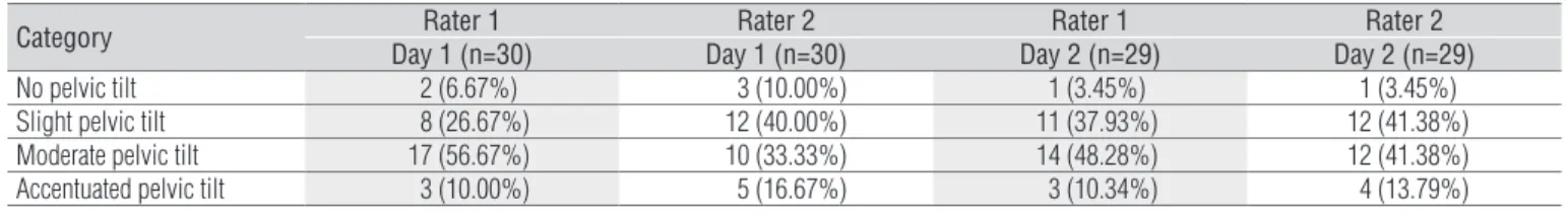
Categorical data are presented on Table 2. Weighted kappa coeicient of the qualitative analysis were classiied as fair for the intra-rater reliability of rater 1 (kw=0.32; 95%CI=0.05 to
0.59); moderate for the intra-rater reliability of rater 2 (kw=0.58;
95%CI=0.30 to 0.85) and substantial for the inter-rater reliabil-ity (kw=0.80; 95%CI=0.68 to 0.92).
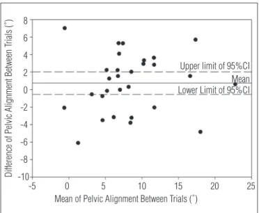
In the quantitative analysis the peak of pelvic tilt was 8.65±5.74º on the first collection day and 7.91±5.47º on the second collection day. The ICC for the analysis of greater degree of pelvic tilt between the two collection days was 0.82 (95%CI=0.65 to 0.91) showing an excellent intra-test reliability. In relation to the analysis of change in data mean, the 95% confidence interval of the mean difference (95%CI d) were -0.51º to 1.99º. The Bland-Altman plot is presented on Figure 3. The statistical measures of variability
demonstrated that the combined SEM was 2.38º, the CVTE
was 28.75%. The MDC95 was 6.59º.
Discussion
his study examined the intra- and inter-rater reliabilities of a qualitative analysis of judgment about transverse plane pelvic alignment during the bridge test with unilateral knee extension, as well as the intra-test reliability of a quantitative analysis of this alignment. Intra-rater reliability ranged from fair to moder-ate, and the rater with the shortest time of professional practice had lower reliability. his result might indicate that the reliability of the test is dependent on the rater’s experience, despite prior training. In addition, the coeicients found in the qualitative analysis may have been inluenced by the various possibilities of judgment (no pelvic tilt, slight, moderate and accentuated pelvic tilt) for a small range of motion on the transverse plane observed in the participants, which may have increased the chance of dis-sagrements between the evaluators22. Inter-rater reliability was
found to be superior than the intra-rater reliability, indicating a higher agreement between raters at one point in time when compared to the agreement of one rater at diferent points in time. his fact may indicate that the examiner’s judgment from diferent points in time may have sufered interference from a change that can be random or systematic, i.e. the chance of disagreement on two diferent days may have been inluenced, for example, by the excessive amount of categories or by a learn-ing efect from the evaluators24,32,31. his interpretation becomes
speculative, since the kappa index does not allow diferentiation between the random and systematic errors31. One factor that
might have contributed positively to the results of qualitative analysis was the use of relective markers due to the better visu-alization of the bony prominences.
he quantitative analysis through the use of ICC showed excellent intra-test reliability demonstrating the consistency of the test in measuring pelvic alignment in the transverse plane through a 2D analysis program. he presence of sys-tematic error was not conirmed since zero was included in the 95%CI d and the points were symmetrically distributed around the zero on the Bland-Altman plot (Figure 3)29,33. he
Category Rater 1 Rater 2 Rater 1 Rater 2
Day 1 (n=30) Day 1 (n=30) Day 2 (n=29) Day 2 (n=29)
No pelvic tilt 2 (6.67%) 3 (10.00%) 1 (3.45%) 1 (3.45%)
Slight pelvic tilt 8 (26.67%) 12 (40.00%) 11 (37.93%) 12 (41.38%)
Moderate pelvic tilt 17 (56.67%) 10 (33.33%) 14 (48.28%) 12 (41.38%)
Accentuated pelvic tilt 3 (10.00%) 5 (16.67%) 3 (10.34%) 4 (13.79%)
273
Bland-Altman plot also demonstrated an absence of random error as there was no tendency of an increase or decrease in the dispersion of points with the increase in mean values29,33. he
absence of systematic error indicates that the volunteers did not perform the test better or worse on the second day and were not inluenced by factors such as change in behavior and learning efect. he absence of random error indicates that a change did not occur because of the method or analysis used24.
he measure demonstrated a small SEM and therefore, it is expected that a measure conducted on the same person at diferent points in time would have a variation of 2.38º that is related to the measure error rather than to an improvement or worsening of the patient in the test23,24. One of the
advan-tages of SEM is that it is highly independent of the population in which it was determined and can be considered as a ixed characteristic of a measure34. In addition, by informing the
vari-ability of a measure in percentage, the CVTE found can be used
for comparisons with other independent measures or scales, facilitating the comparison with other studies24,32.
he MDC95 found indicates that a change in pelvic alignment
between the two occasions above or below 6.59° the original measure has a chance of less than 5% to be due to random varia-tion or a random error of measure25. hus, this index can be used
to indicate whether a real change has occurred in pelvic alignment in a particular patient over time25. he MDC
95 is one of the indexes
that infer about clinically signiicant diferences. For a better un-derstanding of this attribute, it is recommended that future stud-ies consider the combination of MDC with the minimal clinically important diference (MCID), the index that takes into account the individual self-report, for example, if the observed change is important for the patient or physical therapist24,25.
Figure 3. Bland-Altman plot for pelvic alignment.
Difference of Pelvic Alignment Between T
rials (˚)
Mean of Pelvic Alignment Between Trials (˚) -5
-10 -8 -6 -4 -2 0 2 4 6 8
0 5 10 15 20 25
Upper limit of 95%CI
Lower Limit of 95%CI Mean
95%CI=95% Confidence interval of mean differences between trials.
A comparison between the reliability coeicients found in this study with other studies is limited as this is the irst study to examine the pelvic alignment in the transverse plane during the bridge test with unilateral knee extension. Other studies have investigated similar tests that intended to assess core sta-bility. Tidstrand and Horneij13 investigated the reproducibility
of the unilateral pelvic tilt test, in which the participant elevated the pelvis from the assessment bed with one leg supported and the other elevated, with the hip and the knee lexed to 90º. his test was judged to be positive if the patient was unable to maintain the position, or negative, if the patient was able to do it. he unilateral pelvic tilt for maintaining hip and knee lexed probably generates a lower rotational torque on the pel-vic transverse plane than the bridge test with unilateral knee extension. In addition, although the test had less categories for judgment than in this study, the study demonstrated an inter-rater reliability coeicient classiied, according to Landis and Koch27, as moderate to substantial (k=0.47 to 0.61), while the
reliability found in the current study was substantial (kw=0.80).
However, methodological diferences between the aforemen-tioned study and the current do not allow comparison between reliability coeicients. Schellenberg et al.11 investigated the
intra-test reliability of a measure of fatigue time on a supine bridge test. To perform this test the volunteer elevated the hip and if he could stay in this position for two minutes then he was requested to extend the knee of the dominant leg. he time to fatigue was shown to have good reliability, determined by a strong correlation coeicient (r=0.84). Although similar to the bridge test with unilateral knee extension, this study did not provide similar analysis, since it evaluated the core stability through time until fatigue.
274
References
1. Zazulak BT, Hewett TE, Reeves NP, Goldberg B, Cholewicki J. The effects of core proprioception on knee injury: a prospective biomechanical-epidemiological study. Am J Sports Med. 2007;35(3):368-73.
2. Kibler WB, Press J, Sciascia A. The role of core stability in athletic function. Sports Med. 2006;36(3):189-98.
3. Willson JD, Dougherty CP, Ireland ML, Davis IM. Core stability and its relationship to lower extremity function and injury. J Am Acad Orthop Surg. 2005;13(5):316-25.
4. Leetun DT, Ireland ML, Willson JD, Ballantyne BT, Davis IM. Core stability measures as risk factors for lower extremity injury in athletes. Med Sci Sports Exerc. 2004;36(6):926-34.
5. Nadler SF, Malanga GA, Bartoli LA, Feinberg JH, Prybicien M, Deprince M. Hip muscle imbalance and low back pain in athletes: influence of core strengthening. Med Sci Sports Exerc. 2002;34(1):9-16.
6. McConnell J. The physical therapist’s approach to patellofemoral disorders. Clin Sports Med. 2002;21(3):363-87.
7. Ireland ML, Willson JD, Ballantyne BT, Davis IM. Hip strength in females with and without patellofemoral pain. J Orthop Sports Phys Ther. 2003;33(11):671-6.
8. Snyder KR, Earl JE, O’Connor KM, Ebersole KT. Resistance training is accompanied by increases in hip strength and changes in lower extremity biomechanics during running. Clin Biomech (Bristol, Avon). 2009;24(1):26-34.
9. Barr KP, Griggs M, Cadby T. Lumbar stabilization: a review of core concepts and current literature, part 2. Am J Phys Med Rehabil. 2007;86(1):72-80.
10. McGill SM, Grenier S, Kavcic N, Cholewicki J. Coordination of muscle activity to assure stability of the lumbar spine. J Electromyogr Kinesiol. 2003;13(4):353-9.
11. Schellenberg KL, Lang JM, Chan KM, Burnham RS. A clinical tool for office assessment of lumbar spine stabilization endurance: prone and supine bridge maneuvers. Am J Phys Med Rehabil. 2007;86(5):380-6.
12. Bohannon RW. Measuring knee extensor muscle strength. Am J Phys Med Rehabil. 2001;80(1):13-8.
13. Tidstrand J, Horneij E. Inter-rater reliability of three standardized functional tests in patients with low back pain. BMC Musculoskelet Disord. 2009;10:58-65.
14. Hicks GE, Fritz JM, Delitto A, McGill SM. Preliminary development of a clinical prediction rule for determining which patients with low back pain will respond to a stabilization exercise program. Arch Phys Med Rehabil. 2005;86(9):1753-62.
15. Akuthota V, Ferreiro A, Moore T, Fredericson M. Core stability exercise principles. Curr Sports Med Rep. 2008;7(1):39-44.
16. Rogers RG. Research-based rehabilitation of the lower back. Strength Cond J. 2006;28(1):30-5.
17. Hadala M, Barrios C. Different strategies for sports injury prevention in an America’s Cup Yachting Crew. Med Sci Sports Exerc. 2009;41(8):1587-96.
18. Stevens VK, Bouche KG, Mahieu NN, Coorevits PL, Vanderstraeten GG, Danneels LA. Trunk muscle activity in healthy subjects during bridging stabilization exercises. BMC Musculoskelet Disord. 2006;7:75-82.
19. Powers CM. The influence of abnormal hip mechanics on knee injury: a biomechanical perspective. J Orthop Sports Phys Ther. 2010;40(2):42-51.
20. Portney LG, Watkins MP. Foundations of clinical research: applications to practice. 2nd ed. Upper
Saddle River (NJ): Prentice Hall; 2000.
21. Rankin G, Stokes M. Reliability of assessment tools in rehabilitation: an illustration of appropriate statistical analyses. Clin Rehabil. 1998;12(3):187-99.
22. Sim J, Wright CC. The kappa statistic in reliability studies: use, interpretation, and sample size requirements. Phys Ther. 2005;85(3):257-68.
23. Bruton A, Conway JH, Holgate ST. Reliability: what is it, and how is it measured? Physiotherapy. 2000;86(2):94-9.
24. Lexell JE, Downham DY. How to assess the reliability of measurements in rehabilitation. Am J Phys Med Rehabil. 2005;84(9):719-23.
25. Haley SM, Fragala-Pinkham MA. Interpreting change scores of tests and measures used in physical therapy. Phys Ther. 2006;86(5):735-43.
26. Cohen J. Weighted kappa: nominal scale agreement with provision for scaled disagreement or partial credit. Psychol Bull. 1968;70(4):213-20.
27. Landis JR, Koch GG. The measurement of observer agreement for categorical data. Biometrics. 1977;33(1):159-74.
28. Fleiss JL. Reliability of measurement. In: The desing and analysis of clinical experiments. New York: John Wiley & Sons; 1986. p. 1-32.
29. Bland JM, Altman DG. Statistical methods for assessing agreement between two methods of clinical measurement. Lancet. 1986;327(8476):307-10.
30. Bland JM, Altman DG. Measuring agreement in method comparison studies. Stat Methods Med Res. 1999;8(2):135-60.
31. Hartmann DP. Considerations in the choice of interobserver reliability estimates. J Appl Behav Anal. 1977;10(1):103-16.
32. Hopkins WG. Measures of reliability in sports medicine and science. Sports Med. 2000;30(1): 1-15.
33. Atkinson G, Nevill AM. Statistical methods for assessing measurement error (reliability) in variables relevant to sports medicine. Sports Med. 1998;26(4):217-38.