1
Arquivos Brasileiros de Cardiologia - Volume 85, Nº 1, Julho 2005
Case Report
Prevailing Right Ventricular Myocardiopathy for
Previous Myocarditis or Arrhythmogenic Dysplasia?
Edmar Atik, Carlos Eduardo Rochitte, Luis Francisco R. de Ávila, Luiz J. Kajita,
Renata Bacic Palhares
Instituto do Coração do Hospital das Clínicas - FMUSP - São Paulo, SP - Brazil
Mailing address: Edmar Atik - InCor - Av. Dr. Enéas C. Aguiar 44 05403-000 - São Paulo, SP - Brazil - E-mail: [email protected] Received for publishing on 10/14/2004
Accepted on 02/09/2005
A clinical case of a 10-year-old male patient is reported. His dilated and prevailing right ventricular myocardiopathy shows diagnostic difficulties between previous myocarditis etiology and arrhythmogenic dysplasia. As the elements are not pathogno-monic of one or other cause, the increase of cardiac enzymes in subacute stage maybe tends to the supposition of previous myo-carditis. Hence, the questioning that many cases labeled as arrhythmogenic dysplasia can truly correspond to the possibility of evolutional myocarditis. The controversial clinic management is disputable.
In most of myocardiopathies both ventricles are ill-taken, and in dilated way, left ventricle involvement prevails and in arrhythmo-genic dysplasia, is the right ventricle1. However, as there are no
pathognomonic elements from one or the other entity, the prevai-ling right ventricular dilation may correspond to dilated myocardio-pathy or to arrhythmogenic dysplasia of right ventricle. Such doubt may persist even after a careful analysis of the elements that characte-rize them, according to established diagnostic criteria2.
Diagnostic difficulty between those two entities was the reason for this report, in which there was an intense and prevailing ill-taking of right ventricle.
Case Report
A 10-year-old, white male infant, with a 5-month history of tiredness when laying down, without temporal aggravation. A re-cent thoracic radiography had shown cardiomegaly, which moti-vates hospitalization for 13 days, having confirmed the diagnosis of “right ventricular myocarditis”, especially due to the verification of rise of creatine-phosphokinase in blood serum. In that hospita-lization, laboratory exams of interest revealed that inflammatory markers were normal, as 11 mm hemosedimentation, reactive protein C of 0.17 mg/dl, leucocytes 5600/mm3, and negative
rheu-matoid factor. However, the myocardial injury markers showed a rise troponin I of 37.5 ng/ml, on the first day and of 57.4 ng/dl, on the fourth day, and with maximum normal values of creatine phos-phokinase of 166 U/l, glutamic-oxalacetic transaminase of 40 U/l
and pyruvic glutamic transaminase of 20 U/l. Family history em-phasized fatherly sudden death, at 36 years of age, in a football match, in whose previous routine assessment, carried out a year before, there was no indications of the presence of any cardiopathy. At the physical exam, the patient was in a good general condition, eupneic, rosy-cheeked and with normal pulse. Weight: 56.5 kg, height: 153 cm, blood pressure (BP): 100/60 mmHg, heart rate (HR): 78 bpm. Aorta was not palpated at furcula. In the precordium there were discreet impulsions on the left sternal edge and the
ictus cordis was not palpated. Sounds were normophonetic, being the second sound extended changeable and with the two compo-nents equal in intensity. Discreet and mild systolic murmur was auscultated in the 4th and 5th left intercostal spaces, without
irra-diations. The liver was not palpated.
The electrocardiogram showed sinus rhythm and final disorder of conduction through the right ramus of Hiss bundle. There was a widened Q wave at D1 and at aVL, indicating inactive zone of high lateral wall. T wave was negative from V1 to V4. There were no signs of overload of cardiac cavities (fig. 1).
2
Arquivos Brasileiros de Cardiologia - Volume 85, Nº 1, Julho 2005
Prevailing Right Ventricular Myocardiopathy, for Previous Myocarditis or Arrhythmogenic Dysplasia?
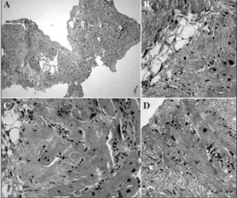
inferior and septal portions. Furthermore, right ventricular wall slendering was observed, which was regarded as being of stressed level. A hemodynamic study showed normal pressures in right cavities (RA: 10, RV: 30/10, PT: 30/15-17, PC: 10 mmHg) and the angiography (fig. 4) showed a stressed right ventricular hypo-kinesia with moderate tricuspid insufficiency. A myocardial biopsy was planned for diagnostic confirmation and showed, in seven tissue specimens removed from the right ventricular septum and free wall, myocardium atrophy, with replacement of myocardio-cytes for fibroadipose tissue, myocardial hypertrophy and moderate level fibrosis, allied to fat deposition and with discreet histiocytary and lymphocite reaction (fig. 5).
Regarding the diagnostic thought of the case, clinical elements initially guided to the possibility of a discreet level tricuspid insuf-ficiency. For its turn, the electrocardiographic change of the high lateral wall with final disorder of conduction led to the hypothesis of dilated myocardiopathy, probably of viral origin. The previous finding, on left ventricular preponderant compromising through many complementary exams, led to the diagnosis of right ventri-cular arrhythmogenic dysplasia, as a premise, given the greater involvement of the ventricle, and the endomyocardial biopsy showed histological changes compatible with that entity. But the endo-myocardial biopsy itself place some questions on the other possi-bility, that such patient could also has had a previous myocarditis with preferential ill-taking of right ventricle, in view of those biopsy elements can be found either in right ventricle arrhythmogenic dysplasia or in previous myocarditis. The increase of myocardial enzymes, verified in the initial stage of the investigation, was added to that finding, in favor of myocarditis. Concerning the established management, in view of the disease being at a stable stage and without tardive potentials present in the high-resolution electrocardiogram, the placement of cardiac defibrillator was dis-charged and the patient remained under clinical observation.
Discussion
Right ventricular dysplasia is defined as an entity in which the histology, the morphologic features, consists of a severe atrophy of the myocardium, with evidence of death of myocytes and repo-sition through adipose tissue, as a repairing process that starts in the epicardium and extends towards the endocardium. A percentage of fibroadipose tissue exceeding 43% of the area of biopsy sample3
has been calculated as a diagnosis. The fibroadipose variant shows, in two thirds of the cases, an inflammatory cellular infiltrate, sometimes with focal myocyte necrosis, findings that would be in accordance with the characteristics of a truly myocarditis. Whe-never present, the infiltrate consists of positive T CD 43 lympho-cytes, also considering the features as a chronic myocarditis. Myocarditis can lead to apoptosis through the release of pro-apoptotic proteins and pro-inflammatory cytokines3.
Other parameters, obtained through histomorphometry4, are
indicated as suggestive for the diagnosis of that pathology, cor-responding to the percentage of adipose tissue greater than 3% and of fibrous tissue greater than 40%, with myocardiocyte atrophy lower than 45%.
That entity clinically makes clear the presence of cardiac arrhythmias that put in jeopardy the life of apparently healthy young people1,5. It constitutes in a myocardial disease of unknown
etiology, whose electric instability arises from the right ventricular fibroadipose atrophy. It causes right heart failure, stressed
ven-Fig. 1 – Electrocardiogram shows “q” wave thickened in I and aVL, which is an electric characteristic of inactive zone of left ventricular lateral wall and negative T wave from V1 to V4, due to probable right ventricular dilatation.
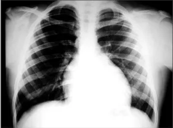
Fig. 2 – Thoracic radiography emphasizes moderate increase of right heart cavities with diminished pulmonary vascular bed.
Fig. 3 – Magnetic resonance imaging shows, in many sections, right ventricular dilatation. Slendering of right ventricular anterior wall is clear in left upper quadrant and the its filling up after gadolinium injection, in the other sections, expression of extensive fibrosis. RD = right atrium, LA = left atrium, RV = right ventricle, LV = left ventricle.
LV LV
RD RV
LV LA
RV RV
RV
3
Arquivos Brasileiros de Cardiologia - Volume 85, Nº 1, Julho 2005
Prevailing Right Ventricular Myocardiopathy, for Previous Myocarditis or Arrhythmogenic Dysplasia?
added and strengthened by enzymatic increase at the initial stage of the case investigation. For this reason, separation of those entities became a very complex task.
Such difficulty is shown even clearer when it is noted that, in the literature, there is a prevalence of biventricular involvement6,7
in right ventricular arrhythmogenic dysplasia over the isolated ill-taking of right ventricle or even, exclusively, of the left ventricle8,9.
So, from 21 cases analyzed in Spain7, in 13 of them there was
biventricular onset, in 4, isolated right ventricular onset and in the other 4 only left ventricle onset. In another assessment6, in Italy,
being 27 for necropsy among 30 patients, the onset of the left ventricle along with the right ventricle took place in 20 patients, being 6 in ventricular septum and 14 on the free wall. In the last ones, the differentiation with dilated cardiomyopathy is difficult, in which there is, in most cases, a preponderant left ventricular involvement. So, left ventricular compromising in right ventricular arrhythmogenic dysplasia is well-known and its long-term evolution can be unfavorable to the point of the features be mistaken with was found in dilated cardiomyopathy8,9. Such difficulty grows when
reports of myocarditis compromising exclusively the right ventricle are verified in the literature, which makes even impossible to separate such entities10,11, especially that in right ventricular
ar-rhythmogenic dysplasia the lymphocytic infiltrate may come from reaction to cellular death6.
So, what is left to us is give a greater importance to the most suitable management, which is not different in either one or ano-ther situation.
Since there is no arrhythmia-predisposing elements, such as multifocus ventricular extra-systoles, tardive potentials in the high-resolution electrocardiogram, history of faints and/or syncope and reinforced by absence of right cardiac insufficiency signs, the ex-pectant management could prevail.
Sudden death in those patients is related to a physical strain and the related arrhythmias depend on the catecholamina action, which is released through exercise and, for that reason, they are known as exercise-dependent1.
tricular arrhythmias in adults and sudden death in youngsters6.
Family occurrence has been documented and the responsible gene was found in the chromosome 14q23-q246. The criteria
establi-shed by the European Society of Cardiology and the Scientific Council on Cardiomyopathies of the World Heart Federation2
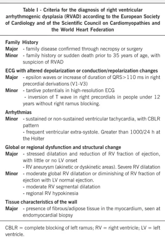
de-termine that, to confirm the diagnosis of right ventricular arrhy-thmogenic dysplasia, it is necessary the presence of two elements considered as major or 4 elements considered as minor or even the presence of a major and two minor elements (tab. I). The analysis of our case under study allowed for the counting of 2 major elements and a minor element, with which the diagnosis of this entity could be possible to establish, by following those same criteria. The major would be the severe dilatation with reduction of right ventricular fraction of ejection, with little ill-taking of the left ventricle and the presence of fibrous and adipose tissues in the myocardium seen at the endomyocardial biopsy and the minor, the inversion of T wave in the right precordial deriva-tions, from V1 to V3, without blocking of right ramus.
However, such elements could be present in previous myocar-ditis, with cicatricial changes, as demonstrated by the biopsy,
Fig. 4 – Right ventricle (RV) angiography highlights ventricular dysfunction in view of great enlargement of that cavity and discreet variation between systole in B and diastole in A, in addition to right atrial (RA) filling up due to tricuspid insufficiency.
RA
RV
RA
RV
Fig. 5 - Photomicrography demonstrating the histological features of endomyocardial biopsy. In A we observed the myocardium is greatly replaced for fibro-adipose tissue. Hematoxylin-eosin (HE) 100x. In B, C and D, myocardiocytes atrophy is observed, with replacement for fibro-adipose tissue, associated to interstitial mononuclear inflammatory infiltrate. Hematoxylin-eosin (HE) 400x
A B
4
Arquivos Brasileiros de Cardiologia - Volume 85, Nº 1, Julho 2005
Prevailing Right Ventricular Myocardiopathy, for Previous Myocarditis or Arrhythmogenic Dysplasia?
1. Gemayel C, Pelliccia A, Thompson PD. Arrhythmogenic right ventricular cardiomy-opathy. J Am Coll Cardiol 2001; 38: 1773-81.
2. Corrado D, Fontaine G, Marcus FI, et al. Arrhythmogenic right ventricular dyspla-sia/cardiomyopathy: need for an international registr y. Study Group on Arrhythmogenic Right Ventricular Dysplasia/Cardiomyopathy of the Working Groups on Myocardial and Pericardial Disease and Arrhythmias of the European Society of Cardiology and of the Scientific Council on Cardiomyopathies of the World Heart Federation. Circulation 2000; 101: 101-6.
3. Thiene G, Basso C. Arrhythmogenic rigth ventricular cardiomyopathy. An updated. Cardiovasc Pathol 2001; 10: 109-17.
4. Angelini A, Basso C, Nava A, Thiene G. Endomyocardial biopsy in arrhythmogenic right ventricular cardiomyopathy. Am Heart J 1996; 132: 203-206; 123: 640-7. 5. Nishikawa T, Ishiyama S, Nagata M, et al. Programmed cell death in the myocar-dium of arrhythmogenic right ventricular cardiomyopathy in children and adults. Cardiovasc Pathol 1999; 8: 185-9.
6. Basso C, Thiene G, Corrado D, Angelini A, Nava A, Valente M. Arrhythmogenic right ventricular cardiomyopathy. Dysplasia, distrophy or myocarditis? Circulation 1996; 94: 983-91.
References
7. Aguilera B, Suarez Mier MP, Morentin B. Arrhythmogenic cardiomyopathy as cause of sudden death in Spain. Report of 21 cases. Rev Esp Cardiol 1999; 52: 656-62. 8. Horimoto M, Akino M, Takenaka T, Igarashi K, Inoue H, Kawakami Y. Evolution of left ventricular involvement in arrhythmogenic right ventricular cardiomyopathy. Cardiology 2000; 93: 197-200.
9. Patel VV, Ferrari VA, Narula N, Wiegers SE, St John Sutton MG. Right ventricular dysplasia in an asymptomatic man: an uncommon case with biventricular involve-ment and no known family history. J Am Soc Echocardiogr 2001; 14: 317-20. 10. Mc Falls EO, van Suylen RJ. Myocarditis as a cause of primary right ventricular
failure. Chest 1993; 103: 1607-8.
11. Michaels PJ, Kobashigawa JA, Child JS, Fishbein MC. Chronic right-sided myocarditis mimicking arrhythmogenic right ventricular dysplasia. Hum Pathol 2000; 31: 618-21. 12. Fagundes MLA, Maia IG, Cruz Filho FES, et al. Arrhythmogenic cardiomyopathy of the right ventricle. Predictive value of QT interval dispersion to assess arrhythmogenic risk and sudden death. Arq Bras Cardiol 2000; 75: 115-24. 13. Sano S, Ishino K, Kawada M, et al. Total right ventricular exclusion procedure: an
operation for isolated congestive right ventricular failure. J Thorac Cardiovasc Surg 2002; 123: 640-7.
dependent on it took place7. In another study6, sudden death
oc-curred in 24 from 27 patients. In the other 3, it was due to congestive heart failure. The ages varied from 15 to 65 years old, with an average age of 28 years old.
In a Brazilian study12, the sudden death of 5 patients, among
26 studied, was correlated with right ventricular arrhythmogenic dysplasia, to the extension of QT interval, which constituted in an important predictive factor in that group of patients. QT interval corresponded to 62±17.8 in the risk group and to 51.9±12.8 in the other group.
Once the patient is aware of the necessary physical limitation, allied to an accurate psycho-emotional guidance, by comprising in this context the family action as a constant support, the expec-tant management can be adopted in the absence of arrhythmias and right heart failure. On the other hand, indications for the placement of defibrillators1, heart transplantation8,9 or
cavopul-monary-type operation13 are inherent to many clinical situations
shown. So, the presence of previous ventricular tachycardia or numerous and multifocus ventricular extra-systoles makes necessary the indication for defibrillators able to be implanted, as the stressed right heart failure requires cavopulmonary operation, and as the congestive heart failure needs heart transplantation.
In those managements, the unusual procedure of total exclusion of right ventricle for the treatment of isolated and refractory right heart failure is distinguished, which can be called upon in the absence of compromising of left ventricle. For such, the tricuspid orifice is closed, the right ventricular free wall is totally dissected and covered with a patch and, then, the total cavopulmonary anastomosis reconstitutes the flow afterwards. The evolution of that procedure can be favorable since the criteria already known for the correct indications are complied with13.
By following that thinking, sudden death in youngsters with right ventricular arrhythmogenic dysplasia, related to physical ac-tivity, occurred in 53% of them, and in other 8 (38%) symptoms
Table I - Criteria for the diagnosis of right ventricular arrhythmogenic dysplasia (RVAD) according to the European Society of Cardiology and of the Scientific Council on Cardiomyopathies and
the World Heart Federation
Family History
Major - family disease confirmed through necropsy or surgery
Minor - family history or sudden death prior to 35 years of age, with suspicion of RVAD
ECG with altered depolarization or conduction/repolarization changes Major - epsilon waves or increase of duration of QRS>110 ms in right
precordial derivations (V1-V3)
Minor - tardive potentials in high-resolution ECG
- inversion of T wave in right precordials in people under 12 years without right ramus blocking.
Arrhythmias
Minor - sustained or non-sustained ventricular tachycardia, with CBLR pattern
- frequent ventricular extra-systole. Greater than 1000/24 h at the Holter
Global or regional dysfunction and structural change
Major - stressed dilatation and reduction of RV fraction of ejection, with little or no LV onset
-RV aneurysm (akinetic or dyskinetic areas). Severe RV dilatation
Minor - moderate global RV dilatation or diminishing of RV fraction of ejection with LV normal ejection.
-moderate RV segmental dilatation
-regional RV hypokinesia
Tissue characteristics of the wall
Major - presence of fibrous/adipose tissue in the myocardium, seen at endomyocardial biopsy