1
Arquivos Brasileiros de Cardiologia - Volume 85, Nº 2, Agosto 2005
Case Report
Surgical Treatment Without Extracorporeal Circulation
of Artery Disease Related to Takayasu’s Arteritis
Involving the Aorta and Great Vessels
Rodrigo Milani, Paulo Brofman, Tayse Sandri, Alexandre Varela, José Augusto Souza,
Luiz Gustavo Emed, Stefan da Silveira, Marcelo Dantas, Maximiliano Guimarães,
Rafael Pontarolli, Francisco Maia
Santa Casa de Misericórdia - PUC - Curitiba, PR - Brazil
Serviço de Cirurgia Cardiovascular - Santa Casa de Misericórdia - PUC/PR Mailing address: Rodrigo Mussi Milani - Rua Sergio Pereira da Silva, 74 - Casa 2 - 82100-210 - Curitiba, PR - Brazil
E-mail: [email protected] Sent for publication: 03/30/2004 Accepted for publication: 11/23/2004 English version by Stela Maris Costalonga
We report the cases of 2 female patients with Takayasu’s arteritis referred to our service with lesions affecting the des-cending thoracic aorta and great vessels. One of the patients had a critical obstructive lesion in the left coronary ostium. Both patients underwent surgery without extracorporeal circu-lation, with full heparinization and autotransfusion.
Takayasu’s arteritis is a chronic vasculitis of unknown etiology, whose first case report dates back to 19081. Women account for
80 to 90% of the cases, and the ages of the affected patients range from 10 and 40 years2,3. Takayasu’s arteritis affects initially
the aorta and its primary4. The inflammation may be restricted to
the thoracic or abdominal aorta, or both, and its, and, sometimes, the manifestations are related to impairment of the coronary ostia. As the disease progresses, all great vessels are commonly affected, and the impairment may range from local lesions to extensive segmentary stenoses. The abdominal aorta and pulmonary arteries are involved in approximately 50% of the patients. The degree of activity of the inflammatory process varies with time, with apparent exacerbations and reductions or remissions.
The indication for surgical treatment is often related to severe circulatory complications, such as refractory hypertension and de-compensated heart failure, in addition to signs of myocardial is-chemia in cases with lesions involving the coronary ostia.
The surgical technique varies from case to case, because the number of vessels affected and the extent of the impairment vary, which requires the use of all surgical resources developed for cardiac surgery.
Case Report
Case 1 - A 32-year-old white female, who sought our service due to arterial hypertension of difficult control, headache, and claudication of the left lower limb. The patient reported that she began to experience intense headache approximately 8 months
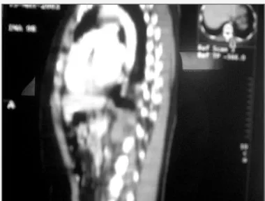
before, being then referred to the cardiologist, who diagnosed severe hypertension. Clinical treatment was initiated, and the symptoms improved slightly. Forty-five days before admission, she reported pain in the lower limbs, mainly in the left side, triggered by physical activity, which evolved to pain at rest. On admission, the physical examination revealed arterial hypertension and a significant decrease in the pulses in the lower limbs. Transtho-racic echocardiography showed a significant myocardial hyper-trophy. Thoracoabdominal tomography showed extensive segmen-tary stenosis of the descending aorta, with a critical point (95%) at the level of the thoracoabdominal transition (fig. 1). The patient underwent angiography of the ascending aorta, aortic arch, and descending and abdominal aorta. Not only were the tomographic findings confirmed, but a critical obstructive lesion was detected in the celiac trunk, as was an extensive segmentary lesion in the left carotid artery. Neither the pulmonary artery, nor its branches had any lesion.
The tests of inflammatory activity were within the normal range. Only VHS was slightly altered. Once the possibility of a new active episode of the disease was ruled out, surgery was indicated with the objective to re-establish the flow between the descending aorta and the abdominal aorta and to correct the lesion in the left carotid artery.
2
Arquivos Brasileiros de Cardiologia - Volume 85, Nº 2, Agosto 2005
Surgical Treatment Without Extracorporeal Circulation of Artery Disease Related to Takayasu’s Arteritis Involving the Aorta and Great Vessels
portion of that tube was led through the cervical region, passing below the musculature, until it reached the greater tube emerging from the ascending aorta. At this point, the 2 tubes were anastomosed. At the end of this anastomosis, heparin was reverted in 75% of its initial dose. The entire procedure was performed without the aid of extracorporeal circulation. The postoperative period was uneventful. The patient was discharged from the intensive care unit on the second postoperative day and from the hospital on the seventh pos-toperative day.
Case 2 - The patient was a 25-year-old female of Asian heritage, who complained of effort-related chest pain for approximately 120 days, which irradiated to the left upper limb, accompanied by sweating. She reported a significant worsening of the symptoms 15 days before admission, and evolution to pain at rest. She also reported having undergone surgical myocardial revascularization at the age of 17 years, in which a bypass of the left internal thoracic artery was performed to the anterior descending branch, and another bypass of the right internal thoracic artery was per-formed to the marginal branch of the circumflex artery. On hospi-tal admission, the electrocardiogram showed extensive ischemia of the entire anterior wall. The exercise test, performed 30 days before admission, was positive for ischemia. The transthoracic echocardiogram showed moderate hypocontractility of the ante-roseptal wall, with preserved ventricular function. Tomography of the thorax and abdomen showed an extensive segmentary lesion in the descending aorta with an 80-mmHg pressure gradient
between the ascending aorta and the abdominal aorta. Then, cardiac catheterization and angiography of the ascending aorta, aortic arch, and descending and abdominal aorta were performed. Coronary angiography showed a critical obstructive lesion of the left coronary ostium with total occlusion of both thoracic arteries. The angiography showed total occlusion of the left carotid artery with filling through collateral circulation through the vertebral arteries, and total occlusion of the left subclavian artery. Angio-graphy confirmed the tomographic finding, the presence of an extensive segmentary lesion in the descending aorta. As the tests of inflammatory activity were normal, an acute event was ruled out. Based on the findings, a new surgery was indicated, aiming at correcting the coronary lesions and improving the blood flow between the ascending aorta and the abdominal aorta.
The access route, like that in the first patient, was sternotomy in association with median laparotomy, extending from the manu-brium of the sternum to the pubic symphysis. Because of the inflammatory process, the heart had numerous adhesions, whose release was extremely difficult. Contrary to the first patient, whose ascending aorta had no inflammatory process, the walls of this patient’s ascending aorta were extremely thickened, which made the anastomosis difficult. Similarly to the first patient, the second patient received 4 mg/kg of heparin, and her ascending aorta was partially clamped. Then, aortotomy was performed in the form of an ellipse in the lateral face of the aorta, and a 20 mm diameter Dacron tube was anastomosed with a continuous 4-0 Prolene
Fig. 2 - Dacron tube anastomosed to the ascending aorta. Fig. 1 - Extensive segmentary lesion of the descending aorta.
3
Arquivos Brasileiros de Cardiologia - Volume 85, Nº 2, Agosto 2005 Surgical Treatment Without Extracorporeal Circulation of Artery Disease Related to Takayasu’s Arteritis Involving the Aorta and Great Vessels
suture. The distal part of the tube was led to the abdomen as reported in the first case, and the anastomosis was performed close to the bifurcation with a continuous 4-0 Prolene suture. When the correction of the aorta was finished, the patient was put in the Trendelemburg position, and the surgical table was rotated to the right side. In that position and with the aid of a suction tissue stabilizer (Octopuss – Medtronic), myocardial re-vascularization was performed with saphenous vein bypass graft to the marginal branch of the circumflex artery and saphenous vein bypass graft to the anterior descending branch. Because the aorta was extremely thick, the proximal anastomoses were per-formed in the Dacron tube in its origin from the ascending aorta. The postoperative period was uneventful, and the patient stayed 2 days in the ICU and 8 more days at the hospital. On hospital discharge, the electrocardiogram showed an improvement in is-chemia as compared with that in the preoperative period. As in the first patient, this surgery was performed without extracorpo-real circulation. Both patients underwent anticoagulation before hospital discharge.
Discussion
In the inflammatory diseases of the aorta, aorticbypass with tubular prosthesis is usually the method selected for treating seg-mentary stenoses, avoiding, therefore, the manipulation of the most impaired segments of the aorta5. Involvement of the great vessels
is relatively common, and, although revascularization of those vessels is the ideal procedure, it is not always possible, due to the extensive fibrosis of those vessels and the extent of the lesion, sometimes involving the intracranial segment of the carotid artery. The coronary lesions, when localized, may be treated by use of angioplasty6, and
most cases may be treated by surgical myocardial revascularization7.
When the coronary lesion is limited to the ostia, some authors recommend correction through ostioplasty8.
In case 1, the disease was limited to the descending aorta, a localized lesion in the left carotid artery and the celiac trunk. Aortic bypass, performed in an inflammation-free area, proved to be a relatively simple surgery, and some technical aspects should be considered in the thoracoabdominal transition through the diaphragm, avoiding the formation of hernias and the erosion of abdominal organs by the presence of the tube. The presence of a disease-free inferior mesenteric artery guaranteed good collateral circulation to the region of the celiac trunk. Due to its proximity
to the aortic bifurcation, much attention is required in performing the abdominal anastomosis. This type of operation proved to be very efficient in alleviating the patient’s symptoms, hypertension and claudication. The carotid lesion, limited to a proximal segment, may also be treated through the implantation of a prosthesis, with good short-term results.
In case 2, the impairment of the great vessels was more important, leading to total occlusion of the carotid and left sub-clavian arteries, whose territories were irrigated through collateral circulation, which was efficient. The occluded vessels had im-portant fibrosis, and their revascularization was difficult. In addition, the patient had already undergone surgery due to impairment of the left coronary ostium 7 years before, which showed the evolu-tionary character of the disease. One of the factors responsible for the occlusion of the left internal thoracic artery may have been the total obstruction of the left subclavian artery, leading to an interrogation in regard to the use of that type of graft in that special subgroup of patients. An alternative to the traditional re-vascularization is ostioplasty, a procedure with good results in patients without impairment of the ascending aorta9. In this case,
however, that segment of the aorta was extremely thick, hin-dering the performance of that procedure, in which case, tradi-tional myocardial revascularization was chosen.
Both patients evolved in a very satisfactory way, showing that complex surgeries without extracorporeal circulation may be greatly beneficial for that group of patients. Because the amount of prosthetic material used in both cases was large, full anticoa-gulation was required, and, due to the evolutionary character of the disease, corticoid therapy was used at low doses to prevent new episodes of the disease. In cases of exacerbation, recurrent corticoid therapy may be necessary.
In conclusion, inflammatory diseases of the aorta, such as Takayasu’s arteritis, have several anatomic variations in regard to the presence of stenoses, and surgery is reserved for the cases with an important impairment in the arteries without the presence of a satisfactory collateral circulation, causing symptoms of is-chemia. Therefore, surgical planning should consider the impro-vement of symptoms, as in the cases reported. In case 1, circulation in the thoracoabdominal aorta was improved, with a reduction in the ischemia of the lower limbs, and a simultaneous attempt to correct hypertension occurred. In case 2, in addition to those 2 symptoms, myocardial ischemia was also corrected.
1. Takayasu M. A case with unusual changes of the central vessels in the retina. Nip-pon Ganka Gakkai Zasshi 1908; 12:554-7.
2. Lupi-Herrera E, Sanchez-Torres G, Marcushamer J et al. Takayasu’s arteritis. Clini-cal study of 107 cases. Am Heart J 1977; 93:4.
3. Arend WP, Michel BA, Block DA et al. The American College of Rheumatology 1990. Criteria for the classification of Takayasu arteritis. Arthritis Rheum 1990; 33:1129. 4. Hata A, Noda M, Moriwaki R, Numano F. Angiographic findings of Takayasu
arte-ritis: New classification. Int J Cardiol 1997; 54(Suppl):155.
5. Hall S, Barr W, Lie JT et al. Takayasu arteritis: A study of 32 North American pa-tients. Medicine 1985; 64:89.
References
6. Rao SA, Mandalam KR, Rao VR et al. Takayasu arteritis: Initial and long-term fol-low-up in 16 patients after percutaneous transluminal angioplasty of the descen-ding thoracic and abdominal aorta. Radiology 1993; 189:173.
7. Tanaka K, Mizutani T, Yada I et al. Aorta-coronary bypass grafting for bilateral ostial stenosis caused by Takayasu’s aortitis. J Thorac Cardiovasc Surg 1990; 99: 948-9. 8. Nakano S, Shimazaki Y, Kaneko M et al. Transaortic patch angioplasty for left co-ronary ostial stenosis in a patient with Takayasu’s aortitis. Ann Thorac Surg 1992; 53: 694-6.