1
Original Article
Correction of Thoracic and Thoracoabdominal Aortic
Aneurysms. Central Cannulation Technique
Salomón Soriano Ordinola Rojas, Viviane Cordeiro Veiga, Januário Manoel de Souza,
Marcos Fassheber Berlinck, Dante Fanganiello Senra, Reinaldo Wilson Vieira,
Luiz Alberto Magna, Domingo Marcolino Braile, Sérgio Almeida de Oliveira
São Paulo, SP and São José do Rio Preto, SP - Brazil
Real e Benemérita Associação Portuguesa de Beneficência and Faculdade de Medicina de São José do Rio Preto
Mailing address: Viviane Cordeiro Veiga Alameda Hungria, 89 -06474-140 – Barueri, SP, Brazil – E-mail: [email protected] Received for publication: 07/31/2003
Accepted for publication: 07/15/2004 English version by Stela Maris Costalonga
Objective
To demonstrate the viability of the use of extracorporeal cir-culation established between the left atrium and ascending aorta to induce deep hypothermia for correcting thoracic and thora-coabdominal aortic aneurysms.
Method
From January 1994 to July 2001, 38 patients (mean age, 54.6 ± 12.7 years) were operated on as follows: 12 (31.6%) patients underwent repair of thoracoabdominal aortic aneurysm, and 26 patients underwent repair of descending thoracic aneu-rysms. Deep hypothermia was induced by use of extracorporeal circulation, with pharyngeal temperature ranging from 15 to 25ºC (20.6 ± 3.2ºC).
Results
Of the neurological complications, paraplegia occurred in 2 (5.3%) patients. One patient developed paraparesis, and another evolved with convulsion. Twelve (31.6%) patients had respiratory complications, and 2 (16.7%) patients died. Two other patients were operated on on an emergency basis and ended up dying. Total mortality was 18.4% (7 patients).
Conclusion
Repair of descending thoracic and thoracoabdominal aortic aneurysms with deep hypothermia by use of extracorporeal circu-lation established between the left atrium and ascending aorta proved to be a viable method for correcting those aneurysms.
Key words
thoracic aortic aneurysms; thoracoabdominal aortic aneurysms; hypothermia
The surgical treatment of arterial aneurysms began with Matas, in 1903, who described the technique of obliterating endoaneu-rysmorrhaphy, which consisted of suturing the proximal and distal orifices, without restoring blood flow continuity. In 1920, Matas introduced the restorative and reconstructive endoaneurysmorr-haphy to maintain blood flow through the aorta 1.
The first study on repair of descending thoracic aortic aneurysms by insertion of a tube in the aneurysm site was reported by Lam and Aram 2 in 1951. Etheredge et al 3, in 1955, reported for the
first time the successful treatment of thoracoabdominal aortic aneurysm by use of a homograft, a temporary aorta-aorta shunt, and anastomosis of the celiac trunk and mesenteric artery, sepa-rately, with the graft.
The etiology of the aneurysms, according to Coselli and LeMaire4, is degenerative in approximately 80% of the patients.
The second most common cause is aortic dissection. The less frequent causes are Marfan’s syndrome, Ehlers-Danlos syndrome, infection, Takayasu’s disease, and trauma.
The risk factors for rupture are the presence of pain, dissection, diameter greater than 5cm, growth of the aneurysm, smoking, chronic obstructive pulmonary disease, systemic arterial hypertension, chronic renal failure, and degenerative diseases of the aorta 5-8.
The major causes of morbidity and mortality are acute myo-cardial infarction, respiratory failure, and stroke. Antecedents of smoking and chronic obstructive pulmonary disease are risk factors for the development of respiratory failure. Patients with previous renal failure are at a higher risk of developing acute renal failure in the postoperative period 6.
In addition, according to Kazui et al 9, the emergency surgery,
the dissections of the aorta, the time of aortic clamping, and the time of extracorporeal circulation are risk factors that increase postoperative mortality.
Thoracoabdominal aneurysms were classified by Crawford et al10 as follows: type I, extending from the left subclavian artery
until right after the visceral branches; type II, extending from the left subclavian artery until the aortic bifurcation; type III, extending from the sixth rib until the renal arteries; and type IV, beginning right below the diaphragm and extending to the renal arteries.
The complementary methods used for diagnosing the aneu-rysms are as follows: chest X-rays, echocardiography, computed tomography, magnetic resonance imaging, and aortography.
2
and the conventional treatment, characterized by interposition of prostheses after resection of the aneurysm.
Palma et al 11 reported a case of saccular aneurysm treated
with the introduction of 2 stents visualized with endoscopy through a thoracotomy and with deep hypothermia and circulatory arrest. Palma 12 used the intravascular treatment for repairing acute
descending thoracic aortic dissections in 27 patients, and obtained an 80% survival in 32 months.
Knowing the anatomy of the spinal cord irrigation is funda-mental for understanding the neurological lesions (paraplegia or paraparesis) consequent to the surgery of thoracic or thoracoab-dominal aorta. Three spinal arteries, one anterior and 2 posterior, exist. The major artery is the anterior artery that irrigates the anterior two thirds of the spinal cord, including the motor area. That artery runs through the thoracolumbar region and receives the intercostal and lumbar arteries; it is the great radicular artery or the Adamkiewicz artery, which usually originates between the ninth and twelfth thoracic vertebrae in 75% of patients, between the fifth and eighth thoracic vertebrae in 15% of patients, and between the first and second lumbar vertebrae in 10% of patients. If the great radicular artery is between the fifth and ninth thoracic vertebrae, a complementary artery may exist, the artery of the conus medullaris, which, at that level, anastomoses with the Adamkiewicz artery.
Currently, several methods of spinal cord protection exist, such as temporary bypass 13,14, reimplantation of the intercostal
arteries15,16, drainage of the cerebrospinal fluid 17-19, deep
hypo-thermia20-26, selective cooling of the spinal cord 21, and
pharma-cological agents 27.
This study aimed at demonstrating the viability of the use of the extracorporeal circulation established between the left atrium and ascending aorta combined with deep hypothermia for correcting thoracic or thoracoabdominal aortic aneurysms. The results were analyzed.
Methods
From January 1994 to July 2001, of 860 patients with aortic disease (555 aneurysms and 305 dissections) operated on in our service, 38 patients with descending thoracic or thoracoabdominal aortic aneurysms were studied. The surgical technique comprised reimplantation of the visceral vessels, elective deep hypothermia with extracorporeal circulation established between the left atrium and the ascending aorta, followed by total circulatory arrest for performing the proximal anastomosis close to the left subclavian artery, without aortic clamping.
The patients’ ages ranged from 22 to 72 (54.6 ± 12.7) years; 26 (68.4%) were men and 12 (31.6%) were women.
Of the 38 patients studied, 12 (31.6%) underwent repair of thoracoabdominal aneurysms, and the remaining 26 (68.4%) underwent repair of thoracic aneurysms (tab. I), which were approa-ched through thoracophrenolaparotomy and posterolateral thora-cotomy, respectively. Seventeen (44.7%) patients had aneurysms without dissection, 13 of which were thoracic (50% of the tho-racic) and 4 (33.3%) were thoracoabdominal. Of the total number of patients, 4 (10.5%) had coarctation of the aorta, 3 (7.9%) of whom had already undergone previous surgery.
The clinical findings comprised pain in 29 (76.3%) patients,
hoarseness in 6 (15.8%), and 3 were asymptomatic on the occasion of diagnosis.
Thirty-one (81.6%) patients had systemic arterial hypertension, and 2 (5.3%) patients had chronic renal failure.
The methods used for diagnostic confirmation and indication of surgical treatment of the aneurysms were chest X-ray, echo-cardiography, aortography, computed tomography, and magnetic resonance imaging. In the patients older than 40 years, cine coro-nary angiography was performed to rule out corocoro-nary lesions.
The etiological diagnoses were as follows: post-trauma (one pa-tient); coarctation of the aorta, repaired or not (4); syphilitic aortitis (2); atherosclerotic (10); and aortic dissection (21) (tab. II).
The patients were induced to deep hypothermia through extra-corporeal circulation, with pharyngeal temperature ranging from 15 to 25°C (20.6 ± 3.2°C) (tab. III).
The duration of the circulatory arrest for the surgical repair ranged from 9 to 36 min (21.3 ± 6.1 min).
The criteria for indicating the surgical technique were based on the size, diameter, and location of the aneurysms in regard to the left subclavian artery. The diameter of the aorta of the patients ranged from 4 to 10.5 cm (8.1 ± 1.5 cm).
All patients were prepared according to our service’s protocol for surgical repair of thoracic and thoracoabdominal aortic aneu-rysms as follows: monitoring of the cardiac rhythm and mean blood pressure by use of catheterization (dissection or puncture) of the right radial artery; catheterization of a peripheral vein with abocat # 16 for anesthetic induction; pulse oximetry; control of urinary output by use of vesical catheterization with a Foley cathe-ter; nasopharyngeal temperature measurement with a tele-ther-mometer, 43 – TA model (Yellow Springs Instruments Co. Inc.
Table I - Location of the aneurysms
Location Number of patients (%)
Thoracic 26 (68.4%)
Thoracoabdominal 12 (31.6%)
Total 38 (.100%)
Table II - Etiology of the thoracic and thoracoabdominal aortic aneurysms
Etiology Number of patients (%)
Trauma 1 (02.6%)
Coarctation of the aorta 4 (10.5%)
Syphilitic aortitis 2 (05.3%)
Atherosclerotic 10 (26.3%)
Dissection of the aorta 21 (55.3%)
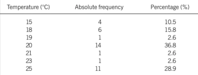
Table III – Degrees of deep hypothermia used for repairing the thoracic and thoracoabdominal aortic aneurysms
Temperature (°C) Absolute frequency Percentage (%)
15 4 10.5
18 6 15.8
19 1 2.6
20 14 36.8
21 1 2.6
23 1 2.6
25 11 28.9
3
Yellow Springs, OH, USA); central venous access through puncture of the subclavian vein or right internal jugular vein; and thermal mattress to maintain body temperature according to surgical needs. The patients underwent selective orotracheal bronchial intu-bation, which allows stopping the ventilation of the left lung during the procedure of dissection and release of the left lung, which is usually adhered to the aneurysm.
All patients were positioned in the right lateral decubitus po-sition, and asepsis was performed with 1% polyvinyl pyrrolidone-iodine (PVPI) complex followed by antisepsis with iodide alcohol, and, later, topic PVPI. The surgical fields were placed. Then, posterolateral thoracotomy was performed in the fourth left inter-costal space from the inter-scapulo-vertebral space until the left internal thoracic artery for the thoracic aneurysms. In those thora-cic aneurysms with distal extremity difficult to expose due to proxi-mity to the diaphragm, one could perform an additional thoraco-tomy in the sixth intercostal space, without the need for a new cutaneous incision. In thoracoabdominal aneurysms, thoracophre-nolaparatomy was performed, beginning in the thorax, following along the middle abdominal line, and sectioning the diaphragm in a circular manner to preserve the phrenic center.
All abdominal viscera were pulled aside, allowing access to the retroperitoneal space until the bifurcation of the abdominal aorta, which was exposed to allow its longitudinal incision, begin-ning in its proximal portion. The pericardium was opened, and the left auricle and ascending aorta were exposed.
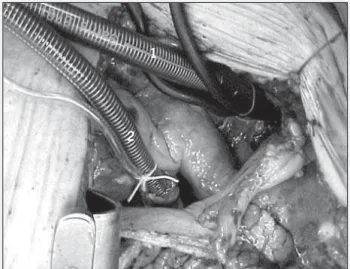
Anticoagulation with sodium heparin (5000 IU per mL) was used at the dosage of 4 mg/kg of body weight. Two pouch sutures were performed in the ascending aorta with “mersilene” 2–0 thread passed through latex tubes aiming at guaranteeing hemos-tasia and fixation around the cannula. Pouch suture was performed in the left auricle with 4-0 polypropylene thread, which was also repaired with a PVC tube. A 20- or 22-F wired arterial cannula, Baxter R.M.I. model, was introduced in the delimited spaces and adequately fixed with the aid of pouch sutures. Then, the cannula was placed in the left atrium (venous cannula, Baxter R.M.I. # 3651 model), in the space delimited by the pouch suture, which served as drainage for the membrane oxygenator and later return to the ascending aorta with an arterial flow of 2.2 L/m2 per minute,
with an adequate oxygen flow of 2 liters and one liter of compressed air in a Macchi-Edwards compressor to maintain the gas parameters at physiological levels (fig. 1).
After beginning extracorporeal circulation, the patient was induced to deep hypothermia, which could reach 15°C. During the procedure of deep hypothermia induction, ice bags wrapped in gauze were placed on the patient’s head. When a temperature between 20 and 18°C was reached, circulation was stopped, blood was drained to the oxygenator, and the aneurysm was ope-ned. Due to a phenomenon of thermal inertia, the nasopharyngeal temperature of the patient reached values around 16°C. Surgical repair was initiated through a longitudinal opening of the thoracic aortic aneurysm close to the subclavian artery; depending on the aneurysmatic impairment of the aorta, the incision was prolonged towards the other great vessels and reconstruction of the aorta or its arch, or both, was initiated with a tubular graft of appropriate diameter (pericardium or Dacron).
Later, the intercostal arteries were ligated and proximal anas-tomosis of the prosthesis was performed with 3 “0” polypropylene thread and a 2.5-cm curved needle reinforced with a Teflon band.
Once the anastomosis was finished, clamping of the tubular graft was performed below the anastomosis, and extracorporeal circulation was reestablished with a 600mL/min flow, which allowed cerebral reperfusion. In the treatment of long thoracic or thoracoabdominal aortic aneurysms, the intercostal arteries located between the eighth and eleventh thoracic vertebrae were reimplanted (fig. 2).
In the cases of thoracoabdominal aneurysms, the diaphragm muscle was sectioned to preserve the phrenic center, reaching the retroperitoneum. After exposure and adequate opening of the aorta, the celiac trunk, the superior mesenteric artery, and the renal arteries, isolated or together, were reimplanted in the tubular graft according to the anatomic detail.
During this sequence of anastomotic procedures, aortic clamping was shifted so that blood flow could increase, reperfusing the struc-tures, depending on the reanastomosed arteries. After performing distal anastomosis between the tubular graft and the abdominal aorta, patient warming was initiated according to adequate rewar-ming criteria until reaching an esophageal temperature of 37°C.
Adequate hemodynamic values were reestablished and termi-nation of the extracorporeal circulation was initiated. Heparin was neutralized with protamine sulfate at the 1:1 ratio. Hemostasia
Fig. 2 – Surgical technique for repairing thoracic and thoracoabdominal aneurysm. Fig. 1 – Surgical technique for repairing thoracic and thoracoabdominal aneurysm: cannulation of the ascending aorta and left atrium. 1) aortic cannula; 2) left atrial cannula.
1
4
was revised, and drainage of the retroperitoneum and thorax was performed with tubular drains. The surgical incision was closed by planes. On thorax closure, the intercostal spaces were appro-ximated, as were the ribs that had been sectioned, with # 5 steel thread. Once the surgical incision was closed, the patient was placed in the dorsal decubitus position, and the oroendobron-chial tube was replaced by a common orotracheal cannula to allow adequate ventilation in the postoperative period. The patient remained sedated for 24 hours on mechanical ventilation. Once the adequate criteria of ventilation and functional tests were achie-ved, the intubation tube was removed from the patients, according to our service’s pre-established protocol.
Statistical analysis was performed by comparing the means by using the Student t test for independent samples and the nonpara-metric Mann-Whitney test. The assessment of the association bet-ween the attribute variables was performed through the chi-square test in tables of contingence or though the Fisher exact test, de-pending on the case. The association between quantitative variables was measured through the analysis of the simple Pearson correlation. In all cases, the proportional error of 5% (P < 0.05) was adopted as the significance level.
Results
All patients were placed under systemic deep hypothermia with temperatures ranging from 15 to 25°C, and this variable was not associated with mortality, because the mean temperature, in hypothermia, of the 31 patients discharged from the hospital was 20.9±3.3°C. The values of the 7 patients who died were similar, 19.6±3.0°C (P = 0.323).
The complications that occurred in the postoperative period of the surgical correction of the thoracic and thoracoabdominal aortic aneurysms are shown in table IV.
Regarding the neurological complications, 2 (5.3%) patients had paraplegia, one of whom also had ischemic stroke; one (2.6%) patient evolved with paraparesis, with later recovery of the move-ments; and one (2.6%) had a convulsive crisis in the postoperative period (tab. V).
Of the 12 patients with thoracoabdominal aneurysms, 27% had neurological complications (95% CI = 6-61%); in the 26 patients with thoracic aneurysms, the incidence of neurological complications was 0% (95% CI = 0-13.2%). Thus, an association between neurological lesion and the thoracoabdominal location of the aneurysm was observed (P = 0.02).
Table IV - Postoperative complications of the surgical repair of thoracic and thoracoabdominal aneurysms
Complications Number of patients (%)
Neurological 4 (10.5%)
Respiratory 12 (31.6%)
Renal 2 (05.3%)
Table V - Neurological complications after surgical repair of thoracic and thoracoabdominal aneurysms
Diagnosis Complications Evolution
Type I thoracoabdominal Paraplegia
aneurysm (dissection) Stroke Death
Type II thoracoabdominal Paraplegia Discharge
aneurysm (dissection)
Type II thoracoabdominal Paraparesis Discharge
aneurysm (dissection)
Type I thoracoabdominal Convulsion Discharge
aneurysm (dissection)
Table VI - Respiratory complications after surgical repair of thoracic and thoracoabdominal aortic aneurysms
Diagnosis Complication Evolution
Aneurysm of the descending thoracic aorta (dissection) Respiratory failure Death
Type I thoracoabdominal aortic aneurysm Respiratory infection Death
Aneurysm of the aortic arch and descending aorta Pleural effusion Discharge
Descending aorta aneurysm Pleural effusion, respiratory infection Discharge
Descending aorta aneurysm Atelectasis Discharge
Descending aorta aneurysm Discharge Discharge
Descending aorta aneurysm (dissection) Atelectasis Discharge
Descending aorta aneurysm Atelectasis Discharge
Type III thoracoabdominal aortic aneurysm Bronchospasm, respiratory infection Discharge
Type III thoracoabdominal aortic aneurysm Respiratory infection Discharge
Descending aorta aneurysm (dissection) Atelectasis and pleural effusion Discharge
Descending aorta aneurysm (dissection) Atelectasis Discharge
Respiratory complications were observed in 12 (31.6%) patients, who had atelectasis, pleural effusion, infection with clinical manifesta-tion of respiratory failure, in an isolated or associated manner. Two (5.3%) patients required tracheostomy for prolonged mechanical ventilation, one of whom died on the 25th postoperative day due to respiratory failure, and the other evolved with tracheal stenosis, requiring tracheoplasty. Of the patients with respiratory complica-tions, 2 died, which resulted in a 16.7% mortality, which was not significantly different from that observed among patients who died due to other causes (19.2%; P = 0.85) (tab. VI).
Two patients, required reimplantation of the renal arteries and had transient elevation in the creatinine serum levels, but required no dialysis.
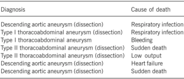
In our case series, 7 patients died (total mortality, 18.4%): 3 (11.5%) patients had thoracic aneurysms and 4 (33.3%) had thoracoabdominal aneurysms (P = 0.11). The causes of death were as follows: one (14.3%) due to intraoperative bleeding; 2 (28.6%) due to respiratory complications; 2 (28.6%) due to sudden death on the seventh and tenth postoperative days; one (14.3%) due to heart failure; and one (14.3%) due to myocardial failure when extracorporeal circulation was withdrawn. Two (5.3%) pa-tients were operated on on an emergency basis due to aortic dissection and died, one because of bleeding resulting from coa-gulopathy, and the other due to late respiratory failure.
5
the emergency character of the surgery (13.9% of the deaths occurred in elective surgeries, and 100% in emergency surgeries, P = 0.03) (tab. VII).
Preoperative pain was not significantly associated with the occurrence of death, although it was observed in 100% of the patients who died; but it was also present in most patients who were discharged from the hospital (74.2%), and no significant difference was observed between both percentages (P = 0.31).
The diameter of the aneurysm, on the other hand, was greater among the patients who died (9.3±1.1 cm) than among those who were discharged (mean, 7.9±1.5 cm) (P = 0.02).
Similarly, a significant association was observed between pa-tients’ death and the presence of aortic dissection, because all 7 patients who died had aortic dissection (P = 0.01).
Most patients (78.9%) had arterial hypertension. Twenty per-cent of the hypertensive patients died, while 12.5% of the nonhy-pertensive patients died; no association was found between arte-rial hypertension and death (P = 1.00).
Discussion
Currently, the methods of spinal cord protection used in surgery for correcting thoracic and thoracoabdominal aortic aneurysms are as follows: temporary bypass, reimplantation of the intercostal arteries, cerebrospinal fluid drainage, deep hypothermia, selective cooling of the spinal cord, and use of pharmacological agents.
Olivier et al 14, in 1984, began to use the temporary bypass
established between the left atrium and the femoral artery. Despite the measures for preventing spinal cord ischemia, Kazui et al 9
and Coselli and LeMaire 4 reported that patients treated with left
bypass had a lower incidence of paraplegia. Coselli and LeMaire reported a 6% incidence.
Reimplantation of the intercostal arteries between the ninth and twelfth thoracic vertebrae is another alternative proposed for spinal cord protection. The location of those arteries through angiography or evoked potential and their reimplantation may decrease the incidence of paraplegia 15,16. Kieffer et al 20 reported
a 5% incidence of paraplegia in patients who underwent that surgical technique. We do not use angiography to identify the intercostal arteries; however, we perform the implantation of the arteries between the ninth and twelfth thoracic vertebrae.
Cerebrospinal fluid drainage has been recommended by Miyamoto et al 17. Safi et al 19, who stated that the cerebrospinal
fluid pressure should be close to 10 mmHg, reported a 9% inci-dence of paraplegia. In our study, we did not use cerebrospinal fluid drainage, because of the risk of a puncture accident during
the procedure, because, as the patient is heparinized, an extradural hematoma may form.
Deep hypothermia, with or without cardiocirculatory arrest, has been used for treating thoracic and thoracoabdominal aortic aneurysms and has been performed by establishing extracorporeal circulation between the aortic arch and right atrium 22,23,25,26.
In this study, deep internal hypothermia was induced by establi-shing extracorporeal circulation between the left atrium and the ascending aorta through the left auricle, because it provides more space for acting on the aneurysms of the aortic arch, mainly when those aneurysms are close to the subclavian artery, or yet, due to the large diameter of the descending aorta. After the proximal anas-tomosis of the prosthesis in the aorta, the aorta is clamped and the extracorporeal circulation is reinitiated, which allowed us to perfuse the cerebral tissue, reducing the time of cardiocirculatory arrest.
Despite the care to avoid paraplegia, it remains a postoperative risk in patients with Crawford’s types I and II aneurysms. In our study, the patients received extracorporeal circulation with deep hypothermia and circulatory arrest; 2 of them had paraplegia (incidence of 5.2%), and one (2.6%) had paraparesis.
Pharmacological agents have been used to prevent reperfusion lesions. Acher et al 27 used naloxone in association with
cerebros-pinal fluid drainage and had a 2% incidence of paraplegia (1/49). According to Kazui et al 9, the postoperative complications
found in 95 patients undergoing repair of thoracic aortic aneurysms were as follows: renal failure in 7 (7.8%) patients, 2 (28.5%) of whom required hemodialysis. Two (2.1%) patients had paraplegia and one experienced bleeding, requiring revision of hemostasia.
Of the 46 patients who survived one month, one (2.2%) patient required dialysis due to renal failure 28. Acher et al 27 reported
renal complications in 2.7% of the patients.
In our study, the occurrence of renal complications was tran-sitory and affected only 2 (5.3%) patients, who required neither dialysis nor any other special treatment.
Westaby 23 reported that 2 type I patients classified according
to Crawford (8%) died, representing 33.3% (2/6) mortality. Acher et al 27 reported that 12 (10.9%) patients in their case series had
a neurological deficit. Kouchoukos et al 28 reported that of 51
patients, 5 (9.8%) died. Their complications were as follows: paraplegia in 2 (Crawford I 8%), and paraparesis in one (2%). In a case series of 94 patients undergoing correction for thoracoabdo-minal aortic aneurysms, Yamashita et al 29 reported 15% mortality.
Anacleto et al 1 reported 13% in-hospital mortality, 33.3%
due to multiple organ failure, 33.3% due to coagulopathy, 8.3% due to anaphylactic shock to protamine, 8.3% due to stroke of the cerebral trunk, 8.3% due to acute liver failure, and 8.3% due to acute myocardial infarction. Mortality in emergency surgeries was 3 times greater than that in elective surgeries. The incidence of paraplegia was 7%.
In our case series, 7 patients died (mortality, 18.4%), 3 (7.9%) due to respiratory failure, one (2.6%) due to sudden death, one (2.6%) due to acute myocardial infarction, one (2.6%) due to low cardiac output, and one (2.6%) due to bleeding.
In conclusion, correction of descending thoracic and thoraco-abdominal aortic aneurysms by using left-side thoracotomy or tho-racophrenolaparotomy with deep hypothermia and extracorporeal circulation established between the left atrium and the ascending aorta proved to be an adequate surgical treatment.
Table VII - Postoperative mortality of the surgical repair of thoracic and thoracoabdominal aneurysms
Diagnosis Cause of death
Descending aortic aneurysm (dissection) Respiratory infection Type I thoracoabdominal aneurysm (dissection) Respiratory infection Type I thoracoabdominal aneurysm Bleeding
6
1. Anacleto A & Anacleto JC. Aneurismas da aorta torácica e toracoabdominal. In: Brito, CJ. Cirurgia Vascular. Rio de Janeiro: Revinter; 2002:439-63.
2. Lam CR, Aram HH. Resection of descending thoracic aorta for aneurysm. Report of the use of the homograft in case and experimental study. Ann Surg. 1951; 134:743-52.
3. Etheredge SN, Yee J, Smith JV. Successful resection of a large aneurysm of the upper abdominal aorta and replacement with homograft. Surgery. 1955; 138:1071-81. 4. Coselli JS, LeMaire AS. Left heart bypass reduces paraplegia rates after
thoraco-abdominal aortic aneurysm repair. Ann Thorac Surg. 1999; 67:1931-4. 5. Juvonen T, Ergin MA, Galla JD, et al. Prospective study of the natural history of
thoracic aortic aneurysms. Ann Thorac Surg. 1997; 63:1533-45.
6. Griepp RB, Ergin MA, Galla JD, et al. Natural history of descending thoracic and thoracoabdominal aneurysms. Ann Thorac Surg. 1999; 67: 1927-30. 7. Fikar CR, Koch S. Etiologic factors of acute aortic dissection in children and young
adults. Clin Pediatr. 2000; 39:71-80.
8. Davies RR, Goldstein LJ, Coady MA, et al. Yearly rupture or dissection rates for thoracic aortic aneurysms: simple prediction based on size. Ann Thorac Surg. 2002; 73:17-28.
9. Kazui T, Komatsu S, Yokoyama H. Surgical treatment of aneurysms of the thoracic aorta with the aid of partial cardiopulmonary bypass: an analysis of 95 patients. Ann Thorac Surg. 1987; 43:622-7.
10. Crawford ES, Crawford JL, Safi JH. Thoracoabdominal aortic aneurysms: preo-perative and intraopreo-perative factors determining immediate and long-term results of operations on 605 patients. J Vasc Surg. 1986; 3:389-404.
11. Palma JH, Geisthovel N, Brasil LA, et al. Tratamento de aneurismas da parte torácica da aorta pela introdução de “stents” sob via endoscópica. Rev Bras Cir Cardiovasc. 1998;13:8-12.
12. Palma JH. Tratamento das dissecções agudas da aorta descendente, utilizando stents aórticos introduzidos pela artéria femoral. Tese de doutorado. Escola Paulista de Medicina, 1999.
13. Berendes JN, Bredee JJ, Schipperheyn JJ, Mashhour YAS. Mechanisms of spinal cord injury after cross-clamping of the descending thoracic aorta. Circulation. 1982; 66(suppl 1):112-116.
14. Olivier H, Maher T, Liebler G, Park S, Burkholder J, Magovern G. Use of the BioMe-dicus centrifugal pump in traumatic tears of the thoracic aorta. Ann Thorac Surg. 1984; 38: 586-91.
15. DiChio G, Doppman J, Ommaya AK. Selective arteriography of the arteriovenous aneurysms of the spinal cord. Radiology. 1967; 88:1065-77.
References
16. Doppman JL, DiChio G, Morton DL. Arteriographic identification of spinal cord blood supply prior to aortic surgery. JAMA. 1968; 204:172-3.
17. Miyamoto K, Veno A, Wada T, Kimoto S. A new and simple method of preventing spinal cord damage following temporary occlusion of the thoracic aorta by with-drawing cerebrospinal fluid. J Cardiovasc Surg. 1960; 16:188-97.
18. Crawford ES, Svensson LG, Hess KR. A prospective randomized study of cerebros-pinal fluid drainage to prevent paraplegia after high-risk surgery on the thoraco-abdominal aorta. J Vasc Surg. 1991; 13:36-45.
19. Safi HJ, Bartoli S, Hess KR. Neurologic deficit in patients at high risk with thora-coabdominal aortic aneurysms: the role of cerebral spinal fluid drainage and distal aortic perfusion. J Vasc Surg. 1994; 20:434-44.
20. Kieffer E, Richard T, Chivas C. Preoperative spinal cord arteriography in aneurysmal disease of the descending thoracic and thoracoabdominal aorta: preliminary re-sults in 45 patients. Ann Vasc Surg. 1989; 3:34-46.
21. Davison JK, Cambria RP, Vierra DJ. Epidural cooling for regional spinal cord hypo-thermia during thoracoabdominal aneurysm repair. J Vasc Surg. 1994; 20:304-10. 22. Kouchoukos NT, Wareing TH, Izumoto H, Klausing W, Abboud N. Elective hypo-thermic cardiopulmonary bypass and circulatory arrest for spinal cord protection during operations on the thoracoabdominal aorta. J Tthorac Cardiovasc Surg. 1990; 99:659-64.
23. Westaby S. Hypothermic thoracic and thoracoabdominal aneurysm operation: a central cannulation technique. Ann Thorac Surg. 1992; 54:253-8.
24. Kieffer E, Koskas F, Walden R et al. Hypothermic circulatory arrest for thoracic aneurysmectomy through left-side thoracotomy. J Vasc Surg. 1994; 19:457-64. 25. Caramutti VM, Dantur JR, Favaloro MR, Weinshcelbaum EE, Favaloro RG. Deep hypothermia and circulatory arrest as na elective technique in the treatment of type B dissecting aneurysm of the aorta. J Card Surg. 1989; 4:206-15. 26. Berlinck MF, Brito JOR, Rojas SSO, Souza JM, Almeida AS. Tratamento cirúrgico
da dissecção da aorta. Rev Bras Cir Cardiovasc. 1990; 5:61-5.
27. Acher CW, Wynn MM, Hoch JR, Popic P, Archibald J, Turnipseed WD. Combined use of cerebral spinal fluid drainage and naloxone reduces the risk of paraplegia in thoracoabdominal aneurysm repair. J Vasc Surg. 1994; 19:236-48.
28. Kouchoukos NT, Daily BB, Rokkas CK, Murphy SF, Bauer S, Abboud N. Hypother-mic bypass and circulatory arrest for operations on the descending thoracic and thoracoabdominal aorta. Ann Thorac Surg. 1995; 60:67-77.