Arq Bras Cardiol volume 75, (nº 6), 2000
Bordignon et al Pregnancy after cardiac transplantation
519
Instituto de Cardiologia/Fundação Universitária de Cardiologia - Porto Alegre Mailing address: Solange Bordignon - Rua Gal. Ibá Mesquita Ilha Moreira, 40/ 602 - 91340-180 - Porto Alegre, RS, Brazil
Solange Bordignon, Anna Marcela Aramayo, Daniel Nunes e Silva, Cíntia Gründler, Ivo Nesralla
Porto Alegre - Brazil
Pregnancy after Cardiac Transplantation. Report of one Case
and Review
Relato de Caso
A 14-year-old female patient became pregnant 6 years after heart transplantation. The pregnancy evolved uneventfully, and the newborn infant was healthy. Five months after delivery, the mother was in good condition with preserved ventricular function, and the baby had normal neuro-psychomotor development. Even though the case reported here was a success, pregnancy following cardiac transplantation is considered a high-risk condi-tion and remains contraindicated.
Cardiac transplantation is a widely accepted techni-que, constituting a treatment for patients with end-stage cardiac disease. The number of recipients surviving trans-plantation keeps growing with improvement in quality of life, including sexuality and delivery, and obtainment of sig-nificant results. Many female patients with damage in target organs such as the heart, lungs, kidneys, liver, bone marrow, and pancreas are infertile. Restoration of the normal functi-on of any of these organs through transplantatifuncti-on has lead to conception and pregnancy. On the basis of data available since the first pregnancy in a renal transplant recipient in 1958 1, it has become evident that reproduction after organ
transplantation is possible. The desire to become pregnant is common and normal in women of childbearing age, inclu-ding recipients of cardiac transplants.
In the United States, a mean of 2,500 transplanta-tions is performed per year, with an expected survival rate of 80% in one year and 65% in 5 years 2,3. The female
car-diac transplant recipient population accounts for 21% to 38% of the cases, a great percentage of which is of chil-bearing age. Since 1958, more than 2,400 pregnancies ha-ve occurred in transplant recipients. Successiha-ve preg-nancies have been reported in kidney, liver, and bone marrow transplant recipients.
Pregnancy after cardiac transplantation involves a licate circumstance and should not be encouraged. The de-nervated heart may respond to hemodynamic changes as-sociated with pregnancy, and a rejection-free graft requires
freeserial endomyocardial biopsies and invasive hemody-namic monitoring.
The objective of this study is to report the case of a 14-year-old female patient became pregnant 6 years after heart transplantation.
Case report
520
Bordignon et al
Pregnancy after cardiac transplantation
Arq Bras Cardiol volume 75, (nº 6), 2000
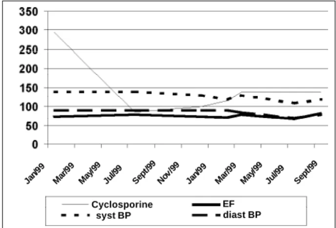
increase in the cyclosporine dose was required during preg-nancy. Blood pressure levels assessed on medical visits we-re never higher than 140/90mmHg; the ejection fraction evi-denced by serial echocardiograms remained stable (fig. 1). Pregnancy was followed up with monthly fetal echography, and no abnormalities were observed. Cardiac function was regularly monitored two-dimensional echocardiography with color Doppler, and all parameters were normal. Renal function did not significantly change during the gestational period. In July of ’98, at the 36th week of pregnancy, a
cesare-an section under general cesare-anesthesia was performed as indi-cated by the obstetrician. The female newborn infant wei-ghed 2,300g and measured 46 cm. Her Apgar score was 8 at the first minute and 8 at the 5th minute. Mother and
daugh-ter evolved satisfactorily. Five months afdaugh-ter birth, the child had no abnormalities, and her neuropsychomotor develop-ment was adequate for her age. The mother was in good con-dition, her blood pressure was 130/80mmHg, her heart rate was 86bpm, her ejection fraction was 74.7%, and she was in NYHA functional class I, with no rejection episode. Curren-tly the patient is taking 200mg/day of cyclosporine.
Discussion
From the pioneering days of heterotopic cardiac trans-plantations in animals at the beginning of the century to the first experiences with human beings in the ‘60s, several obs-tacles have been overcome. Currently, survival for 5 years is estimated to be between 60% and 82% 1-3. Approximately
30% of cardiac transplantations are performed in women, many of whom are of childbearing age 4. The first pregnancy
after cardiac transplantation was reported by Löwenstein et al 5 in 1988. In July ’98, Branch et al 6 reported the existence
of reports in the international literature of 47 pregnancies af-ter cardiac transplantation in 35 women, as 12 of them had had more than one pregnancy. From the 35 first pregnancies, 26 live births, a twin delivery, 4 spontaneous abortions, and 6 therapeutic abortions resulted. From a second pregnancy after cardiac transplantation, 11 births and 2 spontaneous
abortions resulted. In Brazil, the first report published on pregnancy after cardiac transplantation was by Almeida et al 7 in 1995.
Pregnancy after cardiac transplantation has been contraindicated because of the risks to the mother and con-ceptus 8. In the pregnant woman, the risks relate mainly to
hemodynamic alterations and immunosuppressive therapy, determining an elevation in maternal morbidity but not in mortality 8. During pregnancy, blood volume increases
sig-nificantly, an average of 50%. This increase begins in the 6th
week of pregnancy and reaches a peak in the 32nd week of
pregnancy. Cardiac output increases from 30% to 50% and heart rate from 10 to 20bpm. Systemic blood pressure de-creases in the first and second trimesters, reaching preges-tational levels at the end of pregnancy. The most evident change is elevation in end-diastolic volume 9-11. In the
transplanted patient, cardiac output is usually reduced, ventricular compliance is smaller than that in the normal ventricle, and this is maintained by the high central venous pressure 10,11. Intracardiac pressures are normal at rest, but
during exertion, ventricular diastolic pressure drastically in-creases 11. In cardiac transplantation, the heart is
denerva-ted and loses the vagal stimulus, determining an elevation in heart rate at rest, which is usually between 95 and 115bpm. Hypersensitivity to circulating catecholamines al-so occurs 10. If these changes occur in a transplanted
preg-nant woman, they can adversely affect the heart, with an in-crease in cardiac contractility, in central venous pressure, in blood volume, and in cardiac output 7,10. Therefore, cardiac
overload occurs, which in its natural evolution results in an increase in the diameter of the right ventricle with tricuspid regurgitation 7. These hemodynamic changes are usually
well tolerated by transplanted patients during pregnancy. However, hypertension and preeclampsia are more prevalent in these women 13,14. The major risks in regard to
immunosuppres-sive therapy in pregnant women are high indices of gestational diabetes, premature rupture of the membranes, hypertension, adrenal insufficiency, and infection 3,4,15. Other alterations
in-clude a higher incidence of postpartum depression, anemia, and cholestatic jaundice 8, 14,16.
In regard to the conceptus, the major complication is intrauterine growth retardation, accompanied by sponta-neous abortion, prematurity, adrenal insufficiency, low-birth-weight, and teratogenicity 6,11,16. Prematurity is
fre-quent because of the premature rupture of membranes4.
Abortion is more frequent because of the increased risk of infections caused mainly by Listeria, cytomegalovirus, her-pesvirus, and rubella virus 17. Factors related to
hyperten-sion cause a higher prevalence both of low-birth-weight in-fants and intrauterine growth retardation 18-20. The drugs
used in immunosuppressive therapy cross the placental barrier and may have a teratogenic effect 4,7. Lymphopenia,
hypogammaglobulinemia, thrombocytopenia, and thymus hypoplasia are reported in children of women who used aza-thioprine associated with prednisone; these alterations, however, were reversed 4. Cyclosporine is found in fetal
cir-culation in concentrations similar to that found in the
mo-Fig. 1- Evolution of the laboratory tests and blood pressure. Cyclosporine – serum levels of cyclosporine in ng/mL; EF – ejection fraction in %, assessed on echocardio-gram; syst BP – systolic blood pressure in mm Hg; diast BP – diastolic blood pressu-re in mm Hg
Jan/ 99
Mar /99
May /99
Jul/9 9
Sep t/99
Nov /99
Jan/ 99
Mar /99
May /99
Jul/9 9
Sep t/99
Cyclosporine syst BP
Arq Bras Cardiol volume 75, (nº 6), 2000
Bordignon et al Pregnancy after cardiac transplantation
521
ther 21, exerting a possible immunosuppressive effect on the
fetus 7. In experiments with animals, the immunosuppressive
therapy had a teratogenic and fetotoxic effect 7; however, no
appropriate and well-controlled studies on pregnant women are available. Scott et al 16, reviewing 30 cases, found no
congenital anomalies, or fetal and neonatal death.
Rejection of the transplanted organ is an important complication of cardiac transplantation, even under immu-nosuppressive therapy, but this risk is not increased during pregnancy 6,16,22. The most used immunosuppressive
thera-py in transplanted patients is the triple protocol comprising cyclosporin A, azathioprine, and corticotherapy with me-thylprednisolone or prednisone 1,15, which are also
indica-ted in pregnant patients 7,13. Cyclosporine is a lipophilic
cyclic oligopeptide isolated from the fungus Tolypocladium
inflatum Gams. It has a potent immunosuppressive effect,
inhibiting the synthesis of interleukin-2 by the helper T lymphocyte and the proliferation of activated T cells, as well as other lymphokines 23, decreasing the immune response
without a significant mielotoxicity 4. The main side effects in
transplanted patients are as follows: hypertension; renal, hepatic and neurologic damage; gingival hyperplasia; lymphoproliferative disorder; myocardial fibrosis; and hirsutism 24-26. Hypertension is caused by afferent
glo-merular vasoconstriction that decreases gloglo-merular fil-tration and withholds sodium, resulting in a nephrotoxic effect 14. Cyclosporine may be used during pregnancy only
if the benefits justify the potential damage to the fetus 4.
Breast-feeding must be contraindicated because cyclos-porine is present in maternal milk 7. During the third
gesta-tional trimester, due to hemodilution, the levels of serum cyclosporine are significantly reduced and an increase in the daily dose is required 23. Azathioprine is a derivative of
6-mercaptopurine, whose action is mediated by inhibition of the synthesis of the purines, reaching DNA and R-NA15. The adverse effects in transplanted patients include
bone marrow suppression, with leukopenia, thrombocy-topenia, and macrocytic anemia, increased susceptibility to neoplasias and infections, hepatotoxicity, pancreatitis, alopecia, and skin fragility 7. Corticoids act upon
macro-phages and T lymphocytes, and also have a nonspecific immunosuppressive action. Prednisone is more used in the triple protocol and methylprednisolone is preferred in episodes of acute rejection 27.
The contraindication of pregnancy after cardiac trans-plantation is based on the already cited complications, and in some foreign centers therapeutic abortion is recom-mended 4,7,16. No consensus exists about the ideal
contra-ceptive method for female transplanted patients of child-bearing age 28. Contraceptive methods, such as condoms
and vaginal diaphragms, have the smallest adverse effects, but due to their low effectiveness they are not widely re-commended 4. Intrauterine devices have low effectiveness
in immunocompromised patients and have a higher risk of infection 4. Combined oral contraceptives should only be
considered for women without hypertension, and hepatic and thromboembolic diseases, because when associated with immunosuppressive agents they potentiate the occur-rence of hypertension, hepatotoxicity, thromboembolism, cerebral stroke, cholestasis, edema, and gastrointestinal di-sorders 7,27,29. Progestins are the current option because
they can be used in the presence of hypertension and thromboembolism, are effective, and have low levels of ad-verse effects 28. All hormone contraceptive methods require
strict monitoring of the serum levels of the immunosuppres-sive agents because they may influence their metabolism 28.
Permanent contraceptive methods, such as tubal ligation and vasectomy, are a good option because they are safe and have no complications like the hormonal methods do; they, however, are difficult to reverse 4, 7,16.
During pregnancy, the prenatal follow-up is fundamental and should be more strict and frequent when compared with that of normal gestations. The following examinations are indis-pensable during pregnancy: serum levels of cyclosporine, leu-kocyte and platelet counts, routine hemograms, echocardio-graphy, and monitoring of renal function 14,22. Radiographic
examinations and myocardial biopsies are not routinely re-commended 7.
The cardiac transplanted female usually tolerates vagi-nal delivery well, and a cesarean section should only be in-dicated for obstetric reasons 16; however, the cesarean
sec-tion rate in transplanted women is high 30. During childbirth,
the cardiac output is elevated in up to 25% by uterine con-tractions, which cause an increase in venous return; there-fore, strict monitoring is required. In the early postpartum period, an increase in preload occurs due to blood drainage from the uterus to the peripheral circulation, which may re-quire medical intervention 11. In the puerperium,
cyclospori-ne levels tend to increase, requiring a decrease in the daily doses of this drug 7.
522
Bordignon et al
Pregnancy after cardiac transplantation
Arq Bras Cardiol volume 75, (nº 6), 2000
1. Nesralla I, Sant’Anna JRM. Transplante Cardíaco. In: Nesralla I, ed. Cardiologia Cirúrgica: Perspectivas para o Ano 2000. São Paulo: Fundo Editorial Bynk 1994: 617.
2. Kirk EP. Organ transplantation and pregnancy: a case report and review. Am J Obstet Gynecol 1991; 164: 1629-34.
3. Reitz BA. Heart and heart-lung transplantation. In: Braunwald E. Heart Disease: A Textbook of Cardiovascular Medicine. 5th ed. Philadelphia: WB Saunders Co., 1992: 520.
4. Alami WS, Young JB. Pregnancy after cardiac transplantation. In: Elkayam U, Gleicher N, eds. Cardiac Problems in Pregnancy. 3rd ed. 1998: 327. 5. Löwestein BR, Vain N, Perrone S, et al. Successful pregnancy and vaginal
delive-ry after heart transplantation. Am J Obstet Gynecol 1988; 158: 589-90. 6. Branch KR, Wagoner LE, McGrory, et al. Risks of subsequent pregnancies on
mother and newborn in female heart transplantation recipients. J Heart Lung Transplant 1998; 17: 6899-02.
7. Almeida DR, Carvalho AC, Branco JN, Buffolo N, Martinez E. Gravidez após transplante cardíaco. Arq Bras Cardiol 1995; 65: 237-42.
8. Frohlisch ED,Ventura HO, Ochsner JL. Artetial hypertension after orthotopic cardiac transplantation. J Am Coll Cardiol 1990; 15: 1102-3.
9. Reid CL. Pregnancy and heart disease. In:Crawford MH, ed. Current: Diagnosis and Treatment in Cardiology. Connecticut: Appleton and Lange, 1995: 400. 10. Elkayan U. Pregnancy and Cardiovascular Diasese. In: Braunwald E. Heart
Di-sease. 5th ed. Philadelphia: WB Saunders Co., 1997: 1843.
11. Borges JHK, Behr PEB, Barbosa ECD. Cardiopatia e gestação. In: Gomes MF, Azevedo MAV, Frison LI, eds. Rotinas em Cardiologia. Porto Alegre: 1996: 263.
12. Young JB, Winters W, Bourge R, Uretsky B. Task force 4: function of the heart transplant recipient. In: Hunt AS, ed. American College of Cardiology 24th Be-thesda Conference: Cardiac Transplantation. J Am Coll Cardiol 1993; 22: 31. 13. Radomski JS, Ahlswede BA, Jarrell BE, et al. Outcomes of 500 pregnancies in 335
female kidney, liver and heart transplant recipients. Transplant Proc 1995; 27: 1089-90.
14. Delforge C, Kartheuser R, De Plaen JF, Goenen M, Hubinont C. Pregnancy after cardiac transplantation. Transplant Proc 1997; 29: 2481-3.
15. Santos AF, Bittar AE, Keitel E, Garcia VD. Medicação imunossupressora em pa-cientes transplantados: paraefeitos e interações madicamentosas. Rev Med Sta Casa 1996; 8: 1570-4.
16. Scott JR, Wagoner LE, Olsen SL, Taylor DO, Renlund DG. Pregnancy in
heart transplant recipients: managment and outcome. Obstet Gynecol 1993; 82: 324-7.
17. Dick J, Paltramann A, Hamilton D. Listenosis and recurrent abortion in a renal transplant recipient. J Infect 1988; 16: 274-6.
18. Knight M, Redman CWG, Linton EA, Sargent IL. Shedding of syncytiotropho-blast microvilli in to the maternal circulation in pre-eclamptic pregnancies. Br J Obstet Gynecol 1998; 105: 632-40.
19. Babawale MO, Noorden SV, Pignatelli M, Stamp GWH, Elder MG, Sullivan MHF- Morfological interactions of human first trimester placental villi co-cultu-red with decidual explants. Hum Reprod 1996; 11: 444-50.
20. Groot CJM, O’Brien TJ, Taylor RN. Biochemical evidence of impaired tropho-blastic invasion of decidual stroma in women destined to have preeclampsia. Am J Obstet Gynecol 1996; 175: 24-9.
21. Venkataramanan R, Koneru B, Wang CCP, Burckart GJ, Caritis SN, Starzl TE. Cyclosporine and its metabolites in mother and baby. Transplantantion 1988; 46: 468-9.
22. Ohler L, Klein L. Pregnancy after Heart Transplantation. In: Emery RW, Miller LW, eds. Handbook of cardiac transplantation. : Hanley e Beltus 1996: 273. 23. Morris PJ. Cycloporine. In: Morris PJ, ed. Kidney Transplantation: Principles
and Practice. Philadelphia: WB Saunders Co., 1995: 179.
24. Frist WH, Stinson EB, Oyer PE, Baldwin JSC, Shumway NE. Long-term hemodynamic results after cardiac transplantation. J Thorac Cardivasc Surg 1987; 94: 685-93. 25. Austen WG, Cosimi AB. Heart transplantation after 16 years. N Engl J Med
1984; 311: 1436-8.
26. Edwards BS, Loyd MA, Anderson LM. The synergistic effects of cyclosporine and endothelin - demonstration of na important cardiopressor action. Transplan-tation 1993; 55: 8-11.
27. Danovitch GM. Immunosuppressive medications and protocols for kidney transplantation. In: Danovitch GM, ed. Handbook of Kidney Transplantation. Brown and Co., 1992: 67.
28. Casele HL, Laifer AS. Pregnancy after liver transplantation. Semin-Perinatol 1998; 22: 149-55.
29. Kaplan NM, Lieberman E, Neal WW. Hypertension with pregancy and the pill. In: Kaplan NM, Lieberman E, Neal WW, eds. Clinical Hypertesion. 6th ed. Balti-more: Willians and Wilkins, 1994: 343.
30. Wagoner L, Taylor D, Olson S, et al. Immunosuppressive therapy, management and outcomes of heart transplant recipients during pregnancy. J Heart Lung Transplant 1993; 12: 993-1000.