Session editor: Alfredo José Mansur
Associate editors: Desiderio Favarato Vera Demarchi Aiello
Mailing address: Alfredo José Mansur InCor Av. Dr. Enéas C. Aguiar, 44 -05403-000 - São Paulo, SP, Brazil
English version by Stela Maris C. Gandour
Case 4/ 2000 – Heart failure in a 9-year-old child after treatment with anthracycline for W ilms’ tumor (Instituto do Coração of Hospital das Clínicas - FMUSP - São Paulo)
Clinicopathologic Session
A 9-year-old male was referred to our hospital for treat-ment of heart failure after chemotherapy for Wilms’ tumor.
At the age of 6 years, the child was diagnosed with stage III nephroblastoma and underwent nephrectomy. Then, the patient underwent chemotherapy, which included anthracycline at an undisclosed dosage, for one year in ad-dition to radiotherapy. The tumor was considered cured.
One year later, the patient experienced enlargement in abdominal volume and dyspnea. He was diagnosed with ascites due to heart failure, and multiple hospitalizations were necessary for symptom relief. Medical assessment in-cluded angiography with technetium-99m-labeled red blood cells, which revealed a right ventricular ejection fraction of 18% and a left ventricular ejection fraction of 17%. The open liver biopsy showed centrolobular passive conges-tion, adjacent sinusoidal dilaconges-tion, and a mild fibrosis around the central vein. The characteristics of this centrolobular congestion were considered suggestive of hypertension due to postsinusoidal obstruction (heart failure or obstruc-tion of the hepatic veins).
From July ’97 on, the dyspnea evolved to dyspnea on minimum exertion, the abdominal volume enlargement beca-me aggravated, and edema in the lower limbs appeared. The patient was on 0.25mg of digoxin, 60mg of furosemide, and 37.5mg of captopril. Because no clinical improvement oc-curred, the patient was referred to our hospital for heart fai-lure treatment.
The physical examination revealed a malnourished child with facial edema and an increased jugular venous pres-sure (++++/4+). His heart rate was 90bpm, and his blood pressure was 80/60mmHg in all limbs. His thoracic antero-posterior diameter was enlarged, and the lung examination was normal. The heart examination revealed heart ictus in the 4th left intercostal space in the midclavicular line, with
he-art sounds of decreased intensity, and a mild systolic mur-mur in the mitral and tricuspid areas. The abdomen was pro-tuberant with ascites and the liver was palpable 6cm from the right costal margin and 8cm from the xiphoid process.
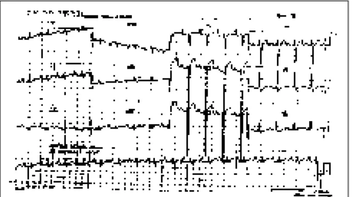
The electrocardiogram showed sinus rhythm, heart rate of 100 bpm, P axis +60° forward, PR interval of 0.24 s,
QRS axis undetermined in the frontal plane, backward, and biatrial and left ventricular overload (fig. 1).
The chest X-ray in the posteroanterior view showed increased lung vascularization, and cardiomegaly (+++/4+) with bulging of the left middle arch.
The echocardiogram revealed diffuse marked dilation and hypokinesia of both ventricles and dilation of the pul-monary artery (table I).
Radiocardiography with technetium-99m-labeled red blood cells synchronized with the electrocardiogram re-vealed a pulmonary transit time above 15 s (normal <7s). The right atrium was enlarged, and both ventricles exhibited marked dilation and hypokinesia. In phase analysis, no change in the normal sequence of chamber movements was observed. Left ventricular ejection fraction was 13%.
During follow-up, the patient required hospitalization due to worsening of the heart failure, and the diagnosis of digitalis intoxication was cogitated. Digoxin was then sus-pended for 15 days, and the dosages of the other medica-tions were increased to 37.5mg of captopril, 80mg of furo-semide, and 25mg of spironolactone. Cardiac scintigraphy with gallium showed no signs of myocardial inflammation.
Oncological assessment considered the tumor cured and with no participation in the patient’s current clinical findings.
Four months later, the patient was admitted to the hos-pital once again because of heart failure. On physical exami-nation, his pulse was 112 bpm, his respiratory rate 44 ipm, and his blood pressure 80/60mmHg. His jugular venous pressure was increased (++++/4+). Lung examination revea-led wheezes and crepitant rales in the pulmonary bases. Heart examination revealed muffled cardiac sounds and a systolic murmur (++/4+) in the mitral area. The abdomen was tense because of ascites, which made liver palpation diffi-cult. Edema was visible in the lower limbs. Stasis dermatitis and ulcers existed in both legs.
Dopamine, dobutamine, furosemide, and albumin were administered by intravenous via, and the 37.5mg of capto-pril already being used was maintained. The patient used all these medications during the entire hospitalization, and a partial improvement of the dyspnea and the lower limb ede-ma was observed. During hospitalization, potassium chlo-ride was also administered for correcting hypokalemia on several occasions.
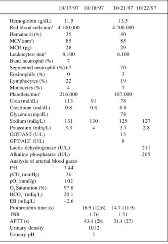
Table II shows the laboratory tests.
The abdominal ultrasound depicted a great amount of ascites, a small left pleural effusion, and a normal-sized left kidney (10cm). The hepatic veins were dilated. The ascitis was drained, and the biochemical examination of the ascitic fluid is shown in table III. Cytology of this fluid revealed innumerable reactive mesothelial cells, histiocytes, red blood cells, and innumerable neutrophils and lymphocytes, but no neoplastic cells.
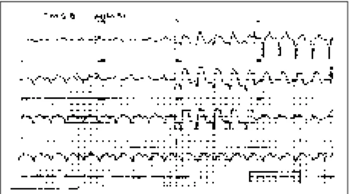
The patient had sustained ventricular tachycardia, and repeated electric cardioversions were unsuccessful. Ventri-cular fibrillation occurred and was reversed to atrial rhythm with widened QRS and elevation of the ST segment (fig. 3), but death followed.
Discussion
Clinical considerations – The findings are compatible
with secondary dilated cardiomyopathy. The patient’s his-tory reveals a well-defined cause: chemotherapy for treat-ment of nephroblastoma. The diagnostic examinations (electrocardiogram, chest X-ray, and radiocardiography wi-th technetium-99m-labeled red blood cells) showed a dila-ted pattern and diffuse hypokinesia of the myocardium, marked left ventricular dysfunction with no signs or fin-dings of hypertrophic or restrictive forms.
The echocardiogram showed no left ventricular hy-pertrophy and no data suggesting restrictive or storage disease.
At first, we should differentiate cardiomyopathy from specific cardiac muscle disease or secondary cardiomyo-pathy. Cardiomyopathy is an idiopathic or primary disease of the cardiac muscle of unknown cause, but secondary car-diomyopathy is a disease of the cardiac muscle of known cause. Secondary cardiomyopathy is clinically similar to primary cardiomyopathy, but its cause is not congenital, valvar, coronary artery, pericardial, or hypertensive disease. Therefore, we should begin our considerations of the patient’s differential diagnoses with idiopathic dilated car-diomyopathy.
Idiopathic dilated cardiomyopathy is a syndrome cha-racterized by cardiac enlargement and a reduction in the systolic function of one or both ventricles. Even though the cause is not very well defined, more than 75 specific disea-ses of the cardiac muscle may cause manifestations similar to those of dilated cardiomyopathy. This condition may
Table I – Echocardiographic findings
Cardiac structure /dimension 9/30/97 10/20/97 Interventricular septum (mm) 6
-Posterior wall (mm) 6
-Aorta (mm) 23
-Left atrium (mm) 37 Enlarged
Left ventricular diastolic diameter 50 56 Left ventricular systolic diameter 44 51 Percentage variation in LV diameter 12 9 Mitral insufficiency Mild Intense Right ventricular diameter 31 Dilated Tricuspid insufficiency Mild Intense Fig. 1 – Electrocardiogram. Biatrial and left ventricular overload.
Table II – Blood laboratory tests at hospital admission
10/17/97 10/18/97 10/21/97 10/22/97 Hemoglobin (g/dL) 11.5 13.5
Red blood cells/mm3 4.100.000 4.700.000
Hematocrit (%) 35 40
MCV (mm3) 85 85
MCH (pg) 28 29
Leukocytes/ mm3 8.100 6.100
Band neutrophil (%) 7
Segmented neutrophil (%) 67 70
Eosinophils (%) 0 3
Lymphocytes (%) 22 19
Monocytes (%) 4 7
Platellets/mm3 216.000 187.000
Urea (md/dL) 113 91 78
Creatinine (md/dL) 0.8 0.8 0.8
Glycemia (mg/dL) 78
Sodium (mEq/L) 131 130 129 127 Potassium (mEq/L) 3.3 4 3.7 2.8
GOT/AST (U/L) 15
GPT/ALT (U/L) 8
Lactic dehydrogenase (U/L) 211 Alkaline phosphatase (U/L) 205 Analysis of arterial blood gases
P H 7.44
pCO2 (mmHg) 30 pO
2 (mmHg) 102
O2 Saturation (%) 97.6 HCO
3
- (mEq/L) 20.1
EB (mEq/L) - 2.6
Prothrombin time (s) 16.9 (12.6) 14.7 (11.9)
INR 1.76 1.51
APTT (s) 43.4 (28) 31.4 (27) Urinary density 1012
Urinary pH 5
Table III - Characteristics of the ascitic fluid
Ascitic fluid Blood
Total protein (g/dL) 4.4 6.6
Albumin (g/dL) 2.9 4.2
Glucose (mg/dL) 108 109
Lactic dehydrogenase (mg/dL) 211 101
sion is less frequent and is characterized by myocardial fi-brosis with or without endocardial fifi-brosis. Left or right or both ventricular dysfunction(s) at rest or during exercise seems to be a common asymptomatic finding occurring 5 to 20 years after radiotherapy 3. Thus the clinical features of
heart disease caused by radiation is not compatible with that of rapidly progressive heart failure refractory to clinical treatment. In addition, it depends on the directly absorbed irradiation by heart, and in the reported case with stage III nephroblastoma, radiotherapy was abdominal.
Another differential diagnosis relates to metastases. Cardiac metastases may result in pericardial involvement (pericarditis or tamponade), superior vena cava syndrome, arrhythmias, cardiomegaly without heart failure, or even no-ninfectious/marantic endocarditis.
In case of myocardial or endocardial metastases, they may be detected on echocardiography, computed tomogra-phy, or nuclear magnetic resonance, or both. In our case, the echocardiogram showed no lesion that could suggest me-tastases. Most of the metastatic cardiac lesions are clinica-lly silent and found only at autopsy 4.
Tumors producing cardiac metastases are in order of frequency: 1) lung cancer; 2) breast cancer; 3) malignant me-lanoma; 4) lymphomas; 5) leukemias 5.
Another point to be considered is that Wilms’ tumor (or nephroblastoma) preferentially metastasizes to the lungs, liver, and contralateral kidney, and not to the heart. Its survi-val rate with appropriate treatment is good in more than 90% of patients, and in our case the tumor was assessed and considered cured 6.
Another differential diagnosis is the anomalous origin of the left coronary artery from the pulmonary artery. In patients with this anomaly, the left ventricle is severely impaired and may develop generalized hypokinesia because of ischemia or acute myocardial infarction. The echocardio-gram may show abnormalities in segmental wall motion and in some cases identify the left coronary artery originating from the pulmonary trunk. The diagnosis is established through angiographic demonstration. If not surgically treated, this malformation can be fatal. Eighty to 90% of the patients with this malformation die in the first year of life; the disorder, however, may manifest itself as heart failure, angina, syncope, or death only at adulthood. Thus the general sce-nario does not fit our patient’s case, both in regard to the age range and echocardiographic findings.
Cardiotoxicity related to chemotherapy (anthracycli-ne) is another differential diagnosis to be considered. Even though most of the complications related to this drug are limited to rapidly proliferating tissues, such as the bone marrow and the gastrointestinal tract, and include myelo-suppression, early and late cardiotoxicity has also been re-cognized as a complication. Cardiotoxicity is an important side effect and has a poor prognosis. Therefore, surveillan-ce of left ventricular systolic function is part of the medical assessment during the use of anthracycline 7.
The causal mechanism of the cardiotoxicity of anthra-cycline-induced cardiomyopathy is unknown. Major evi-result from a common final via of myocardial lesion by a
number of mechanisms, such as cytotoxic, metabolic, immu-nologic, genetic, or infectious mechanisms.
Idiopathic dilated cardiomyopathy usually affects middle-aged people, which is not our case. Our patient is a child with symptoms beginning after treatment for Wilms’ tumor. However, we should remember that among the major mechanisms of primary cardiomyopathy we could cite gene-tic factors, immunologic abnormalities, and viral/cytotoxic myocarditis.
Our patient underwent chemotherapy and radiothera-py in his treatment. One initial complication of this type of treatment is myelosuppression that may facilitate clinical or subclinical viral infection and myocarditis, one of the proba-ble origins of idiopathic cardiomyopathy.
According to my understanding, this is not the patient’s diagnosis because a well-defined cause exists in the history for the onset and severe evolution of the heart failure.
Another differential diagnosis is myocarditis, and the most frequent one is viral myocarditis caused by Coxsackie B virus. In children, the evolution may not be as benign as in adults, in whom myocarditis is usually limited. We would like to stress that the patient was not only immunosup-pressed by the tumor but also by the treatment performed. Consequently, he had a greater predisposition to severe infections; however, cardiac scintigraphy with gallium showed no cardiac inflammation compatible with myocardi-tis, which is found in most children with dilated cardiomyo-pathy 1.
Another differential diagnosis involves radiotherapy, which may result in a variety of chronic, occasionally acute, cardiac complications, such as pericarditis with effusion, cardiac tamponade or constriction, fibrosis of the coronary arteries, myocardial infarction, valvar abnormalities, myo-cardial fibrosis, and disorders in heart conduction.
Even though radiation causes a certain degree of tissu-lar lesion, clinically significant cardiac impairment occurs in a minority of patients long after radiotherapy is over.
dence suggests a correlation with the capacity to produce free radicals, and the resulting oxidative stress 8. Another
cause could be the capacity of anthracycline to interfere with the sodium-potassium pump of the sarcolemma.
Risk factors for the development of anthracycline-in-duced cardiomyopathy exist and some of them will be consi-dered below. The total cumulative dose of anthracycline ad-ministered is one of these risk factors. In our case, however, we do not know the total dosage of anthracycline adminis-tered. Cases of severe heart failure 9,10 developing with
ad-ministration of low doses of anthracycline have also been reported.
Others risk factors are the patient’s age in regard to the period of exposure. Our patient was 6-years-old (Wilms’ tu-mor occurs in children, and its greatest incidence is at the age of 3 years); combined chemotherapy, which is the basis of the treatment of nephroblastoma. It should be performed in patients with stage III Wilms’ tumor; cardiac or mediasti-nal radiotherapy is another risk factor. The patient under-went radiotherapy, and in patients with stage III nephroblas-toma radiation application should be latus-abdominal; hypertension are also risk factors. Wilms’ tumor is usually associated with an abdominal mass, anemia, hypertension, abdominal pain, and fever; previous heart disease, espe-cially coronary artery disease, is a risk factor; and hepatic disease.
The cardiomyopathy due to anthracycline is detected by physical examination, electrocardiogram, and echo-cardiographic measurements. The ejection fraction measu-rement is a sensitive noninvasive method for detecting and following cardiomyopathy. Angiocardiography and en-domyocardial biopsy may also be used. The latter, despite being very sensitive and specific, is also the most invasive and least available of them all.
Cardiomyopathy due to anthracycline may have a good evolution with conventional treatment or may evol-ve with refractory heart failure, and cardiac transplantation may even be considered. Our patient was apparently healthy in regard to his cardiovascular condition, but evolved with severe cardiomyopathy after chemotherapy with anthracycline for nephroblastoma in the presence of many risk factors for the development of secondary cardiomyopathy, such as age, combined chemotherapy, etc. The patient had a severe clinical evolution, which is in accordance with several cases reported of this important specific cardiac muscle disease.
The main diagnostic hypothesis in this case is that of the natural evolution of a cardiomyopathy secondary to the use of anthracycline, emphasizing that the patient had an anatomical substrate ventricular dysfunction and hypoka-lemia/electrolytic alteration (K=2.8)] for the development of ventricular tachycardia. The pulseless electric activity in cardiopulmonary arrest15 is very common in patients with
severe left ventricular dysfunction, the final event of this disease and, in a few cases, it may be associated with car-diac rupture.
The second diagnostic hypothesis would be
cardio-myopathy secondary to anthracycline and complicated with pulmonary thromboembolism, resulting in worsening of the functional class and aggravation of the patient’s condition, leading to death.
(Dr. Otávio Andrade Carneiro da Silva)
Diagnostic hypothesis - Toxic heart disease
seconda-ry to the use of anthracycline
Urologist’s comment - Survival of children with
Wilms’ tumor has drastically increased in the last 25 years mainly resulting from cooperative study groups, such as the National Wilms’ Tumor Study (NWTS), the Interna-tional Society of Pediatric Oncology (SIOP), and in Brazil, the Cooperative Group for Treatment of Wilms’ Tumor (GCTTW). Currently, the overall survival of these patients reaches 85%, progressing from “cure at any cost” to “cure with lower morbidity”. All patients with Wilms’ tumor are treated aiming at a cure 11,12.
The patient studied was diagnosed with stage III Wilms’ tumor (residual tumor confined to the abdomen: in-volved lymph nodes, peritoneal dissemination, peritoneal implants, not completely resected tumor, tumor rupture before surgery). Treatment for stage III comprises, in addi-tion to surgical resecaddi-tion, chemotherapy with 3 drugs (ac-tinomycin, vincristine, and adriamycin) and local radio-therapy. The correct distinction between stages II and III is of major importance, because stage II children undergo chemotherapy with 2 drugs (actinomycin and vincristine), do not undergo radiotherapy, and have a much lower mortality.
The heart may be injured by radiotherapy or chemo-therapy.
Adriamycin, an antibiotic of the anthracycline class, binds to DNA and inhibits nucleic acid synthesis, causing cardiomyopathy with a lower contractility that may evolve to congestive heart failure. Results of the NWTS II and III show development of congestive heart failure in 1.7% of the patients in the long-term, with symptom onset from 1.3 to 11.7 years after treatment. Patients receiving radiotherapy on the chest developed congestive heart failure in 5.4% of
the cases (adriamycin potentiates the effects of radiothe-rapy).
Some authors recommend cardiac assessment and Holter monitoring every 3 years after treatment, mainly in stress situations such as pregnancy.
(Dr. Wladimir Alfer)
Autopsy
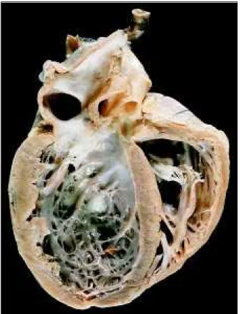
The heart weighed 200g (normal for the age is up to 115g). Grossly, a mild straw-colored pericardial effusion was observed, as well as global dilation of the four cardiac chambers with thinning of the ventricular and atrial walls (fig. 4). An organized thrombus was evidenced in the right auricle. Histologic examination of the myocardium revealed diffuse hypertrophy of the cardiomyocytes, absence of inflammatory infiltrate, and focal areas of interstitial and subendocardial fibrosis. Areas of microinfarcts of up to 24-hour evolution were observed in diverse sites of the ventricular walls, from the outer third to the inner third, the right ventricle being the most affected. These areas even formed transmural ischemic areas and vacuolization of cardiomyocytes (fig. 5). The valves and the coronary, epicardial, and intramural arteries showed no changes. The liver and lungs showed chronic passive congestion with focal areas of acute pulmonary hemorrhage. Surgical absence of the left kidney was noted and no metastatic
lesion or recidivation of the primary tumor was evidenced. Mild fibrous adhesions between the abdominal viscera were observed (surgical bridles).
(Dr. Glacy Sabra Vieira)
Anatomicopathological diagnoses – 1) dilated
cardio-myopathy; 2) histological myocardial changes secondary to hypopotassemia; 3) morphological changes of congesti-ve heart failure.
Comments
In 1994 the patient underwent to a surgery for Wilms’ tumor in his left kidney, and after that he underwent chemo-therapy for one year. One year later he developed conges-tive heart failure secondary to cardiotoxicity caused by the anthracycline.
Anthracycline is a class of chemotherapy drugs that are cardiotoxic and may have caused the patient’s cardio-megaly. Clinically, the patient had refractory congestive heart failure preceded by an enlargement in the cardiac chambers. These changes are related to an individual sen-sitivity to the administered dose 13 and depend on the
ac-cumulation of the doses 14. In our case, the heart showed a
pattern of dilated cardiomyopathy involving the 4 cham-bers.
The pathogenesis of the lesion caused by anthra-cycline administration is not well established. Some propo-sals include the generation of free radicals and toxic meta-bolites, changes in calcium levels and prostaglandins. The most characteristic findings are those of electron micros-copy and comprise sarcoplasmatic dilation and myofi-brillar loss with no inflammatory infiltrate. In a study 14 of
11 patients with cardiotoxicity induced by doxorubicin during antineoplastic treatment, right ventricular endo-myocardial biopsies showed those lesions in all patients, but in 6 patients myocarditis occurred with a predominan-tly CD8+ lymphocytic infiltrate.
Fig. 5 - Hypopotassemia: necrosis in the contraction band (arrows) and vacuoliza-tion of the cytoplasm in myocardiocytes from the right ventricle, the most suscep-tible site (HE, 160x).
1. Camargo PR, Mazzieri R, Snitcowsky R. Endomyocardial biopsy and myocar-dial imaging with 67-gallium in the diagnosis of active myocarditis in children with dilated myocardiopathy. Arq Bras Cardiol 1990; 54: 27-31.
2. Benoff LJ, Schweitzer P. Radiation therapy-induced cardiac injury. Am Heart J 1995; 129: 1193-6.
3. Constine LS, Schwartz RG, Savage DE, King V, Muhs A. Cardiac function, perfu-sion, and morbidity in irradiated long-term survivors of Hodgkin’s disease. Int J Radiat Oncol Biol Phys 1997; 39: 897-906.
4. Majano-Lainez RA. Cardiac tumors: a current clinical and pathological perspec-tive. Crit Rev Oncog 1997; 8: 293-303.
5. Abraham KP, Reddy V, Gattuso P. Neoplasms metastatic to the heart: review of 3314 consecutive autopsies. Am J Cardiovasc Pathol 1990; 3: 195-8. 6. Thomas PR. Wilms’ tumor: Changing role of radiation therapy. Semin Radiat
Oncol 1997; 7: 204-211.
7. Sung RY, Huang GY, Shing MK, et al. Echocardiographic evaluation of cardiac function in paediatric oncology patients treated with or without anthracycline. Int J Cardiol 1997; 60: 239-48.
8. Gille L, Nohl H. Analyses of the molecular mechanism of adriamycin-induced car-diotoxicity. Free Radic Biol Med 1997; 23: 775-82.
References
9. Dunn J. Doxorubicin-induced cardiomyopathy. J Pediatr Oncol Nurs 1994; 11: 152-60.
10. Narahara KA, Singer JW, Ritchie JL, Williams DL, Hamilton GW, Kennedy JW. Time- and dose-dependent changes in ejection fraction after anthracycline thera-py. J Cardiovasc Pharmacol 1979; 1: 395-401.
11. Green DM, Thomas PR, Shochat S. The treatment of Wilms tumor. Results of the Na-tional Wilms Tumor Studies. Hematol Oncol Clin North Am 1995; 9: 1267-74. 12. Pritchard J, Imeson J, Barnes J, et al. Results of the United Kingdom Children’s
Cancer Study Group first Wilms’ Tumor Study. J Clin Oncol 1995; 13: 124-33. 13. Fenoglio JJ. The effects of drugs on the cardiovascular system. In: Silver MD, ed. Cardiovascular Pathology. New York: Churchill Livingstone, 1983: 1085-1107. 14. Gaudin PB, et al. Myocarditis associated with doxorubicin cardiotoxicity. Am J
Clin Pathol 1993; 100: 158-63.
15. Lindeman RD. Hypokalemia: causes, consequences and correction. Am J Med Sciences 1976; 272: 5-17.
16. Cowan MJ, Giddens Jr WE, Reichenbach DD. Selective miocardial cell necrosis in nonhuman primates. Arch Pathol Lab Med 1983; 107: 34-9.
17. French JE. A histological study of the heart lesions in potassium-deficient rats. A.M.A. Arch Pathology 1952; 53: 485-96.
Myocarditis is not reported as a characteristic of anth-racycline-induced intoxication, but this study shows that a possible association may exist through an autoimmune me-chanism 2.
On microscopic examination, the most frequently found changes are chronic, consisting of a fibrous scar 13. In our
case, we found focal areas of fibrosis that could correspond to chronic lesion due to anthracycline administration. These findings are common to different processes; therefore, the clinical information about chemotherapy is necessary.
Another microscopic finding was the presence of focal
areas of myocardial microinfarcts of up to 24-hour evolution. These microinfarcts were related to the immediate cause of death. Clinically, the patient developed hypopotassemia, whi-ch is a known cause of necrosis of myocardial fibers with va-cuolar degeneration similar to an infarct in different myocardial areas 15-17. This involves mainly the right ventricle 17, as
obser-ved in our patient. The coronary arteries showed no abnorma-lity on the anatomicopathological study, eliminating a possible coronary cause for these infarcts. Therefore, congestive heart failure aggravated by multiple areas of acute myocardial infarct (due to the hypopotassemia) was the cause of death.