Arq Bras Cardiol volume 75, (nº 6), 2000
Franken et al Pseudo-myocardial infarction during episode of Herpes Zoster
527
Faculdade de Ciências Médicas da Santa Casa de São Paulo
Mailing address: Roberto A. Franken – Rua Dr. Franco da Rocha 163/52 – 05015-040 – São Paulo, SP, Brazil
English version by Stela Maris C. Gandour
Roberto A. Franken, Marcelo Franken
São Paulo, SP - Brazil
Pseudo-Myocardial Infarction During an Episode of
Herpes Zoster
Case Report
The patient arrived at the emergency unit with a his-tory of acute myocardial infarction, for which she was treated. Without improvement in the pain, the patient deve-loped heart failure and underwent a hemodynamic study, which showed normal coronary arteries and extensive ventricular impairment. During evolution, the clinical fin-dings improved and herpes zoster appeared on the right shoulder. In a few months the clinical findings subsided, and the findings of the electrocardiogram, chest X-ray, and ventricular function were normal. The patient is currently asymptomatic.
Clinical manifestation of myocarditis may vary from an asymptomatic state secondary to focal infection to severe heart failure. In some cases, clinical manifestations may si-mulate myocardial infarction1, as electrocardiographic and
laboratory findings do. Approximately 25 viruses may be associated with myocarditis, including the varicella-zoster virus. Myocardial involvement during infection by herpes zoster virus is rare, and when it occurs, it is usually asympto-matic or manifests as heart failure 2.
We report the case of a patient with clinical findings of myocardial infarction and infection by herpes zoster virus.
Case Report
The patient is a white 68-year-old female, who arrived at the emergency unit complaining of precordial pain that ir-radiated to the left upper limb and was accompanied by sweating and weakness for 3 hours. The patient reported malaise in the previous 2 days, and a continuous discomfort in her right shoulder with no alleviating or worsening fac-tors. The patient was hypertensive and had been using 20mg of enalapril for the last 5 years. She also reported ha-ving a family history of longevity.
On physical examination the patient was restless, pale, sweating, and dyspneic. Her blood pressure was 134/80m-mHg in both upper limbs, her heart rate was 116bpm, the lungs showed no rales, and the peripheral pulses were palpable.
The clinical hypothesis of myocardial infarction led the patient to immediately undergo electrocardiography (fig. 1), which confirmed the diagnosis. A thrombolytic (streptokinase) was started, and hypotension occurred during infusion; therefore, the dripping was decreased, dopamine was started followed by dobutamine. After infusion, the patient remained restless, more dyspneic, and still complained of pain. On pulmonary auscultation, crepitant rales could be heard in the inferior 2/3 of the lungs. The patient was given sodium nitroprusside and a diuretic. The chest X-ray (fig. 2) showed the classical pattern of pulmonary edema. As the pain persisted after 4 hours of thrombolytic infusion, the patient underwent coronary angiography, which revealed normal coronary arteries and an increase in left ventricular end-systolic volume due to an extensive anterior and inferior akinetic area, and also apical dyskinesia (fig. 3). Table I shows the levels of the cardiac enzymes.
The echocardiogram of 3/12/96 revealed an ejection fraction of 45%, mild mitral insufficiency, a deficit in left ventricular relaxation, middle apical dyskinesia, and anteroinferior and septal akinesia. The remaining walls were hyperkinetic.
The patient gradually improved. Twenty-four hours after admission, the patient reported aggravation of the pain in her right shoulder and upper limb, and vesicles with a clear content and surrounded by a pink halo appeared on her right shoulder. The clinical diagnosis of herpes zoster was then established. Evolution of the electrocardiogram is shown in figures 4 and 5 and the chest X-ray in figure 6. The echocardiogram of 3/19/96 revealed akinesia in the apical portion of the septum and anterior wall, with no mitral insufficiency. On 4/9/96, the echocardiogram was normal with no changes in the segmentary contractility and normal relaxation.
Currently, the patient is using a calcium channel blo-cker (diltiazem) and is clinically well, with normal cardiac function and no deficit.
Discussion
528
Franken et al
Pseudo-myocardial infarction during episode of Herpes Zoster
Arq Bras Cardiol volume 75, (nº 6), 2000
extension of the myocardial impairment was notable, the latter being clinically observed and angiographically and echocardiographically confirmed. During evolution, some vesicles appeared allowing the diagnosis of herpes zoster, which had already been manifesting as a discomfort in the right shoulder a few days prior to the episode of chest pain. According to the report by Franken et al1, the new Q
wave of the electrocardiogram should be interpreted and the findings referred to as an electrically inactive zone and not as a necrotic area.
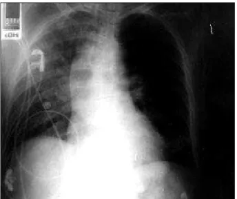
Fig. 2 – Chest X-ray (3/10/96). Pattern of pulmonary edema.
Fig. 1 – Electrocardiogram on hospital admission: elevation of the ST segment in D1, AV1, V2, V3, V4, V5, and V6; QS pattern in D2, D3, AVf, V2, V3, V4; and QR pattern in V5, and V6. Conclusion: electrically inactive zone in inferior and anterolateral wall, anterolateral lesion.
Fig. 3 – Angiographic study (3/10/96): normal coronary arteries, ventriculography shows an anteroinferior akinetic zone and an apical dyskinetic zone: a) end-diastole, b) end-systole.
A
B
Table I – Evolution of the cardiac enzymes
CPK CKMB
Admission 140 18
6 hours 173 32
12 hours 261 64
Arq Bras Cardiol volume 75, (nº 6), 2000
Franken et al Pseudo-myocardial infarction during episode of Herpes Zoster
529
Fig. 4 - Electrocardiogram (3/30/96). Reappearance of R in D2 D3, AVf, V4, V5, and V6; diffusely negative T wave. Conclusion: anterior electrically inactive zone, diffuse alterations of ventricular repolarization.
Fig. 5 - Electrocardiogram (6/10/96). Normal electrocardiogram.
Fig. 6 – Chest X-ray (6/10/96). Normal.
terized by transient myocardial impairment after an acute ischemic event, and results from cytoplasmic accumulation of calcium and free radicals. From the electrophysiological point of view, the stunned area becomes unresponsive to the electric stimulus and, consequently, noncontractile, perfectly mimicking an area of necrosis. The region stunned by electrolytic changes becomes incompletely repolarized (rest potential above –20 mv) and, therefore, unresponsive. We found reports in the literature of myocarditis asso-ciated with herpes zoster virus, in which the clinical mani-festation is heart failure 5. However, we found no report in
the literature on stunned myocardium related to acute myo-carditis, which would be perfectly acceptable from the pa-thophysiologic point of view. Du Bois et al 6 reported a
simi-lar case induced by the use of interleukin-2 in the treatment of neoplasia.
We cannot, however, discard viral impairment of the coronary arteries, as has already been observed in epide-miological evidence relating acute coronary artery episodes to infectious disease 7.
Heyndrick et al 3 reported the entity related to
charac-530
Franken et al
Pseudo-myocardial infarction during episode of Herpes Zoster
Arq Bras Cardiol volume 75, (nº 6), 2000
1. Franken RA, Assef MAS, Stella FP, Gandra SMA, Rivetti AL. Onda Q do eletro-cardiograma: Necrose ou zona eletricamente inativa? O pseudo-infarto do mio-cárdio. Arq Bras Cardiol 1992; 59: 297-301.
2. Guess H. Population – based studies of Varicella complications. Paediatrics 1986; 78: 723-7.
3. Heyndrickx GR, Millardd RW, Mc.Ritchie RJ, et al. Regional myocardial functio-nal and electrophysiological alterations after brief coronary occlusion in cons-cious dogs. J Clin Invest 1975; 56: 978-85.
References
4. Braunwald E, Kloner RA. The stunned myocardium: prolongued post ischemic ventricular disfunction Circulation 1982; 66: 1146-9.
5. Tsintsof A, Delprado WJ, Keogh AM. Varicella zoster myocarditis progressing to cardiomyopathy and cardiac transplantation. Br Heart J 1993; 70: 93-5. 6. Du Bois JS, Udelson JE, Atkins MB. Severe reversible global and regional
ven-tricular disfunction associated with high dose interleukin-2 immunotherapy. J Immunother Emphasis Tumor Immunol 1995; 18: 119-23.