Case Report
Modified Kraske Procedure with Mid-Sacrectomy
and Coccygectomy for
En Bloc
Excision of Sacral Giant
Cell Tumors
Vítor M. Gonçalves,
1Álvaro Lima,
2João Gíria,
3Nuno Carvalho,
4José Parreira,
5and Manuel Cunha e Sá
11Neurosurgery Department, Garcia de Orta Hospital, Avenida Torrado da Silva, 2801-951 Almada, Portugal
2Orthopedic Department, Beatriz ˆAngelo Hospital, Avenida Carlos Teixeira, 2674-514 Loures, Portugal
3General Surgery Department, CUF Infante Santo Hospital, Travessa do Castro, 1350-070 Lisbon, Portugal
4General Surgery Department, Garcia de Orta Hospital, Avenida Torrado da Silva, 2801-951 Almada, Portugal
5Plastic and Reconstructive Surgery Department, Garcia de Orta Hospital, Avenida Torrado da Silva, 2801-951 Almada, Portugal
Correspondence should be addressed to V´ıtor M. Gonc¸alves; [email protected]
Received 14 July 2014; Accepted 5 October 2014; Published 16 October 2014
Academic Editor: Yoshiharu Kawaguchi
Copyright © 2014 V´ıtor M. Gonc¸alves et al. his is an open access article distributed under the Creative Commons Attribution License, which permits unrestricted use, distribution, and reproduction in any medium, provided the original work is properly cited.
Sacral giant cell tumors are rare neoplasms, histologically benign but potentially very aggressive due to the diiculty in achieving a complete resection, their high recurrence rate, and metastization capability. Although many treatment options have been proposed,
en blocexcision with tumor-free margins seems to be the most efective, being associated with long term tumor control, improved
outcome, and potential cure. An exemplifying case of a 29-year-old female with progressive complaints of pain and paresthesias in the sacral and perianal regions, constipation, and weight loss for 6 months is presented. he surgical technique foren blocexcision of a large sacral giant cell tumor through a modiied Kraske procedure with mid-sacrectomy and coccygectomy is described. Complete resection with wide tumor-free margins was achieved. At 5 years of follow-up the patient is neurologically intact, without evidence of local recurrence on imaging studies. A multidisciplinary surgical procedure is mandatory to completely remove sacral tumors. In the particular case of giant cell tumors, it allows minimizing local recurrence preserving neurovascular function, through a single dorsal and deinitive approach.
1. Introduction
Giant cell tumors (GCT) of bone are rare neoplasms compris-ing 5% of all primary bone tumors in adults [1] and 5 to 10% of all benign bone tumors [2], with a 2% to 8.2% incidence rate [3–5]. hey usually afect metaepiphyseal regions of long bones, most oten in the knee and radius. Sacrum is the third most common site of involvement [2] and the most afected bone of the axial skeleton, accounting for 2–8% of all GCT [6–
8]. his type of neoplasm is the second most frequent primary bone-involved tumor in the sacrum [4].
GCT are histologically benign, presenting a slow growth rate and insidious or clinically silent onset, making early diagnosis diicult. Usually they exhibit a very large size when
diagnosis is made [8]. hey are locally highly aggressive and present a high recurrence rate and the power to metastasize, being associated with high morbidity [2, 9–13]. Although considered benign, they are usually lethal, making them a complex medical disease [14–16]. Distant metastization is unusual. he reported incidence of lung metastases from a histologically proven GCT ranges from 1% to 9% [9, 17–
20]. he local recurrence rate seems to be as high as 33% [4], reaching more than 50% when intralesional curettage excision is performed [2, 8]. his may be explained by diiculties in achieving an early diagnosis, the large tumor volume at initial presentation, aggressive behavior, poorly deined tumor margins, and the diiculty to surgically access these lesions without harming the patient [1, 5, 20]. Local
malignant transformation has also been reported, accounting for 16% of primary cases [8,21].
Magnetic resonance imaging (MRI) and computed to-mography (CT) scans are useful for early diagnosis and preoperative planning [22]. Needle biopsy may be reserved for selected cases [23,24].
Diferent treatment options have been used for sacral GCT [7,14]. hese tumors are relatively resistant to radiation therapy [4, 14, 15, 17], which on the long term may result in radiation-induced sarcoma (3–11%) [15, 21, 25, 26]; no standard chemotherapy protocols are available. his may be the reason why such treatment options remain controversial [14,25].
When located in the sacrum, surgical resection is the pri-mary treatment modality, being advocated by most authors [4, 5, 27–29]. En bloc excision with tumor-free margins, although challenging, is the procedure of choice, once this constitutes the most efective method for local disease control and recurrence prevention, improving outcome and provid-ing the best chance for cure [3,4,14,17,28].
We present an exemplifying case of a patient harboring a lesion, which was surgically treated through a modiied Kraske procedure with mid-sacrectomy and coccygectomy, for en bloc excision of the tumor, with wide tumor-free margins. A detailed and comprehensive step-by-step surgical technique overview is presented.
2. Case Presentation
A 29-year-old female, without known past medical his-tory, was admitted with progressive complaints of severe pain and paresthesias in the sacral and perianal regions-for 6 months. In this period of time she also presented constipation and 5 Kg weight loss. hese symptoms were refractory to medical therapy. Pain exacerbated in the night and by Valsalva maneuvers, causing severe functional disability.
Physical examination revealed severe pain on palpation and percussion of the sacral region, without a visible or palpable lesion or other signs of inlammation. Digital rec-tal examination revealed a large midline presacral mass, ixed to the sacrum, with irm consistency and irregular surface.
he lumbosacral CT and MRI scans showed a large, expansive, and osteolytic lower and mid-sacral lesion, with poorly deined margins, extending up to the inferior half of S2 vertebra. he mass comprised both intra- and extracanalar components, a ventral extension displacing the rectum ante-riorly, and dorsal expansion out of the sacral hiatus and dorsal foramina with sot tissue compromise. It was located in the midline, slightly more pronounced on the right side, in between the inferior half of S2 vertebra and the sacrococcygeal junction. S2 nerve roots were spared but all nerve roots distal to that were involved by the tumor. he coccyx was not afected (Figure 1).
Further diagnostic workup was performed including laboratory studies with tumor markers, chest, abdomen,
and pelvis CT-scan and positron emission tomography. No abnormalities or other lesions were detected.
Ater complete characterization of the boundaries of this sacral solitary lesion, a multidisciplinary elective and deinitive surgery was scheduled with the collaboration of general and plastic surgeons. No previous biopsy was performed.
2.1. Operative Technique. he patient was electively operated by one of the senior authors (A.L.).En blocexcision of the tumor with wide tumor-free margins through a modiied Kraske procedure with mid-sacrectomy and coccygectomy was achieved requiring sacriice of the nerve roots and thecal sac below the level of S2 nerve roots (Figure 2).
Anesthesia and Positioning. Under general anesthesia the patient was intubated. Arterial line was placed for blood pres-sure monitoring. Intravenous dexamethasone and antibiotic prophylaxis (cefazolin 1 g) were administered preoperatively. he patient was positioned prone. All pressure points were covered with padding. Care was taken to avoid elevated abdominal and airway pressures because this would lead to inconvenient bleeding. he posterior lumbosacral area, low back buttock, and posterior thighs were subsequently sterilized and draped ater the skin was dried, giving the plastic surgeons many options for sot tissue reconstruction and wound closure at the inal step of the surgery. Extra care was taken at this stage due to the proximity of the anal oriice to the surgical ield, increasing the risk of wound contamination.
Modified Kraske Procedure. A midline longitudinal skin incision was carried out posteriorly extending from the lumbosacral junction to the coccyx. he sacral fascia was exposed from L5-S1 level to the tip of the coccyx (Figure 3).
hese limits were, respectively, superior and inferior to the tumor and not involved by it. Initially, the fascia opening and sot tissue dissection were performed inferiorly exposing the tip of the coccyx and then coming around anterior to it (Figure 4).
he anococcygeal ligament was transected at a distance from the anal sphincter and, working ventral to the coc-cyx, the levator ani muscles were detached from it and retracted laterally allowing the approach to the presacral space.
Kraske approach was then performed. Finger dissection was used to mobilize the rectum creating a plane between the posterior aspect of the rectum and the ventral part of the sacrum including the anterior surface of the tumor (Figure 5).
(a) (b)
(c)
Figure 1: Preoperative T2-weighted contrast enhanced MRI showing an expansive and osteolytic lower and mid-sacral lesion, extending up to the inferior half of S2 vertebra, with both intra- and extracanalar components and a ventral extension displacing the rectum anteriorly. (a) Coronal, (b) sagittal, and (c) axial views.
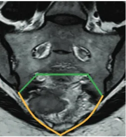
Figure 2: Previeweden blocexcision of the tumor on coronal MRI. Green and yellow lines represent the limits of the specimen to be resected, respectively, corresponding to the 3 sacral osteotomies and the inferior margin around the tumor.
attachments and the inferior portion of the gluteal muscles allowed a full hand to be insinuated into this plane and provide further dissection and palpation of the sacrum above the tumor (Figure 6).
Creation of this space was very important to protect the rectum and provide tactile feedback and guidance during the subsequent osteotomies. he obtained dissection plane was conveniently preserved with large surgical patties with identiication string.
Fascia opening and subperiosteal dissection were then performed superiorly, exposing the tumor-free dorsal surface of the upper sacrum at the S1 and S2 levels (Figure 7).
Figure 3: Exposition of dorsal fascia from lumbosacral junction to the tip of the coccyx.
Figure 4: Inferior sot tissue dissection exposing the tip of the coccyx.
Laterally at the distal sacrum, the gluteus muscle attach-ments and the sacrospinous and sacrotuberous ligaattach-ments were detached exposing the coccygeus and piriformis mus-cles, which were divided revealing the lower elements of the sacral plexus. Subperiosteal dissection was subsequently carried out over the posterior superior iliac spines allowing mobilizing the sot tissues to bilaterally expose the sciatic notches.
At this point the Kraske approach was performed infe-riorly. Superiorly we have exposed the dorsal bony elements including the S1 and S2 lamina, as well as posterior superior iliac spines.
Laminectomy, hecal Sac, and Nerve Root Ligation. Under 2,5x magniication surgical loupes view, S1 and S2 laminec-tomies were accomplished using a ine Kerrison rongeur, allowing exposition of the thecal sac and the tumor-free S1 and S2 nerve roots, going to the respective foramina. At this point it was very important to make sure the correct level was identiied, not to harm the inappropriate nerve roots, as the thecal sac was going to be ligated. S1 and S2 nerve roots were correctly identiied. he S2 nerve roots were dissected and skeletonized on their way to the respective foramen. Tumor was identiied bilaterally in the axilla of the S3 nerve roots
Figure 5: Initial inger dissection used to mobilize the rectum.
Figure 6: Kraske procedure providing access between mesorectum and presacral component of the tumor.
Figure 8: Sot tissues intentionally let behind and included in the specimen to be resected.
which were too intimately involved by the tumor to be spared. Tumor capsule was intact.
hecal sac ligation was then performed with two 2-0 silk ties passed around the thecal sac, distal to S2 nerve roots (Figure 9).
A scalpel blade number 15 was then used to sharply cut the thecal sac, distal to these ties. he distal thecal sac and S3, S4, and S5 nerve roots were compromised by the tumor, being sacriiced and included within the specimen to amputate. A meticulous hemostasis was achieved with coagulation of epidural venous plexus.
Sacral Osteotomies.hree sacral osteotomies were performed, two oblique, executed laterally on each side, between the S2 foramen and the ipsilateral greater sciatic notch, followed by a medial transverse osteotomy, done between the S2 foramina (Figure 10).
Ater thecal sac and nerve root ligation was achieved, bilateral S2 nerve roots were extensively dissected and fol-lowed on their way to the respective S2 foramina. his constitutes an important landmark for the execution of lateral osteotomies, once these extend between the S2 foramina laterally and the greater sciatic notch. Remaining sot tissues at the sciatic notch were dissected with monopolar electro-cautery, enabling a inger to be insinuated superior to the piriformis muscle into the sciatic notch for further dissection, and advanced medially to palpate the ventral S2 foramen. With the inger in place, this important maneuver allowed guiding and safely performing the lateral osteotomies. Once the osteotomy has passed from the S2 foramen out to the sciatic notch, the lateral aspect of the sacrum was cut allowing the lateral osteotomies to be completed. his step was accom-plished bilaterally. Sacroiliac joints were completely spared and safeguarded.
Having completed the lateral osteotomies, some addi-tional dorsal gluteal musculature was taken down laterally with the monopolar electrocautery. he Kraske approach was again used for guidance of the transverse osteotomy. he tactile feedback helped to direct the bone cut. his could be performed with osteotomes oriented in a transverse direction
Figure 9: hecal sac ligation distal to S2 nerve roots (red line).
between the S2 foramina, beginning at one S2 foramen and carrying over to the other. he hand inserted by the Kraske approach into the presacral space protected the dorsal aspect of the rectum and again provided additional tactile feedback for the osteotomy. It helped to guide osteotomes’ trajectory and sense when the anterior bony cortex was perforated and bony cut has been completed. Bleeding from the sacral osteotomies was controlled with bone wax.
En Bloc Resection of the Tumor.At this point, we performed a coccygectomy and the Kraske approach inferiorly, gluteal musculature release laterally, thecal sac and nerve root liga-tion below the level of S2 nerve roots, and lateral and trans-verse osteotomies superiorly. Ater this was accomplished, the specimen was tilted dorsally, stretching the S2 nerve roots so that they could be extensively dissected all the way from their origin at the thecal sac, freeing them from the remaining foramina and tracing them out distally.
With additional mobilization, sot tissue attachments were subsequently released. he remaining deeper muscular and ligamentous (sacrotuberous and sacrospinous ligaments) attachments and the distal ends of the S3, S4, and S5 nerve roots were identiied and cut with the monopolar electro-cautery. his allowed the specimen to be circumferentially freed and removed from the surgical ield, resulting in a satisfactoryen blocresection with wide tumor-free margins.
he specimen included the tumor with its presacral component (Figure 11(a)) and the dorsal paraspinal muscles, let to provide a wide margin posteriorly (Figure 11(b)). A satisfactory superior margin was also achieved. With this technique the tumor capsule was not disrupted and S2 nerve roots were preserved and remained intact along their entire length (Figure 12).
Hemostasis and Closure.A large dead-space cavity resulted from the excision of the specimen. Careful hemostasis of the presacral sot tissue was achieved. Bleeding from the sacral osteotomies was controlled with bone wax.
(a) (b)
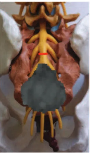
Figure 10: Representation in a model of the 3 osteotomies performed (green lines) and the level of thecal sac ligation (thin red line). (a) Posterior view and (b) anterior view.
(a) (b)
Figure 11:En blocresected specimen. (a) Ventral surface and (b) dorsal surface.
provided sot tissue reconstruction to ill the defect and close the wound. At this time, the skin incision was lengthened incorporating the prior midline incision. Adipomuscular mobilization and rotational laps of the gluteus maximus were used in the reconstruction of the sacral defect (Figure 13). Adequate sot tissue reconstruction was achieved, as well as wound closure in a layered and tensionless fashion. Two suction drains were let in place. Cefazolin was used in prophylactic dose (1 gm IV) 60 minutes before surgery and then every six hours during 24 hour postoperatively.
3. Results
En blocexcision with wide tumor-free margins of this large lower and mid-sacral mass was achieved through a modiied Kraske procedure with mid-sacrectomy and coccygectomy. It required sacriice of the nerve roots and thecal sac below the level of S2 nerve roots.
As previewed on the preoperative MRI scan, the tumor location at the lower and mid-sacrum and its limits (superi-orly: the inferior half of S2 segment; inferi(superi-orly: the superior
half of S5 segment; laterally: sparing the sacroiliac joints; ventrally: in intimate relation with the rectum which was anteriorly displaced; dorsally: invading the posterior surface of the sacrum out through the sacral hiatus and dorsal foramina; inside the sacral canal: afecting the thecal sac and nerve roots below the S2 nerve roots level) were corroborated with the intraoperative indings and allowed to precisely deine the boundaries of the specimen to be removed, keeping distance from the tumor capsule and sacroiliac joints.
here were no procedure-related complications. Histo-pathological analysis of the resected specimen revealed a benign GCT (Figure 14).
Figure 12: S2 nerve roots were preserved and remained intact along their entire length.
Figure 13: Rotational laps of the gluteus maximus provided for sot tissue reconstruction and wound closure.
perianal region, without any other neurological deicits and performing well in all living activities, being inclusively able to sit for long periods of time without pain. he postoperative MRI scan showed complete resection of the distal sacrum and coccyx with no evidence of residual lesion. Last follow-up MRI was performed ive years ater surgery showing no recurrence of the tumor (Figure 15).
4. Discussion
Management of GCT of the sacrum is complex and challeng-ing from diagnosis to treatment. his is due to their rarity and heterogeneous clinical scenarios and because the surgical procedures generally involved are extensive and aggressive, aiming for complete tumor resection and potential cure.
hese procedures may be extra-demanding not only because of the aggressive nature and behavior of GCT, but also once the majority of them are diagnosed in an advanced stage of the disease, exhibiting large volumes and sometimes, poorly deined margins. his may create technical diiculties in surgical access and tumor resection, due to the surrounding anatomical constraints and nearby noble
structures to preserve [1,5,20,30]. For this reason, proper patient selection is paramount.
A detailed clinical and neurological assessment including digital rectal examination is mandatory for helping to estab-lish an early diagnosis. Investigation for metastatic disease should always be performed in order to decide the best treatment option (curative versus palliative).
Tumors’ radiological appearance and their location, accessibility, local extension, and involvement of adjacent neurovascular structures are of paramount importance and should always be kept in mind before considering performing a biopsy, once this is an invasive, noninnocuous procedure, with well-known risks of tissue contamination through the biopsy tract, hemorrhage, and infection [14,31]. CT-guided ine-needle biopsy may be a helpful tool for the histological diagnosis [14] in selected cases: whenever this informa-tion is preoperatively relevant for potentially changing or inluencing the treatment approach; for unresectable lesions; or in patients with signiicant comorbidity, precluding a more aggressive surgery (if required to indicate adjuvant or palliative therapy). Biopsy is useful to reach the diferential diagnosis of sacral lesions (metastases, giant cell tumor, chor-doma, teratoma, and chondrosarcoma). In this particular case, we adopted a direct approach to the lesion, without previous biopsy, because the patient had a good general medical condition; there were no other documented lesions; the tumor was well circumscribed, surgically accessible with the possibility of reachingen blocresection with tumor free margins without further neurological deterioration. Having this in mind, the surgical approach would be the same regardless of the biopsy result.
Preoperatively, it is important to precisely deine the goal of surgery and the anatomical boundaries to be respected intraoperatively. It is crucial to accurately assess several ima-giological parameters such as the level of sacral involvement, iniltrated structures (sacral canal, thecal sac, nerve roots, muscles, ligaments, vascular and visceral structures, and sacroiliac joints), and ventral and dorsal extensions. his is essential to establish a preliminary diagnosis and decide the best surgical approach and for surgical planiication of theen blocresection [22].
Several treatment options have been proposed: intratu-moral curettage plus radiotherapy, possibly aided by preop-erative embolization, the use of osteoclast inhibiting drugs (bisphosphonates), and cryosurgery with liquid nitrogen, limited by the risk of injury to adjacent neural structures in sacral tumors.
According to some authors,en blocexcision is the gold standard procedure for sacral GCT which present radio-logical criteria denoting the potential aggressiveness of the tumor: poorly deined margins, cortical bone destruction, and sot tissue extension by expansive tumor growth [6,30,
32–34].
(a) (b)
Figure 14: (a) Hematoxylin-eosin stained tissue demonstrated a highly cellular, solid neoplasm consisting of mononuclear cells and osteoclast-like giant cells; (b) intense immunohistochemical staining for CD68 (KP1).
(a) (b)
Figure 15: MRI scan evaluation 5 years ater surgery, showing no recurrence of the tumor. (a) Coronal and (b) sagittal views.
and coccygectomy allows maximizing local tumor control, minimizing the risk of local recurrence, and providing possible cure, giving the possibility to preserve neurovascular function, by this mean decreasing morbidity and improving the inal outcome.
During the surgery of lower or mid-sacral tumors, several nuances must be taken into account. Only the afected sacral nerve roots should be sacriiced, and the expected neurological outcome needs to be preoperatively predicted and discussed with the patient [33,35–37]. Also, the sacroiliac joints should be spared to avoid spinal instability [38–42].
Given the complexity of evaluation, treatment, and management of GCT, a coordinated multidisciplinary team approach to the problem, involving neurosurgeons, gen-eral surgeons, and plastic surgeons, working together in specialized units, has proved useful [33]. his collabora-tion is essential helping to select and implement surgical treatment to minimize the risk of perioperative complica-tions. General surgeons are important for the approach and
mobilization of the rectum from the tumor and ventral sacrum.
he major beneits of this surgical technique are the capability to successfully achieve complete surgical resection with wide tumor-free margins, protect the presacral vascular and visceral structures, and accomplish these goals through a single and deinitive dorsal approach. Trying to accomplish complete removal of GCT during the initial surgery is very important and should be, if possible, the main goal. his allows reaching a favorable prognosis, minimizing the risk of recurrence [28]. Nevertheless, careful selection of the patients amenable to this approach is a must. Low or mid-sacral tumors constitute the perfect indication.
5. Conclusion
Surgical treatment of sacral GCT is challenging and tech-nically demanding, due to the complex regional anatomy in this area, and the advanced stage of disease by the time diagnosis is made. A well-coordinated multidisciplinary team approach, working in specialized units, is mandatory.
Early diagnosis, complete (en bloc) surgical resection with tumor-free margins, and a comprehensive treatment are essential for local tumor control, best long-term prognosis, and improved outcome with possible cure.
he diicult conlict between patient’s functional integ-rity and the cure of the disease must be preoperatively well weighted and discussed with the patients and the team.
An accurate preoperative planning must precisely locate the tissues involvement (bone, muscle, nerves, and joints) and delineate the extension of the area to be resected.
Conflict of Interests
he authors declare that there is no conlict of interests regarding the publication of this paper.
References
[1] W. M. Mendenhall, R. A. Zlotecki, M. T. Scarborough, C. P. Gibbs, and N. P. Mendenhall, “Giant cell tumor of bone,”
American Journal of Clinical Oncology: Cancer Clinical Trials,
vol. 29, no. 1, pp. 96–99, 2006.
[2] R. L. Randall, “Giant cell tumor of the sacrum,”Neurosurgical
Focus, vol. 15, no. 2, article E13, 2003.
[3] H. W. Sung, W. P. Shu, H. M. Wang, S. Y. Yuai, and Y. B. Tsai, “Surgical treatment of primary tumors of the sacrum,”Clinical
Orthopaedics and Related Research, vol. 215, pp. 91–98, 1987.
[4] R. E. Turcotte, F. H. Sim, and K. K. Unni, “Giant cell tumor of the sacrum,”Clinical Orthopaedics and Related Research, no. 291, pp. 215–221, 1993.
[5] M. Campanacci, N. Baldini, S. Boriani, and A. Sudanese, “Giant-cell tumor of bone,”Journal of Bone and Joint Surgery—Series A, vol. 69, no. 1, pp. 106–114, 1987.
[6] R. hangaraj, R. J. Grimer, S. R. Carter, A. J. Stirling, J. Spilsbury, and D. Spooner, “Giant cell tumour of the sacrum: a suggested algorithm for treatment,”European Spine Journal, vol. 19, no. 7, pp. 1189–1194, 2010.
[7] C. Martin and E. F. McCarthy, “Giant cell tumor of the sacrum and spine: series of 23 cases and a review of the literature,”he
Iowa Orthopaedic Journal, vol. 30, pp. 69–75, 2010.
[8] W. Guo, T. Ji, X. Tang, and Y. Yang, “Outcome of conservative surgery for giant cell tumor of the sacrum,”Spine, vol. 34, no. 10, pp. 1025–1031, 2009.
[9] M. G. Rock, D. J. Pritchard, and K. K. Unni, “Metastases from histologically benign giant-cell tumor of bone,”Journal of Bone
and Joint Surgery—Series A, vol. 66, no. 2, pp. 269–274, 1984.
[10] R. M. Kay, J. J. Eckardt, L. L. Seeger, J. M. Mirra, and D. J. Hak, “Pulmonary metastasis of benign giant cell tumor of bone: six histologically conirmed cases, including one of spontaneous regression,”Clinical Orthopaedics and Related Research, no. 302, pp. 219–230, 1994.
[11] R. V. Hutter, J. N. Worcester Jr., K. C. Francis et al., “Benign and malignant giant cell tumors of bone. A clinicopathological analysis of the natural history of the disease,”Cancer, vol. 15, pp. 653–690, 1962.
[12] F. Bertoni, D. Present, and W. F. Enneking, “Giant-cell tumor of bone with pulmonary metastases,”Journal of Bone and Joint
Surgery A, vol. 67, no. 6, pp. 890–900, 1985.
[13] M. W¨ulling, C. Engels, N. Jesse, M. Werner, G. Delling, and E. Kaiser, “he nature of giant cell tumor of bone,”Journal of
Cancer Research and Clinical Oncology, vol. 127, no. 8, pp. 467–
474, 2001.
[14] Z. M. Yang, C. C. Shen, H. Li, Z. L. Shi, and H. M. Tao, “Current treatment of sacral giant cell tumour of bone: a review,”Journal
of International Medical Research, vol. 40, no. 2, pp. 415–425,
2012.
[15] R. E. Leggon, R. Zlotecki, J. Reith, and M. T. Scarborough, “Giant cell tumor of the pelvis and sacrum: 17 cases and analysis of the literature,”Clinical Orthopaedics and Related Research, no. 423, pp. 196–207, 2004.
[16] S. E. Larsson, R. Lorentzon, and L. Boquist, “Giant cell tumor of bone. A demographic, clinical, and histopathological study of all cases recorded in the Swedish cancer registry for the years 1958 through 1968,”Journal of Bone and Joint Surgery—Series A, vol. 57, no. 2, pp. 167–173, 1975.
[17] P. L. Althausen, P. D. Schneider, R. J. Bold, M. C. Gupta, J. E. Goodnight Jr., and V. P. Khatri, “Multimodality management of a giant cell tumor arising in the proximal sacrum: case report,”
Spine, vol. 27, no. 15, pp. E361–E365, 2002.
[18] F. Bertoni, D. Present, A. Sudanese, N. Baldini, P. Bacchini, and M. Campanacci, “Giant-cell tumor of bone with pulmonary metastases: six case reports and a review of the literature,”
Clinical Orthopaedics and Related Research, no. 237, pp. 275–285,
1988.
[19] K. A. Siebenrock, K. K. Unni, and M. G. Rock, “Giant-cell tumour of bone metastasising to the lungs,”Journal of Bone and
Joint Surgery—Series B, vol. 80, no. 1, pp. 43–47, 1998.
[20] D. C. Dahlin, “Giant cell tumor of bone: highlights of 407 cases,”
he American Journal of Roentgenology, vol. 144, no. 5, pp. 955–
960, 1985.
[21] S. Boriani, A. Sudanese, N. Baldini, and P. Picci, “Sarcoma-tous degeneration of giant cell tumours,” Italian Journal of
Orthopaedics and Traumatology, vol. 12, no. 2, pp. 191–199, 1986.
[22] W. K. Jong, H. W. Chung, Y. C. Eun et al., “MRI indings of giant cell tumors of the spine,”American Journal of Roentgenology, vol. 189, no. 1, pp. 246–250, 2007.
[24] M. Jain, H. M. Aiyer, M. Singh, and M. Narula, “Fine-needle aspiration diagnosis of giant cell tumour of bone presenting at unusual sites,”Diagnostic Cytopathology, vol. 27, no. 6, pp. 375– 378, 2002.
[25] A. Chakravarti, I. J. Spiro, E. B. Hug, H. J. Mankin, J. T. Eird, and H. D. Suit, “Megavoltage radiation therapy for axial and inoperable giant-cell tumor of bone,”Journal of Bone and Joint
Surgery A, vol. 81, no. 11, pp. 1566–1573, 1999.
[26] I. C. Gibbs and S. D. Chang, “Radiosurgery and radiotherapy for sacral tumors,”Neurosurgical Focus, vol. 15, no. 2, article E8, 2003.
[27] T. Ozaki, U. Liljenqvist, H. Halm, A. Hillmann, G. Gosheger, and W. Winkelmann, “Giant cell tumor of the spine,”Clinical
Orthopaedics and Related Research, no. 401, pp. 194–201, 2002.
[28] R. C. Marcove, D. S. Sheth, E. W. Brien, A. G. Huvos, and J. H. Healey, “Conservative surgery for giant cell tumors of the sacrum. he role of cryosurgery as a supplement to curettage and partial excision,”Cancer, vol. 74, no. 4, pp. 1253–1260, 1994. [29] P. Wuisman, O. Lieshout, S. Sugihara, and M. van Dijk, “Total sacrectomy and reconstruction: oncologic and functional out-come,”Clinical Orthopaedics and Related Research, no. 381, pp. 192–203, 2000.
[30] J. Llauger, J. Palmer, S. Amores, S. Bagu´e, and A. Camins, “Primary tumors of the sacrum: diagnostic imaging,” he
American Journal of Roentgenology, vol. 174, no. 2, pp. 417–424,
2000.
[31] J. M. Aranda-Narv´aez, A. J. Gonz´alez-S´anchez, C. Montiel-Casado et al., “Posterior approach (Kraske procedure) for surgical treatment of presacral tumors,”World Journal of
Gas-trointestinal Surgery, vol. 4, no. 5, pp. 126–130, 2012.
[32] S. Gitelis, B. A. Mallin, P. Piasecki, and F. Turner, “Intralesional excision compared with en bloc resection for giant-cell tumors of bone,”Journal of Bone and Joint Surgery, Series A, vol. 75, no. 11, pp. 1648–1655, 1993.
[33] D. R. Fourney, L. D. Rhines, S. J. Hentschel et al., “En bloc resec-tion of primary sacral tumors: classiicaresec-tion of surgical ap-proaches and outcome,”Journal of Neurosurgery: Spine, vol. 3, no. 2, pp. 111–122, 2005.
[34] R. A. Hart, S. Boriani, R. Biagini, B. Currier, and J. N. Weinstein, “A system for surgical staging and management of spine tumors: a clinical outcome study of giant cell tumors of the spine,”Spine, vol. 22, no. 15, pp. 1773–1783, 1997.
[35] L. T. Todd Jr., M. J. Yaszemski, B. L. Currier, B. Fuchs, C. W. Kim, and F. H. Sim, “Bowel and bladder function ater major sacral resection,”Clinical Orthopaedics and Related Research, no. 397, pp. 36–39, 2002.
[36] S. Nakai, H. Yoshizawa, S. Kobayashi, K. Maeda, and Y. Oku-mura, “Anorectal and bladder function ater sacriice of the sacral nerves,”Spine, vol. 25, no. 17, pp. 2234–2239, 2000. [37] L. van der Heijden, M. A. J. van de Sande, I. C. M. van der Geest
et al., “Giant cell tumors of the sacrum—a nationwide study on midterm results in 26 patients ater intralesional excision,”
European Spine Journal, vol. 23, no. 9, pp. 1949–1962, 2014.
[38] R. L. Randall, J. Bruckner, C. Lloyd, T. H. Pohlman, and E. U. Conrad III, “Sacral resection and reconstruction for tumors and tumor-like conditions,”Orthopedics, vol. 28, no. 3, pp. 307–313, 2005.
[39] M. Doita, T. Harada, T. Iguchi et al., “Total sacrectomy and re-construction for sacral tumors,”Spine, vol. 28, no. 15, pp. E296– E301, 2003.
[40] N. Ohata, T. Ozaki, T. Kunisada, Y. Morimoto, M. Tanaka, and H. Inoue, “Extended total sacrectomy and reconstruction for sacral tumor,”Spine, vol. 29, no. 6, pp. E123–E126, 2004. [41] H. Y. Zhang, I. hongtrangan, R. S. V. Balabhadra, J. A. Murovic,
and D. H. Kim, “Surgical techniques for total sacrectomy and spinopelvic reconstruction,”Neurosurgical Focus, vol. 15, no. 2, p. E5, 2003.
[42] C. Sahakitrungruang, K. Chantra, N. Dusitanond, P. Atittharn-sakul, and A. RojanaAtittharn-sakul, “Sacrectomy for primary sacral tumors,”Diseases of the Colon and Rectum, vol. 52, no. 5, pp. 913–918, 2009.
[43] M. Onaitis, K. Ludwig, A. Perez-Tamayo et al., “he Kraske procedure: a critical analysis of a surgical approach for mid-rectal lesions,”Journal of Surgical Oncology, vol. 94, no. 3, pp. 194–202, 2006.
[44] J. Diaz, W. S. McDonald, M. Armstrong, F. Eismont, M. Hel-linger, and S. haller, “Reconstruction ater extirpation of sacral malignancies,”Annals of Plastic Surgery, vol. 51, no. 2, pp. 126– 129, 2003.
[45] P.-K. Koh, B.-K. Tan, S.-W. Hong et al., “he gluteus maximus muscle lap for reconstruction of sacral chordoma defects,”
Annals of Plastic Surgery, vol. 53, no. 1, pp. 44–49, 2004.
[46] M. Alper, U. Bilkay, Y. Kec¸eci et al., “Transsacral usage of a pure island TRAM lap for a large sacral defect: a case report,”Annals
of Plastic Surgery, vol. 44, no. 4, pp. 417–421, 2000.
[47] D. W. Chang and G. L. Robb, “Recent advances in reconstructive surgery for sot-tissue sarcomas,”Current Oncology Reports, vol. 2, no. 6, pp. 495–501, 2000.
[48] W. K. Miles, D. W. Chang, S. S. Kroll et al., “Reconstruction of large sacral defects following total sacrectomy,”Plastic and
Reconstructive Surgery, vol. 105, no. 7, pp. 2387–2394, 2000.
Submit your manuscripts at
http://www.hindawi.com
Stem Cells
International
Hindawi Publishing Corporationhttp://www.hindawi.com Volume 2014
Hindawi Publishing Corporation
http://www.hindawi.com Volume 2014
INFLAMMATION
Hindawi Publishing Corporation
http://www.hindawi.com Volume 2014
Behavioural
Neurology
Endocrinology
International Journal ofHindawi Publishing Corporation
http://www.hindawi.com Volume 2014
Hindawi Publishing Corporation
http://www.hindawi.com Volume 2014
Disease Markers
Hindawi Publishing Corporation
http://www.hindawi.com Volume 2014
BioMed
Research International
Oncology
Journal of Hindawi Publishing Corporationhttp://www.hindawi.com Volume 2014
Hindawi Publishing Corporation
http://www.hindawi.com Volume 2014
Oxidative Medicine and Cellular Longevity
Hindawi Publishing Corporation
http://www.hindawi.com Volume 2014
PPAR Research
The Scientiic
World Journal
Hindawi Publishing Corporationhttp://www.hindawi.com Volume 2014
Immunology Research
Hindawi Publishing Corporation
http://www.hindawi.com Volume 2014
Journal of
Obesity
Journal ofHindawi Publishing Corporation
http://www.hindawi.com Volume 2014
Hindawi Publishing Corporation
http://www.hindawi.com Volume 2014
Computational and Mathematical Methods in Medicine
Ophthalmology
Journal ofHindawi Publishing Corporation
http://www.hindawi.com Volume 2014
Diabetes Research
Journal ofHindawi Publishing Corporation
http://www.hindawi.com Volume 2014
Hindawi Publishing Corporation
http://www.hindawi.com Volume 2014
Research and Treatment
AIDS
Hindawi Publishing Corporation
http://www.hindawi.com Volume 2014
Gastroenterology Research and Practice
Hindawi Publishing Corporation
http://www.hindawi.com Volume 2014
Parkinson’s
Disease
Evidence-Based Complementary and Alternative Medicine
Volume 2014 Hindawi Publishing Corporation