I
ntroductIonDiabetes mellitus is a metabolic disorder characterized by
hyperglycemia, secretion insuficiency and receptor insensitivity
to endogenous insulin1. Its incidence is associated with high
morbidity and mortality rates. Increased oxidative stress is believed to play an important role in the etiology and pathogen-esis of chronic complications of diabetes2,3, and is mainly
char-acterized by imbalance between organism antioxidant defenses and oxidant molecules, known as free radicals. Free radicals are very reactive species, capable of inducing oxidation of the
biological membrane of phospholipids and proteins, resulting in
modiications of cell function and cellular death4,5,6.
Many animal models have contributed to the elucidation of human diabetic syndromes and associated genetic factors. Among these animal models, there are those that reproduce human diabetes using streptozotocin (STZ). Probably, STZ induces diabetes by generating reactive oxygen radicals (ROS), which leads to islet cell destruction in experimental animals7.
Models for rats presenting severe diabetes (fasting glycemic level higher than 200/360 mg/dL), which reproduce uncon-trolled type-1 diabetes (DM1) in humans, are well established
*Correspondence:
Departamento de Ginecologia e Obstetrícia, FMB – Unesp Distrito de Rubião Júnior, s/n Botucatu - Estado de São Paulo, Brazil
CEP: 18618-000 Tel: 55- 14- 3811-6181 Fax: 55- 14- 38116090 [email protected]
neonatally
-
Induced
dIabetes
:
lIpId
profIle
outcomes
and
oxIdatIve
stress
status
In
adult
rats
yurI Karen sInzato1, paula Helena ortIz lIma2, Kleber eduardode campos1, ana carolIna InHasz KIss1, marIlza vIeIra cunHa rudge2, débora crIstIna damasceno*2
Laboratory of Experimental Research in Gynecology and Obstetrics, Botucatu Medical School, Unesp - São Paulo State University, Brasil
1. Pós-graduação do Laboratório de Pesquisa Experimental em Ginecologia e Obstetrícia, Departamento de Ginecologia e Obstetrícia, Faculdade de Medicina de Botucatu – UNESP – Universidade Estadual Paulista, Botucatu, SP
2. PhD - PhD – Professoras no Laboratório de Pesquisa Experimental em Ginecologia e Obstetrícia, Departamento de Ginecologia e Obstetrícia, Faculdade de Medicina de Botucatu – UNESP – Universidade Estadual Paulista, Botucatu, SP
Summary
bacKground. Experimental models are developed for the purpose of enhancing the understanding of the
pathophysiological mechanisms involved in diabetes. Experimental indings lead to the development of
treatment strategies to maintain metabolic conditions as close to normal as possible. There are several reports about streptozotocin induced mild diabetes to reproduce type 2 diabetes. However, studies
about the interaction among glucose levels, lipid proile, and oxidative stress in these animals remain insuficient. Therefore, this study evaluated these parameters in blood samples from adult Wistar rats
treated neonatally with streptozotocin.
metHods. Female newborn Wistar rats received streptozotocin (70 mg/kg, i.p.) on the 5th day of life
(n5-STZ). Glycemia was measured in the 3rd and 4th month of life. At the end of the 4th month, blood samples were collected and processed for lipid proile and oxidative stress measurements.
results. Glycemia of n5-STZ rats were significantly higher compared to those of control rats (p<0.05). There was no alteration in levels of total cholesterol, triglycerides, lipid peroxidation (TBARS), SOD activity and GSH-t determination (p>0.05) in the n5-STZ animals when compared to control group. However n5-STZ animals showed a significant decreased HDL-cholesterol rate (p<0.05).
conclusIon. This streptozotocin-induced diabetes model in rats caused hyperglycemia (120-360mg/dL), characterizing mild diabetes. This glycemic level led to HDL-lipoprotein alteration, which was not sufficient to impair antioxidant enzyme activities or determination of lipid peroxida-tion in adult life of rats. Further this experimental investigaperoxida-tion contributed to the understanding of different results found in other models for mild/moderate diabetes induction in laboratory animals as well as to a better comprehension of the pathophysiological mechanisms of mild diabetes or hyperglycemia in humans.
utilizing high doses of STZ during the adult life of animals8,9,10,11.
Experimental mild diabetes (fasting glycemic level from 120 to 360 mg/dL) characterizes clinical status of type 2 Diabetes mellitus (DM2). Portha et al.12 were the irst to describe an
animal model for mild diabetes using neonatal STZ. Some authors administered STZ at day of birth (n0-STZ)13,14, 2 days
after birth (n2-STZ)15,16, after 5 days of life (n5-STZ)16,17, and
on the 2nd and 9th days of life18. These studies showed that, at
8 weeks and thereafter the animals presented impaired glucose tolerance and a 50% decrease in pancreatic insulin content with mild hypoinsulinemia. As reviewed by Portha et al, incom-petence of the regenerated beta cells may be due to a reduced GLUT2 content, limiting glucose entry and metabolism and a
decreased glucokinase afinity to glucose19. Experimental ind
-ings lead to the development of treatment strategies to maintain metabolic conditions as close as possible to normal. There are several reports about the use of streptozotocin to induce mild diabetes reproducing type 2 Diabetes mellitus. However, these investigations did not disclose any relation among glucose
toler-ance, insulin content and insulin resistance with lipid proile
and oxidative stress status in adult life of rats. Therefore, this study aimed to analyze the model of mild diabetes induction in
Wistar rats and to evaluate these parameters in the adult life of
rats treated neonatally with streptozotocin.
m
etHodsWistar male and female rats weighing about 180g (90 days of
life) were adapted in our laboratory for seven days. The rats were kept in collective cages, in controlled conditions of temperature of (22 ± 3º C), light (12 h light/dark cycles) and relative humidity
(60 ± 5%). The animals were fed with laboratory chow (Purina®)
tap water ad libitum and cared for in accordance with the prin-ciples of the Guide for Care and Use of Experimental Animals. The local Committee of Ethics in Animal Experimentation approved all experimental procedures of this study.
Female rats were mated overnight with normal male rats (parental generation). Sperm presence in vaginal wet smears in the following morning was considered as day zero (0) of pregnancy. Pregnant rats were kept in individual cages during the pregnancy period (21 days), including vaginal delivery and lactation periods (21 days). The female newborn (NB) received streptozotocin (STZ, 70 mg/kg body weight, intraperitoneally) dissolved in citrate buffer (0.1M, pH 4.5) at day 5 of life, as previ-ously described by Murali & Goyal 17. The terminology n5-STZ
is used here to refer to this version of a model that characterizes clinical status of DM2. In the control group, NB received only citrate buffer, at day 5 of life.
In the month 3 and 4 of life, blood samples were obtained from a cut tail tip for non-fasting glycemic determinations (glucose oxidase) using a glucosimeter (One Touch Ultra - Johnson & Johnson®) in the morning. At the end of 4th month of
life, the glycemia were obtained immediately before anesthesia. All animals were anesthetized with sodium pentobarbital and killed. Blood samples were collected from each animal and
processed for lipid proile and oxidative stress measurements. The chemicals were purchased from Wiener (Rosario, Argen -tina) and Sigma Chemical (St. Louis, MO, USA). Blood samples were collected in anticoagulant-free test tubes and kept at low
temperature for 30 min and then centrifuged at 3,500 rpm for 10 min at 4ºC. The supernatant was collected as serum and stored at -80ºC for lipid determination. Another blood fraction was placed in anticoagulant tubes and centrifuged at 1,200 rpm for 10 min at room temperature for assay of oxidative stress biomarkers, which were estimated in the washed erythrocytes20.
Serum concentrations of total cholesterol, triglycerides and high-density lipoprotein cholesterol (HDL-C) were determined by the enzymatic method21. The absorbance of these parameters
was measured at 505 nm.
Oxidative stress biomarkers evaluated were superoxide dismutase (SOD), glutathione total (GSHt) and thiobarbituric acid reactive substances as the lipid peroxidation index (TBARS). For TBARS concentration, the absorbance was measured at a wavelength of 535 nm and results were expressed as nM of thio-barbituric acid reactive species (TBARS) per gram of hemoglobin
(nM/g Hb). The SOD enzymatic activity unit was deined as SOD
units able to produce 50% of pyrogallol oxidation inhibition. All data were expressed in units of SOD per milligram of hemoglobin. GSHt, which consists of reduced and oxidized glutathiones, was enzymatically determined using 5.5’- dithio-bis (2-nitrobenzoic acid) (DTNB) and glutathione reductase in the presence of a reduced form of nicotinamide adenine dinucleotide phosphate (NADPH), forming 2-nitro-5-thiobenzoic acid. GSHt activity was measured at 412 nm on a spectrophotometer. One unit of its activity was equal to the micromolar of substrate reduced per gram of hemoglobin20.
Data are expressed as mean ± standard error of mean (SEM). The Student t test was used to determine differences between groups22. The limit of statistical signiicance was 5% (p<0.05).
r
esultsGlycemia
In the 3rd month of life, the glycemic means of rats in the n5-STZ group were signiicantly higher than those of rats in the
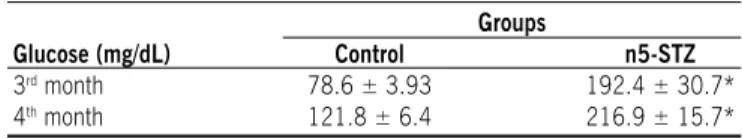
control group (p<0.05). All rats in the control group exhibited glycemia under 100 mg/dL. Among the rats in the n5-STZ group, 50.0% showed glycemia under 120 mg/dL and 50.0% from 120 to 360 mg/dL. In the 4th month of life, glycemic means of rats in the n5-STZ group remained signiicantly higher in relation to the control group (p<0.05). When glycemic levels were indi -vidually analyzed, alteration was observed in the distribution of glycemic ranges of rats in the n5-STZ group, since all the animals presented glycemia between 120 and 360mg/dL. Nevertheless, in the control group, the rats maintained glycemic levels similar to those found in the 3rd month of life. Glucose levels at month
3 and 4 of life are presented in Table 1 and the glucose range distributions are presented in Table 2.
Table 1 - Non-fasting glycemic levels of rats treated with citrate buffer solution (control group) and streptozotocin
(n5-STZ group) in the neonatal period (mean ± SEm)
Groups
Glucose (mg/dL) Control n5-STZ
3rd month 78.6 ± 3.93 192.4 ± 30.7*
4th month 121.8 ± 6.4 216.9 ± 15.7*
Biochemical parameters and oxidative stress biomarkers
The biochemical parameters evaluated in the 4th month of life
are presented in Table 3. Both groups did not exhibit statistical differences (p>0.05) in serum levels of total cholesterol and
triglycerides. However, n5-STZ animals showed a signiicant
decrease in HDL-cholesterol levels as compared to the control group (p<0.05).
The evaluation of oxidative stress parameters is presented in Table 4. Streptozotocin-induced diabetic rats did not present changes in lipid peroxidation (TBARS). Alterations were not observed in SOD activity or GSH-t determination when compared to the control group (p>0.05).
d
IscussIonIn this study, the control animals presented normoglycemia at months 3 and 4. Control animals presented slight glycemia above 120mg/dL at the 4th month of life however, these animals were not considered mild diabetic because glycemia was veriied
in the non-fasting state and our data corroborate those of other authors17,23. Our results showed that STZ administration on the
5th day of neonatal life caused onset of hyperglycemia in the 3rd
month of life in rats, whose postprandial glycemic mean was of approximately 192.4 mg/dL. This model presented glycemic results similar to those found by Murali & Goyal17. The individual
analysis of glycemia in rats showed that 50% did not present mild diabetes at the 3rd month. Nevertheless, after the 4th month
of life, 100% of the n5-STZ rats showed glycemic levels between 120 and 360 mg/dL, similar to the glycemic mean observed by other researchers24,25 thus ensuring the viability of the model for mild diabetes induction. Further, Weir et al.26 demonstrated that n5-STZ rats presented lack of signiicant insulin re-accumulation
in the pancreas, 2 weeks after the b-cell insult.
In patients, Diabetes mellitus increases the risk of deve-loping atherosclerosis and coronary artery disease27,28,29, and
lipids are indicated as some of the major pathogenic biological markers in situations of metabolic dysfunction (such as in insulin resistance, associated type-2 Diabetes mellitus or not)30. This
investigation showed that n5-STZ animals presented a
non-signiicant increase in total cholesterol and triglyceride levels and decreased HDL levels. With regard to the absence of alterations
in the determinations of total cholesterol and triglycerides, our
indings are not in agreement with those described in literature.
Insulin resistance and Diabetes mellitus affect virtually every lipid and lipoprotein and, therefore, dyslipidemia is present in most diabetic patients27,31,32. Dyslipidemia is relected by
elevated levels of triglycerides, VLDL and LDL-cholesterol and
lower HDL-cholesterol levels33. Although our results do not relect
comments in clinical studies, the Brazilian Society of Cardiology acknowledges that decreased HDL (isolated or in association with increased levels of LDL and/or triglycerides) characterizes the
laboratory classiication of dyslipidemia34. HDL cholesterol and
apo AI levels are characteristically reduced in insulin-resistant people. Much of this derives, as in the case of low dense LDL, from the action of CETP (cholesterol ester transferase) mediated transfer of cholesteryl ester from HDL to triglyceride rich
lipo-proteins (chylomicrons and VLDL). A consistent inding is the
inverse relationship between plasma insulin (or C-peptide) concentrations, which are measures of insulin resistance and HDL-cholesterol levels35.
There is increasing evidence, in both experimental and clinical studies, to suggest that oxidative stress plays a major role in the pathogenesis of Diabetes mellitus. In this study, TBARS levels in the n5-STZ rats remained unaltered. Our result suggests that the neonatal STZ-induced diabetes model was
insu-ficient to increase glycemia and so, exacerbate oxidative stress. However, increase of glycemia to a moderate level was veriied. With respect to SOD, the enzyme responsible for neutralizing
Table 4 - Determination of thiobarbituric acid reactive species (TBarS), enzymatic activity of antioxidant dismutase superoxide (SOD) and concentration of total glutathione (GSHt) in the 4th month
of life in rats treated with citrate buffer solution (control group) and streptozotocin (n5-STZ group) in the neonatal period (mean ± SEm)
Groups
Control n5-STZ
TBARS (nM/g Hb) 226.1 ± 71.9 276.2 ± 22.3
SOD (U/mg Hb) 7.1 ± 0.4 7.2 ± 0.4
GSHt (uM/g Hb) 0.032 ± 0.003 0.034 ± 0.010
p>0.05 - non-signiicant
Table 2 - Distribution of rats in relation to non-fasting glycemic levels. rats treated with citrate buffer solution (control group) and streptozotocin (n5-STZ group) in the neonatal period (mean ± SEm)
Groups
3rd month 4th month
Glycemia Control n5-STZ Control n5-STZ
< 120 mg/dL 78.6 ± 3.9 (100.0%a) 91.1 ± 10.6 (50.0%a) 108.5 ± 14.8* (40.0%a) 0 *
120 - 360 mg/dL 0 293.6 ± 26.9 (50.0%a) 130.7 ± 1.5* (60.0%a) 216.9 ± 15.7* (100%a)
a % of rats presenting glycemia with indicated values in the 3rd and 4th months of life.
*p<0.05 - statistically signiicant difference compared to month 3.
Table 3 - Biological lipid proile in the 4th month of life in rats treated
with citrate buffer solution (control group) and streptozotocin (n5-STZ group) in the neonatal period (mean ± SEm)
Groups
Control n5-STZ
Triglycerides (mg/dL) 87.7 ± 20.1 106.0 ± 17.9
Cholesterol (mg/dL) 130.0 ± 36.1 179.3 ± 28.5
HDL-cholesterol (mg/dL) 47.5 ± 6.3 32.4 ± 2.5 *
superoxide anion levels, this study showed no changes in SOD activity in the n5-STZ rats. These results corroborate with
litera-ture, which observed no signiicant changes in SOD activity in
diabetic rats36,37. However, there are also conlicting indings in
literature. There is evidence of reduced SOD activity in alloxan-induced diabetic rats38, and higher activity was observed in DM1
and DM2 subjects39.
The glutathione antioxidant system has a fundamental role in cellular defense against reactive free radicals and other oxidant species 40. GSHt concentration also presented no change in the
n5-STZ rats in this investigation. Damasceno et al.6 observed
reduction in GSH determination in STZ induced-severe diabetic rats. Furthermore, it has been suggested that there may be temporal changes in enzyme activity that are both transitory and biphasic in nature. For instance, after prolonged hyperglycemia in severe diabetes (glycemia>360 mg/dL), the induction of certain antioxidant enzymes or a return to normal values from previously decreased values may occur as a compensatory mechanism in response to constant exposure to increased stress. This could explain the decrease, increase or normality in SOD and GSH observed by different investigators. Thus, the present study showed that the neonatal streptozotocin-induced mild diabetes
model in rats caused lipid proile alteration in the HDL-cholesterol level, but it was not suficient to impair antioxidant enzyme
activity or peroxidation lipid determination sampled from adult
Wistar rats.
c
onclusIonThe present investigation conirmed a mild diabetes status
since the 4th month of life in rats. This study contributed to understanding the relation between lipid proile and oxidative
stress status in the mild/moderate diabetes model for laboratory animals as well as to a better comprehension of the pathophy-siological mechanisms of mild diabetes or hyperglycemia.
a
cKnowledgementsThe authors are grateful to Mariana Takaku and Marisa Akemi Takeno, graduate students at the Botucatu Medical School - Unesp; Mr. Carlos R. Gonçalves Lima, from the Experimental Laboratory of Infectious Diseases, for technical support, and to the Research Support Center (RSC) for their valuable contribution in study design and statistical analysis.
Conlict of interest: none
r
esumodIabete InduzIdo no período neonatal: repercussões no perfIl lIpídIco e avalIação dos marcadores de estresse oxIdatIvo na vIda adulta de ratas
Introdução. Modelos experimentais são desenvolvidos com
propósito de ampliar o entendimento dos mecanismos fisio-patológicos envolvidos no diabete. Os achados experimentais levam ao desenvolvimento de tratamentos alternativos para a manutenção das condições metabólicas normais. Existem vários estudos sobre o diabete induzido por streptozotocin mimeti-zando o quadro clínico do DM2. No entanto, a interação entre os níveis de glicose, perfil lipídico e estresse oxidativo nestes animais são escassos. Portanto, o objetivo do trabalho foi avaliar
estes parâmetros em ratas Wistar adultas com diabete induzido com streptozotocin no período neonatal.
Métodos. Fêmeas recém-nascidas receberam streptozotocin
(70mg/Kg, ip) no 5º dia de vida (n5-STZ). A glicemia foi medida no terceiro e quarto meses de vida dos animais. No final do quarto mês de vida, amostras de sangue foram coletadas e processadas para a dosagem de lipídios e marcadores de estresse oxidativo.
resultados. A glicemia das ratas do grupo n5-STZ foi
signi-ficativamente maior comparada às ratas do grupo controle (p<0,05). Não houve alteração nos níveis de colesterol total e triglicérides, peroxidação lipídica (TBARS), atividade da SOD e determinação da GSH-t (p>0,05) nas ratas n5-STZ em relação às ratas do grupo controle. No entanto, houve diminuição signi-ficativa no HDL-colesterol (p<0,05).
ConClusão. Este modelo de indução de diabete em ratas
causou hiperglicemia (120-360mg/dL), caracterizando o diabete moderado. Essa glicemia levou a alterações no HDL-colesterol, a qual não foi suficiente para prejudicar a atividade das enzimas antioxidantes ou marcadores da peroxidação lipí-dica na vida adulta. Além disso, esta investigação experimental contribuiu para entender os diferentes resultados encontrados em outros modelos de indução do diabete moderado em animais de laboratório, como também para a melhor compreensão dos mecanismos fisiopatológicos do diabete moderado ou da
hiperglicemia em humanos. [Rev Assoc Med Bras 2009; 55(4):
384-8]
uNitermos: Ratas Streptozotocin. Lipídeos. Estresse oxidativo.
Diabetes. Estudo experimental.
r
eferences1. Rolo AP, Palmeira CM. Diabetes and mitochondrial function: Role of hypergly-cemia and oxidative stress. Toxicol Appl Pharmacol. 2006;212(2):167-78. 2. Baynes J. Role of oxidative stress in development of complications in diabetes.
Diabetes. 1991;40(4):405-12.
3. Vincent AM, Brownlee M, Russell JW. Oxidative stress and programmed cell
death in diabetic neuropathy. Ann N Y Acad Sci. 2002;959:368-83. 4. Therond P, Bennefont-Rousselot D, Davit-Spraul A, Conti M, Legrand A.
Biomarkers of oxidative stress: an analytical approach. Currt Op Clin Nutr Metab Care. 2000;3(5):373-84.
5. Droge W. Aging-related changes in the thiol/disulide redox state: implications
for the use of thiol antioxidants. Exp Gerontol. 2002;37(12):1333-45. 6. Damasceno DC, Volpato GT, Calderon IMP, Rudge MVC. Oxidative stress and
diabetes in pregnant rats. Anim Reprod Sci. 2002;72(3-4):235-44.
7. Tavridou A, Unwin NC, Laker MF, White M, Alberti GK. Serum concentra -tions of vitamin A and E in impaired glucose tolerance. Clin Chim Acta. 1997;266(2):129-40.
8. Calderon IMP, Rudge MVC, Brasil MAM, Henry MACA. Diabete e gravidez experimental em ratas I. Indução do diabete, obtenção e evolução da prenhez. Acta Cir Bras. 1992;7(1):142-6.
9. Sinzato YK, Damasceno DC, Lima PHO, Souza MSS, Amorin RL, Calderon IMP,
et al. Effect of maternal smoke exposure in the placental morphometry and maternal-fetal DNA damage. Placenta. 2006;27(Suppl 1):A59.
10. Damasceno DC, Volpato GT, Calderon IMP, Aguilar R, Rudge MVC. Effect of
Bauhinia foricata extract in diabetic pregnant rats: maternal repercussions. Phytomedicine. 2004;11(2-3):196-201.
11. Nash P, Eriksson UJ. Suramin-restricted blood volume in the placenta of normal and diabetic rats is normalized by vitamin E treatment. Placenta. 2007;28(5-6):505-15.
12. Portha B, Levacher C, Picon L, Rosselin G. Diabetogenic effect of streptozoto-cinin on the rat during the perinatal period. Diabetes. 1974;23(11):888-95. 13. Portha B, Picon L, Rosselin G. Chemical diabetes in the adult rat as the spon-taneous evolution of neonatal diabetes. Diabetologia. 1979;17(6):371-7.
14. Portha B, Kergoat M. Dynamics of glucose-induced insulin release during the
15. Bonner-Weir S, Trent DF, Honey RN, Weir GC. Responses of neonatal rat islets
to streptozotocin. Limited B-cell regeneration and hyperglycemia. Diabetes. 1981;30(1):64-9.
16. Wang RN, Bouwens L, Kloppel G. Beta-cell growth in adolescent and adult
rats treated with streptozotocin during the neonatal period. Diabetologia. 1996;39(5):548-57.
17. Murali B, Goyal RK. Improvement in insulin sensitivity by losartan in
non-insulin-dependent diabetic (NIDDM) rats. Pharmacol Res. 1999;44(5):385-9.
18. Ulicná O, Zlatos L, Holzerová J, Kvaszová E, Cársky J, Gvozdjáková A, et al.
The effect of streptozotocin-induced diabetes in the neonatal period on hepatic mitochondrial bioenergetics in adult rats. Bratisl Lek Listy. 1999;100(1):5-11. 19. Shafrir E, Desoye G. Pregnancy in diabetic animals. In: Hod M, Jovanovic L,
Di Renzo GC, Leiva A, Langer O. Textbook of diabetes and pregnancy. United
Kingdon: Informa UK Ltd.; 2003. p. 96-7.
20. Ferreira ALA, Machado PEA, Matsubara LS. Lipid peroxidation, antioxidant enzymes and glutathione levels in human erythrocytes exposed to colloidal iron hydroxide in vitro. Braz J Med Biol Res. 1999;32(6):689-94. 21. Young DS. Effects of drugs on clinical laboratory tests. London: AACC Press;
2000.
22. Zar JH. Bioestatistical analysis. New Jersey: Prentice Hall; 1999.
23. Movassati J, Saulnier C, Portha B. Insulin administration enhances growth of the β-cell mass in streptozotocin-treated newborn rats. Diabetes. 1997;46:1445-52.
24. Fantus IG, Chayoth R, O’Dea L, Marliss E, Yale J-F, Grose M. Insulin binding and glucose transport in adipocytes in neonatal streptozotocin-injected rat model of Diabetes mellitus. Diabetes. 1987;36(5):654-660.
25. Blondel O, Bailbé D, Portha B. Insulin resistance in rats with non-insulin-dependent diabetes induced by neonatal (5 days) streptozotocin: evidence for reversal following phorizin treatment. Metabolism. 1990;39(8):787-93.
26. Weir GC, Leahy JL, Bonner-Weir S. Experimental reduction of b-cell
mass. Implications for the pathogenesis of diabetes. Diabetes Metab Rev. 1986;2(1-2):125-61.
27. Raman M, Nesto RW. Heart disease in Diabetes mellitus. Chronic complica -tions in diabetes. Endocrinol Metab Clin North Am. 1996;25(2):425-38.
28. Feher MD, Ekelles RS. Editorial: Lipid modiication and coronary heart
disease in type 2 diabetes: different from the general population? Heart. 1999;81(1):10-11.
29. American Diabetes Association. Dyslipidemia management in adults with diabetes. Diabetes Care. 2004;27(Suppl 1):S68-71.
30. Ribeiro EB, Marmo MR, Andrade IS, Dolninkoff MS. Effect of fasting on mono-sodium glutamate-obese rats. Braz J Med Biol Res. 1989;22(7):919-21. 31. Cowie CC, Harris MI. Physical and metabolic characteristics of persons with
diabetes. Diabetes in America. 2nd ed. Washington (DC): National Institutes
of Health; 1995. p. 117-64.
32. Syvänne M, Taskinen MR. Lipids and lipoproteins as coronary risk factors in non-insulin-dependent Diabetes mellitus. Lancet. 1997;350(Suppl I):20-3. 33. Howard BV, Ruotolo G, Robbins DC. Obesity and dyslipidemia. Endocrinol
Metab Clin. 2003;32(4):855-67.
34. Damaso AR. Obesidade e regulação endócrina da ingestão alimentar. In: Obesidade. Rio de Janeiro: Medsi; 2003. p.54-63.
35. Horowitz BS, Goldberg IJ, Merab J, Vanni T, Ramakrishnan R, Ginsberg HN. Increased plasma and renal clearance of an exchangeable pool of apolipo-protein A-I in subjects with low levels of high density lipoapolipo-protein cholesterol. J Clin Invest. 1993;91:1743-60.
36. Muruganandan S, Gupta S, Kataria M, Lal J, Gupta PK. Mangiferin protects
the streptozotocin-induced oxidative damage to cardiac and renal tissues in rats. Toxicology. 2002;176(3):165-73.
37. Tormo MA, Romero de Tejada A, Morales I, Paredes S, Sánchez S, Barriga C, et al. Orally administered tryptophan and experimental type 2 diabetes. Mol Cell Biochem. 2004;261(1-2):57-61.
38. Sailaja Devi MM, Suresh Y, Das UN. Preservation of the antioxidant status in chemically-induced Diabetes mellitus by melatonin. J Pineal Res. 2000;29(2):108-15.
39. Seghrouchni I, Drai J, Bannier E, Riviere J, Calmard P, García I, et al. Oxidative stress parameters in type I, type II and insulin-treated type 2 Diabetes mellitus:
insulin treatment eficiency. Clin Chim Acta. 2002;321(1-2):89-96. 40. Mak DH, Ip SP, Li PC, Poon MK, Ko KM. Alterations in tissue glutathione
antioxidant system in streptozotocin-induced diabetic rats. Mol Cell Biochem. 1996;162 (2):153-8.