5 artigo 441
ORIGINAL ARTICLE
INTRODUCTION
Trochanteric fractures are extracapsular fractures of the proximal femur involving the area between the greater and lesser trochanters. They are common fractures in the elderly population because of osteopo-rosis, and are mainly associated with low-energy
trau-ma, such as falling from the individual’s own height.
The incidence of fractures of the proximal femur has increased significantly over recent decades and is expected to double over the next 25 years because
1- Attending Physician in the Hip Group, Orthopedics and Traumatology Service, State Public Servants’ Hospital (HSPE), IAMSPE, São Paulo, Brazil. 2- Resident Physician in the Orthopedics and Traumatology Service, State Public Servants’ Hospital (HSPE), IAMSPE, São Paulo, Brazil.
3- Physician and Head of the Orthopedics and Traumatology Service, State Public Servants’ Hospital (HSPE), IAMSPE, São Paulo, Brazil. Work performed at the State Public Servants’ Hospital (HSPE), IAMSPE, São Paulo.
Correspondence: Rua Borges Lagoa, 755, 1o andar, sala 180 – 04038-034 – São Paulo, SP – E-mail: robdanqueiroz@globo.com / ortopediahspe@gmail.com
Work received for publication: October 26, 2010; accepted for publication: December 23, 2010.
PROSPECTIVE ASSESSMENT OF THE CLINICAL, RADIOGRAPHIC AND
FUNCTIONAL EVOLUTION OF TREATMENT FOR UNSTABLE
TROCHANTERIC FRACTURES OF THE FEMUR
USING A CEPHALOMEDULLARY NAIL
Richard Armelin Borger1, Frederico Araújo Leite2, Rodrigo Pires de Araújo2, Thiago Ferreira Nunes Pereira2, Roberto Dantas Queiroz3
ABSTRACT
Objective: To assess the clinical, radiological and functio-nal evolution of osteosynthesis using a cephalomedullary nail, in unstable trochanteric fractures of the femur, over a one-year postoperative follow-up. Methods: Fourteen men and 23 women of mean age 77.7 years were eva-luated. Twenty-seven of them had fractures classified as AO/ASIF 31A2 and ten as 31A3. The patients were evaluated clinically, radiologically and functionally one week, two weeks, one month, two months, six months and one year after the operation. Results: The clinical complications comprised five cases of death, one case of calcaneal ulcer, one case of acute arterial obstruction and two cases of deep vein thrombosis. The radiogra-phic evaluation showed that the mean cervicodiaphyseal angle in the immediate postoperative period was 132.5°. The mean tip-apex index was 22.8 mm. After one year,
The authors declare that there was no conflict of interest in conducting this work
This article is available online in Portuguese and English at the websites: www.rbo.org.br and www.scielo.br/rbort
the mean cervicodiaphyseal angle was 131.7°. Fracture consolidation was seen in all the patients six months after the operation, except in one case that presented cut-out. There were no cases of fracture below the implant. The functional evaluation using the Harris score after one year showed a mean of 69.3 points. The evaluation of walking progress showed that after one year, 40.6% of the patients had the same ability to walk that they had before the frac-ture. The visual analogue pain scale showed that a signi-ficant decrease in pain complaints occurred, going from 5.19 in the first week to 2.25 after 1 year. Conclusion: Osteosynthesis using a cephalomedullary nail resulted in low rates of clinical and mechanical complications and adequate functional outcomes.
Keywords - Hip Fractures; Fracture Fixation, Internal/me-thods; Femoral Fractures; Bone Nails; Fracture Healing; Postoperative Complications.
of increasing life expectancy among the population(1).
It has been estimated that nine out of every ten tro-chanteric fractures occur in individuals over the age of 65 years(2). Around one in every 1000 inhabitants per
year, in developed countries, is affected by fractures of the proximal femur(3).
381
it diminishes the complications resulting from pro-longed restriction to bed(2).
Because of the abundant blood supply in the tro-chanter region, the rates of osteonecrosis and pseu-darthrosis are low, thus favoring surgical treatment via internal fixation. Osteosynthesis of trochanteric fractures is the principal surgical treatment method, although replacement using a prosthesis is occasio-nally indicated(2).
The result from surgical treatment depends on the fracture pattern, bone quality, reduction and fixation method. Among the mechanical complications from the surgical treatment, varus collapse, uncontrolled shortening and fixation failure (which are more com-mon in unstable fractures) can be highlighted(7).
A variety of fixation devices have been developed to face up to the difficulties in fixation of trochante-ric fractures(8). The implants can be intramedullary
or extramedullary. The extramedullary implant most frequently used is the sliding hip screw (DHS). For stable fractures, this type of screw is the implant of choice for treating unstable fractures, according to many authors(4,9-11).
Cephalomedullary systems are biomechanically better for reducing flexor moment, because of better rotational control and better control over varus collapse
UNSTABLE TROCHANTERIC FRACTURES OF THE FEMUR USING A CEPHALOMEDULLARY NAIL
Figure 1 – AO/ASIF classification.
A 1.1 A 1.2 A 1.3
A 2.1 A 2.2 A 2.3
A 3.1 A 3.2 A 3.3
Figure 2 – Tronzo classification.
2009 to 240 billion in 2040. The mortality rate after six months ranges from 12 to 41% and results mainly from clinical complications such as pulmonary throm-boembolism and sepsis(4).
The classification systems most used in the lite-rature are AO/ASIF(5) (Figure 1) and the Tronzo
sys-tem(6) (Figure 2).
Determination of the fracture pattern in terms of stability is fundamental for assessing the treatment options. Fractures are considered to be unstable in the presence of comminution of the posteromedial cortical bone, reverse obliquity and subtrochanteric extent(2). Unstable fractures are grouped as AO/ASIF
31A2 and 31A3 and Tronzo III, IV and V.
Non-surgical treatment is reserved for patients with comorbidities that put them at unacceptable risk in relation to anesthesia, surgical procedures, or both(2). Through the principle of relative stability,
382
The patients included in this study were over 60 years of age, with unstable fractures classified according to the AO/ASIF system as 31A2 or 31A3. They were included only after they authorized their participation through signing a free and informed consent statement. The exclusion criteria included the presence of femoral fractures with subtrochanteric extent, patho-logical fractures due to tumor lesions, previous inca-pacity to walk and associations with other fractures that would interfere with rehabilitation. Three patients were excluded because they were under 60 years old, because their ages could have caused distortions in the functional assessment of walking. Thus, the final sample was composed of 37 patients.
Fourteen men (37.8%) and 23 women (62.2%) were assessed. The right side was affected in 18 ca-ses (49%) and the left side in 19 caca-ses (51%). The
patients’ mean age was 77.7 years, with a range from
60 to 95 years. The most common trauma mechanism
was a fall from the individual’s own height, in 36
cases (97.3%), There was one case (2.7%) resulting from trauma due to a projectile from a firearm.
All the fractures were classified using the AO/ ASIF and Tronzo classification systems. According to the AO/ASIF system, 27 patients (73%) presented type 31A2 fractures and 10 (27%) had type 31A3 fractures. According to the Tronzo classification, 23 patients (62%) were in Tronzo III, four (11%) were in Tronzo IV and 10 (27%) patients were in Tronzo V.
Comorbidities were found to be present in 31 pa-tients (83.8%). Cardiovascular diseases were seen in 25 patients (67.5%), diabetes in 11 (29.7%), Parkin-son disease in three (8.1%), pulmonary disease in two
(5.4%) and Alzheimer’s disease in two (5.4%). Other
comorbidities present included epilepsy, alcoholism, hypothyroidism and chronic kidney failure. There were no comorbidities in six patients (16.2%). The patients underwent surgical treatment as soon as their clinical conditions allowed this. The mean time elap-sed from hospital admission to the date of the surgery was 7.1 days, with a total mean duration of hospital stay of 9.9 days.
All the patients underwent osteosynthesis using a Targon-PF® cephalomedullary nail, after indirect
reduction of the fracture on the orthopedic table with the aid of fluoroscopy. The nails used had a distal diameter of 10 or 12 mm, single proximal diameter of 17 mm, mediolateral angle of 6° and cervicodia-and shortening, given that their layout is more medial
than extramedullary devices are(12). Several studies
have reported that osteosynthesis using cephalome-dullary devices promotes faster return to walking, shorter duration of surgery and less blood loss(2,13).
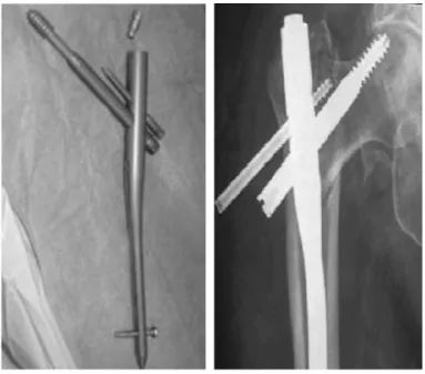
The design of proximal femoral nails has evol-ved and the nails are now in their third generation. The improvements in the design have reduced the occurrences of some complications like intraoperative fractures and fractures below the nail tip (after the operation). The Targon-PF® cephalomedullary nail (Figure 3) presents the differential that the cephalic anti-rotation nail and cephalic jacket of the sliding screw are fixed in the femoral nail itself, thereby avoi-ding the “Z” effect that occurs with other cephalome-dullary nails(14) (Figure 4).
The objective of this study was to prospectively assess the clinical, radiological and functional evolu-tion of osteosynthesis using a cephalomedullary nail (Targon-PF®), in unstable trochanteric fractures of
the femur, over a one-year postoperative follow-up.
Figure 3 – PF-Targon-PF® nail. Figure 4 – Anteroposterior
radio-graph on hip, with “Z” effect.
METHODS
383
physeal angles of 125°, 130° and 135° between the screws in the neck and the axis of the intramedullary nail. The implant was chosen after preoperative plan-ning, according to the cervicodiaphyseal angle of the proximal extremity of the contralateral femur and the diameter of the diaphyseal medullary region.
Drug prophylaxis for deep vein thrombosis (DVT) was administered during the pre and postoperative periods. The patients also received prophylactic an-tibiotic therapy at the time of induction of anesthesia and for 48 hours after the operation, consisting of cefazolin. During the immediate postoperative period, standard analgesia was administered and the patients were instructed to gradually start to walk again ac-cording to their tolerance level.
Through a prospective cohort evaluation with cross-sectional analyses, the patients who underwent osteosynthesis using Targon-PF® to treat unstable
in-tertrochanteric fractures were assessed one week, two weeks, one month, two months, six months and one years after the operation. At each return visit, clinical, radiological and functional assessments were made on the patients.
The clinical assessment was made by means of analyzing occurrences of clinical complications, in-cluding assessment of the mortality rate and its causes. The radiological assessment included an analysis on the maintenance of the quality of reduction, im-plant positioning, fracture consolidation and synthe-sis material failure. The quality of fracture reduction and implant positioning were evaluated by means of radiographic examination during the immediate postoperative period, using anteroposterior (AP) and lateral radiographic views of the proximal femur. The reduction was considered to be ideal when the cervi-codiaphyseal angle on the AP radiograph was between 130° and 135°. If the angle was smaller, the case was considered to be one of varus reduction; and if it was larger, valgus reduction(15). Implant positioning was
assessed by means of the tip-apex index(12). Fracture
consolidation, varus collapse and synthesis material failure were assessed by means of radiographic analy-sis at subsequent returns.
The functional assessment was made through analysis on Harris scores, progression of walking and pain scale evolution. At the return visit one year after the operation, a final functional assessment was made using the Harris score (a functional assessment scale
with a maximum score of 100 points that includes evaluations on four categories: pain, mobility, daily activities and range of motion). Scores lower than 70 are considered to be poor; between 70 and 80, reasonable; 80 to 90, good; and 90 to 100, excellent (Annex 1)(16). During the one-year follow-up,
pro-gression of walking and pain scale evolution were assessed. Walking ability was assessed by dividing the patients into five groups: non-walking, walking with the aid of a stick, with crutches and with a walking frame and walking unaided. Before suffering the fracture, 16.2% of the patients were already using a walking frame, 34.3% were using a stick and 59.5% were walking unaided. For the pain evaluation, a visual analogue scale graded from 0 to 10 was used. This sca-le was directly proportional to the pain sca-level reported by the patient: 0 – free from pain; 1 to 3 – mild pain; 4 to 6 – moderate pain; and 7 to 10 – severe pain.
The statistical analysis was performed using the
SPSS software. Student’s t test was used for normally
distributed independent variables. Differences were considered to be statistically significant when the re-jection level for the nullity hypothesis (P) was 0.05 (significance level of 95%).
RESULTS
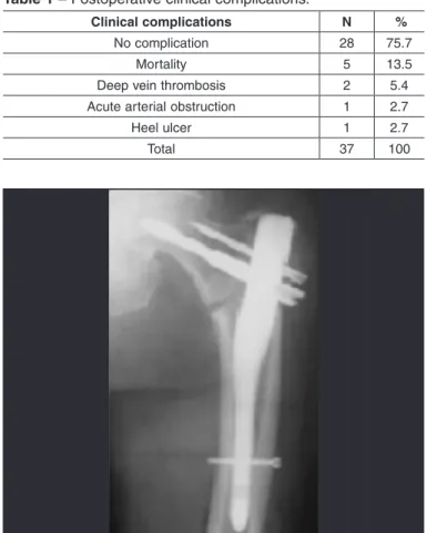
The clinical assessment over the one-year period showed that 28 patients (75.7%) did not have any postoperative clinical complications, while the other nine patients (24.3%) presented complications. The mortality rate over the evaluation period was 13.5% (five patients). Of these, four patients (80%) died due to sepsis resulting from bronchopneumonia and one patient (20%) died due to an episode of upper digesti-ve tract hemorrhage. The other clinical complications comprised two cases (5.4%) of deep vein thrombosis, one case (2.7%) of heel ulcer and one case (2.7%) of acute arterial obstruction (Table 1).
From the radiographic evaluation, the mean cer-vicodiaphyseal angle immediately after the operation was 132.5°, with a standard deviation of 9.8. The re-duction after the operation was considered ideal in 31 patients (83.6%) (Figure 5), while there were three cases of varus reduction (8.2%) and three cases of valgus reduction (8.2%) (Table 2). The mean tip-apex index was 22.8 (standard deviation = 8.3), and 27
384
Hip ID:
Study Hip:Left Right
Examination Date (MM/DD/YY): Subject Initials: _____/_____/______ Medical Record Number:
Interval:
Pain (check one) Enter public transportation
None or ignores it (44) Yes (1)
Slight, occasional, no compromise in activities (40) No (0)
Mild pain, no effect on average activities, rarely moderate Stairs
pain with unusual activity; may take aspirin (30) Normally without using a railing (4)
Moderate Pain, tolerable but makes concession to pain. Normally using a railing (2)
Some limitation of ordinary activity or work. May require In any manner (1)
Occasional pain medication stronger than aspirin (20) Unable to do stairs (0)
Marked pain, serious limitation of activities (10) Put on Shoes and Socks
Totally disabled, crippled, pain in bed, bedridden (0) With ease (4)
Limp With difficulty (2)
None (11) Unable (0)
Slight (8) Absence of Deformity (All yes = 4; Less than 4 =0)
Moderate (5) Less than 30° fixed flexion contracture
Severe (0) Less than 10° fixed abduction
Support Less than 10° fixed internal rotation in extension
None (11) Limb length discrepancy less than 3.2 cm
Cane for long walks (7) Range of Motion (*indicates normal)
Cane most of time (5) Flexion (*140°)
One crutch (3) Abduction (*40°)
Two canes (2) Adduction (*40°)
Two crutches or not able to walk (0) External Rotation (*40°)
Distance Walked Internal Rotation (*40°)
Unlimited (11) Range of Motion Scale
Six blocks (8) 211° - 300° (5) 61° - 100 (2)
Two or three blocks (5) 161° - 210° (4) 31° - 60° (1)
Indoors only (2) 101° - 160° (3) 0° - 30° (0
Bed and chair only (0) Range of Motion Score
Sitting Total Harris Hip Score
Comfortably in ordinary chair for one hour (5) On a high chair for 30 minutes (3)
385
patients (27%) had an index > 25 mm (Table 3). After one year of follow-up, the mean cervicodiaphyseal angle was 131.7°, with a standard deviation of 9.3. There were no cases of varus collapse. Consolidation was confirmed in all the patients six months after the operation, except in one case that presented “cut--out” one month after the operation. This case then underwent resection arthroplasty and consolidation count not be assessed (Annex 2). This was the only case of “cut-out” (2.7%) found in the present study.
Cut-out consists of migration of the proximal screws superolaterally in relation to the head, with extrusion and loss of cephalic fixation (Figure 6). This patient then underwent resection arthroplasty for pain relief. This was the only case that required a new surgical pro-cedure. There were no cases of peri- implant fracture.
The functional assessment using the Harris score one year after the operation showed that the mean was 69.2 with a standard deviation of 9.3. In 16% of the patients, the result presented was excellent, 19% good, 28% reasonable and 38% poor. The Harris scale showed a statistically significant inverse relationship with age (p = 0.023), with a coefficient of -0.0402, i.e. younger ages were associated with higher Harris scores (Figure 7). The Harris score was not influenced by the fracture classification according to the AO/ ASIF system (Table 4).
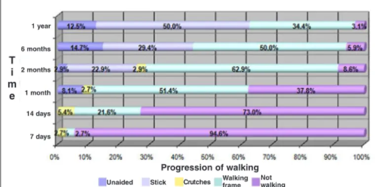
The evaluation of the progression of walking sho-wed that the quality of walking gradually improved over the assessment period. Despite the instructions to gradually start walking early on, as tolerated, 35 patients (94.6%) did not do any active walking du-ring the first week, but only bore weight on the limb.
Table 1 – Postoperative clinical complications.
Clinical complications N %
No complication 28 75.7
Mortality 5 13.5
Deep vein thrombosis 2 5.4
Acute arterial obstruction 1 2.7
Heel ulcer 1 2.7
Total 37 100
Figure 5 – Anteroposterior radiograph on hip, with ideal
re-duction.
Table 2 – Cervicodiaphyseal angle measurement.
Cervicodiaphyseal angle AP N %
Varus (< 130°) 3 8.2
Ideal (130° - 135°) 31 83.6
Valgus (> 135°) 3 8.2
Total 37 100
Table 3 – Tip-apex index measurement.
Tip-apex index N %
≤ 25 mm 27 73
> 25 mm 10 27
Total 37 100
Figure 6 – Anteroposterior and lateral radiographs on right hip
with cut-out.
Age (years)
100
90 80 70
60
50
40
30
20 10 0
50 55 60 65 70 75 80 85 90 95 100
H a r r i s
s c o r e
386
Annex 2 – Clinical, functional and radiographic assessment on the patients who underwent osteosynthesis using a cephalomedullary nail.
Age Tronzo Ao Side Time i Time c Sex Comorbidity Complications  ap  ap f Tai Harris
A 82 III A2 R 12 10 F COPD Death 135 * 24 *
B 94 V A3 L 6 3 F SAH-DM Death 160 * 20 *
C 86 III A2 R 8 6 F Alzheimer Death 130 * 25 *
D 88 IV A2 L 15 13 M SAH-DM-COPD Death 130 * 25 *
E 83 III A2 R 16 11 M SAH-Alzheimer Death 150 * 24 *
F 76 III A2 L 15 13 M AMI-SAH-
Angioplasty 0 125 125 20 95
G 77 III A2 R 8 5 F SAH-DM 0 130 130 20 93
H 63 V A3 R 8 5 F SAH 0 130 130 18 92
I 76 III A2 R 8 4 F SAH 0 130 130 20 91.01
J 25 V A3 R 4 6 M 0 0 115 120 35 90
L 73 V A3 R 18 15 M SAH-DM 0 125 127 30 87
M 65 V A3 R 15 12 F SAH-DM 0 135 135 20 85
N 88 III A2 R 7 5 F DM 0 135 135 27 85
O 89 III A2 R 7 5 F SAH-DM AAO 130 130 25 85
P 70 III A2 L 18 15 F SAH 0 137 137 15 84
Q 61 III A2 R 7 5 M 0 0 110 118 10 80
R 54 III A2 L 7 4 M SAH-Previous
Stroke 0 135 135 40 78.48
S 78 III A2 R 5 3 M Depression-Myeloma 0 150 153 25 78
T 58 V A3 L 6 3 F
SAH-DM-CRF-Hypothyroidism 0 136 136 27 77.85
U 67 V A3 L 8 6 F SAH 0 130 130 30 76.85
V 88 III A2 L 5 3 M SAH-DM 0 130 130 25 75
X 79 III A2 L 18 15 F
DM-SAH-Atrial Flutter 0 125 125 20 74
Z 75 III A2 R 6 3 F
DM-SAH-Alcoholism 0 130 130 35 71.3
A1 78 III A2 L 7 6 F 0 0 130 130 5 71
B2 82 III A2 R 7 5 F 0 0 130 130 10 70
C1 88 III A2 L 7 4 M Parkinson Heel Ulcer-DVT 130 130 18 60.41
D1 82 V A3 R 7 4 M DM 0 135 135 25 60
E1 89 III A2 L 9 6 F SAH-DM 0 130 130 20 59.22
F1 66 III A2 L 6 4 F SAH DVT 130 130 20 57.15
G1 88 III A2 L 7 5 F SAH 0 135 135 25 50.15
H1 70 IV A2 R 20 18 M Parkinson-SAH 0 135 135 30 50
I1 71 III A2 R 7 5 F 0 0 135 135 14 48.57
J1 91 III A2 R 20 14 M 0 Cut-out 130 130 40 48
L1 75 III A2 L 13 4 F Parkinson 0 140 140 21 43
M1 88 IV A3 L 8 4 F
Epilepsy-Hypothyroidism 0 130 130 24 41
N1 91 III A2 L 15 10 F SAH-DM 0 130 130 2 38.57
O1 78 III A3 L 6 4 M Epilepsy 0 140 140 30 20
387
At the assessment two months after the operation, it was observed that only three patients (8.6%) were not walking actively. Six months after the operation, only 3.1% were still not walking, while 50% were walking with the aid of stick, 34.4% with a walking frame and 12.5% without using any aid (Figure 8). Before the fracture, 16.2% of the patients were already using a walking frame, 34.3% were using a stick and 59.5% were walking unaided.
The evaluation on the evolution of pain by means of the visual analogue scale graduated from 0 to 10 showed that there was a progressive diminution over the assessment period, with a mean of 5.19 in the first week and 2.25 one year after the surgery (Figure 9).
The general incidence of mortality after trochan-teric fractures is described in the current literature as 6 to 11% within the first month and 14 to 36% within the first year(16). Mortality typically results
from cardiopulmonary, thromboembolic and septic complications. In our sample, the mortality rate was 13.5% (five patients) over the one-year period. The cause of death was sepsis due to bronchopneumonia in four cases (80%), while one case (20%) was due to upper digestive tract hemorrhage.
Deep vein thrombosis (DVT) is a substantial cause of morbidity and mortality during the follow-up of cases of fracture of the proximal femur(4). Symptomatic
DVT occurs in around 2%, and 85% of these cases are diagnosed during the first five weeks after the fracture. Factors such as prolonged immobility, coagulopathy and delayed surgical treatment favor occurrences of DVT(17). In the present study, antithrombotic drug
pro-phylaxis was instituted in all cases, and two patients (5.4%) were diagnosed with DVT. No cases of pulmo-nary thromboembolism were found among our sample. No cases of superficial or deep infection were iden-tified in the present study. The other clinical complica-tions were one case (2.7%) of acute arterial obstruction and one case (2.7%) of pressure ulcer on the heel.
According to Schipper et al(8), the most common
postoperative complication seen in radiological eva-luations is cut-out. Its incidence has been described in the literature as 0.7 to 10.6%(2). In the present study,
there was only one case of cut-out (2.7%). Inadequate reduction, especially with varus presentation, incor-rect implant positioning and advanced osteoporosis are the factors that determine its occurrence.
Implant positioning should follow the concepts introduced by Baumgaertner et al(12), in which the
distance between the tip of the sliding screw and the center of the femoral head should not be more than 25 mm, as a sum of the AP and lateral-view radiographs (tip-apex index < 25 mm), which facilitates telesco-ping of the dynamic system of the implant and reduces the risk of cut-out(15). Also according to Baumgaertner
et al(12), occurrences of cut-out increase
considera-bly when the tip-apex index is greater than 25 mm, independent of the quality of the fracture reduction. The tip-apex index has been described for osteosyn-thesis using a DHS. Use of this index for assessing the adequacy of positioning of cephalomedullary nails remains controversial, especially with regard to nails
Table 4 – Harris scale for AO/ASIF classification.
AO N Mean Standard deviation p-value*
A2 23 68.9504 16.95327 0.895
A3 9 69.9667 24.86849 0.895
* Student’s t test.
Figure 8 – Time versus progressing of walking.
Figure 9 – Pain scale versus time.
Time
10
8
6
4
2
0
1 week 2 weeks 1 month 2 months 6 months 1 year
P a i n
5.19
4.38
3.59
2.84 2.53 2.25
DISCUSSION
Trochanteric fractures are correlated with high morbidity and mortality rates. The most prevalent and most important clinical complications are deep vein thrombosis, pulmonary thromboembolism, superficial infection and deep infection(4).
T i m
e
1 year
6 months
2 months
1 month
14 days
7 days
Progression of walking
Unaided Stick Crutches Walking frame Notwalking
388
with two screws fixed proximally, because of the diffi-culty in positioning the sliding screw at the center of the femoral head in AP-view radiographs. Thus, there is a greater tendency towards positioning the sliding screw at a lower location on AP radiographs, especially in patients with a short femoral head and neck.
Despite this proviso, this index has been used by some authors for assessing whether implants have been positioned correctly(15). In our study, we found
a mean tip-apex index of 22.8 (standard deviation of 8.3). There were 27 patients (73%) with a tip-apex
index ≤ 25 mm and 10 (27%) with an index > 25 mm.
Even with the presence of the anti-rotation nail in the Targon-PF, we were able to respect the principles recommended by Baumgartner, for most of our pa-tients. The patient in this study who presented cut-out had advanced osteoporosis and inadequate implant positioning (tip-apex index of 40 mm), as risk factors. Werner-Tutschku et al(18) reported that the main
reason for occurrences of cut-out is an initial unsatis-factory reduction, generally with varus presentation. As well as favoring occurrences of cut-out, varus align-ment may result in Trendelenburg gait. In our sample, the mean cervicodiaphyseal angle was 132.5°, with a standard deviation of 9.3. The postoperative reduction was considered to be ideal in 31 patients (83.6%), and there were three cases with varus reduction (8.2%) (Table 2). According to Werner-Tutschku et al, the in-cidence of varus consolidation is 11.2%(18). The angular
deviation did not favor cut-out and did not statistically influence the quality of gait in this sample (p = 0.442).
In a meta-analysis, Kaplan et al(2) presented a mean
time taken to achieve consolidation of four months, independent of the device used. On the other hand, Bride et al10 reported that consolidation occurred
af-ter an average of six months. According to Crawford et al(19), the consolidation rate found among patients
treated with a cephalomedullary nail was 89%(19). In
the present study, consolidation was observed in all the patients after six months, except for one case that presented cut-out one month after the operation, who then underwent resection arthroplasty. Consequently, consolidation could not be assessed. The abundant blood supply in the trochanteric region, associated with stable synthesis, favored this result.
One complication from using cephalomedullary nails is fractures of the femoral diaphysis below the tip of the implant(7). Lack of experience on the part of the
surgeon and the inadequate design of first and second--generation nails have favored this complication(14).
Nails of the latest generation have a radius of curvature that fits the anatomical shape of the femur better. In contrast with older reports, recent studies have reported significant decreases in this complication(2). The rate
of diaphyseal fracture below the nail tip ranges in the literature from zero to 2.1%(2). In the present study,
no cases of fractures distal to the implant were found. With regard to functional evaluation, the Harris score is the scale used by the majority of authors for postoperative functional assessment of trochanteric fractures(16). Schipper et al(8) found a mean score of66.80
(standard deviation = 17.94) with a proximal femoral nail of PFN® type, and 69.50 (standard deviation =
16.00) with a nail of Gamma Nail® type, after one year.
The mean value found in the present study was 69.3 (standard deviation = 9.3) with the Targon-PF®; 16%
of the patients presented excellent results, 19% good, 28% reasonable and 38% poor. The functional result according to the Harris score was considered to be poor or moderate for the majority of the patients who underwent osteosynthesis with a cephalomedullary nail. The great difficulty in functional assessment of fractures of the proximal femurs using the Harris score is that it is impossible to make an assessment before the surgery and thus, no comparisons from before to after the operation can be made, as used in elective surgery. Given the advanced age of these patients, with preexisting limitations, they would possibly present scores that are already compromised. One important indicator is that the Harris score showed a statistically significant correlation with age (Figure 7), and was not influenced by the Tronzo and AO classifications, and not even by the quality of the reduction and positioning of the implants. Thus, the energy of the trauma and the quality of the surgery, evaluated radiographically, did
not influence the functional result, but the patient’s
389
With a new scale, it would be possible to better assess the influence of the trauma energy and the quality of the surgery in functional assessments, after removing
the influence of the patient’s previous state from the
assessment. We emphasize that even though the mean Harris score of 69.3 found in the present study is low, it is similar to values found by other authors(16). For a
better functional assessment on the patients, we analyzed another two important parameters separately: recovery of walking ability and evolution of the pain scale.
According to Pajarinen et al(13), patients who
un-derwent osteosynthesis with a cephalomedullary nail, in unstable trochanteric fractures, presented a signifi-cantly faster return to their previous level of walking(13).
Herrera et al(9) reported on a study involving 250
pa-tients treated with the PFN® and Gamma nail®
cepha-lomedullary nails, in which around 50% of the patients had recovered their previous walking capacity, one year after the surgery(4). In the present study, we
as-sessed the recovery of walking ability over the course of time. We divided the subjects into five groups: no walking; walking with a frame; walking with crutches; walking with a stick; and walking unaided (Figure 8). The greatest evolution in the quality of walking occur-red over the first two months after the operation, such that only 8.6% of the patients were still not walking at that time. One year after the operation, only 3.1%
of the patients were still not walking, while 50% were walking with the aid of a stick, 34.4% with a walking frame and 12.5% without any aid. Thus, one year after the operation, 40.6% of the patients presented same walking capacity as presented previously.
An assessment of pain was made by Nuber et al(20),
through a six-month follow-up with successive sco-res, comparing patients who received a DHS with those who received an intramedullary nail. The pain scores were considerably greater in the group treated with intramedullary nails. In the present study, the patients were assessed using a visual analogue scale for pain and showed progressive diminution of pain at the return visits over the one-year period, as presen-ted in Figure 9. The evolution of the pain scale was not influenced by the trauma energy, age, reduction parameters or implant positioning parameters.
CONCLUSION
Osteosynthesis using a cephalomedullary nail Targon®-PF, used in unstable trochanteric fractures,
resulted in low rates of clinical complications, excel-lent stabilization, few mechanical complications and adequate functional results. The authors consider that this is an appropriate technique for treating unstable trochanteric fractures of the femur.
REFERENCES
1. Parker MJ, Handoll HH. Intramedullary nails for extracapsular hip fractures in
adults.Cochrane Database Syst Rev. 2009;(3):CD004961.
2. Kaplan K, Miyamoto R, Levine BR, Egol KA, Zuckerman JD. Surgical
mana-gementof hip fractures: an evidence-based review of the literature. II.
Intertro-chanteric fractures.J Am Acad Orthop Surg. 2008;16(11):665-73.
3. Haidukewych GJ. Intertrochanteric fractures: ten tips to improve results. J Bone Joint Surg Am. 2009;91(3):712-9.
4. Todd CJ, Freeman CJ, Camilleri-Ferrante C, Palmer CR, Hyder A, Laxton CE, et al. Differences in mortality after fracture of the hip: the east Anglian audit.
Br Med J. 1995;310(6984):904-8.
5. Müller ME.Classification and international AO-Documentation of femur fractu-res.Unfallheilkunde. 1980;83(5):251-9.
6. Tronzo RG. Symposium on fractures of the hip.Special considerations in
ma-nagement.Orthop Clin North Am. 1974;5(3):571-83.
7. Laros G, Moore JF. Complications of fixation in intertrochanteric fractures.Clin Orthop Relat Res. 1974;(101):110-9.
8. Schipper IB, Steyerberg EW, Castelein RM, van der Heijden FH, den Hoed PT, Kerver AJ, et al. Treatmentof unstable trochanteric fractures.Randomised
comparison of gamma nail and the proximal femoral nail. J Bone Joint Surg
Br.2004;86(1):86-94.
9. Herrera A, Domingo LJ, Calvo A, Martinez A, Cuenca J.Comparative study of
trochanteric fractures treated with the Gamma nail or the proximal femoral nail. Int Orthop. 2002;26(6):365-9.
10. Bridle SH, Patel AD, Bircher M, Calvert PT.Fixation of intertrochanteric fractures of the femur. A randomized prospective comparison of the gamma nail and the
dynamic hip screw.J Bone Joint Surg Br. 1991;73(2):330-4.
11. Schipper IB, Bresina S, Wahl D, Linke B, van Vugt AB, Schneider E, et al.
Biomechanical evaluation of the proximal femoral nail.Clin Orthop Related
Res. 2002;(405):277-86.
12. Baumgaertner MR, Curtin SL, Lindskog DM.Intramedullary versus
extrame-dullary fixation for the treatment of intertrochanteric hip fractures. Clin Orthop Related Res. 1998;(348):87-94
13. Pajarinen J, Lindahl J, Michelsson O, Savolaien V, Hirvensalo E.
Pertrochan-teric femoral fractures treated with a dynamic hip screw or a proximal femoral nail. A randomised study comparing post-operative rehabilitation. J bone Joint Surg Br. 2005;87(1):76-81.
14. Helwig P, Faust G, Hindenland U, Hirschmüller A, Konstantinidis L, Bahrs C, et al.Finite element analysis of four different implants inserted in different positions to stabilize an idealized trochanteric femoral fracture.Injury. 2009;40(3):288-95.
15. Guimarães JA, Guimarães AC, Franco JS.Avaliação do emprego da haste
femoral curta na fratura trocantérica instavel do fêmur. Rev Bras Ortop. 2008; 43(9):406-417.
16. Guimarães RP, Alves DP, Silva GB, Bittar ST, Ono NK, Honda E, et al.Tradução e adaptação transcultural do instrumento de avaliação do quadril “Harris Hip Score”. Acta Ortop Bras. 2010;18(3):142-7.
17. Hefley FG, Nelson CL, Puskarich-May CL.Effect of delayed admission to the
hospital on the preoperative prevalence of deep-vein thrombosis associated with fractures about the hip. J Bone Joint Surg Am. 1996;78(4):581-3.
18. Werner-Tutschku W, Lajtai G, Schmiedhuber G, Lang T, Pirkl C, Orthner E.
Intra-and perioperative complications in the stabilization of per-and subtrochan-teric femoral fractures by means of PFN. Unfallchirurg. 2002;105(10):881-5.
19. Crawford CH, Malkani AL, Cordray S, Roberts CS, Sligar W.The trochantheric
nail versus the sliding hip screw for intertrochanteric hip fractures: a review of
93 cases.J Trauma. 2006; 60(2):325-8.
20. Nuber S, Schönweiss T, Rüter A. Stabilisation of unstable trochanteric femoral fractures. Dynamic hip screw (DHS) with trochanteric stabilisation plate vs. proximal femur nail (PFN). Unfallchirurg. 2003;106(1):39-47.