J of Evolution of Med and Dent Sci/ eISSN- 2278-4802, pISSN- 2278-4748/ Vol. 3/ Issue 16/Apr 21, 2014 Page 4316
LEFT AXIS DEVIATION IN ELECTROCARDIOGRAPHY
S. S. Nelson1, V. K. Mehta2, A. C. Nagpal3
HOW TO CITE THIS ARTICLE:
S. S. Nelson, V. K. Mehta, A. C. Nagpal. Left Axis Deviation in Electrocardiography. Journal of Evolution of Medical and Dental Sciences 2014; Vol. 3, Issue 16, April 21; Page: 4316-4324,
DOI: 10.14260/jemds/2014/2437
ABSTRACT: BACKGROUND: The electrocardiographic finding of left axis deviation (LAD) occurs in wide variety of clinical conditions yet its significance has been debated. Paul-Wood stated its presence in 10% of healthy individuals while Pryor has quoted as low as 0.2%. OBJECTIVES: To study the incidence of LAD in various cardiac disorders. Conversely, to study the incidence of various cardiac disorders presenting as LAD. To make an attempt to find out how far it is of organic significance. METHOD: The study is based on the outcome of 70 cases with LAD out of 1180 cases attending the Medicine OPD or admitted in medicine wards of N.S.C.B. Medical College & Hospital, Jabalpur (M.P). RESULTS: LAD was found in only 5.9% of the cases examined. True LAD (-30 to –90 degrees) was found in 3.8%. Among various cardiovascular disorders the highest incidence of LAD was in cardiomyopathy (33.3%). Next being ischemic heart disease (15.5%) followed by hypertension and hypertensive heart disease (6.6%). No organic heart disease (3.6%), pulmonary heart disease (3.3%), congenital heart disease (3.1%), and rheumatic heart disease (2.1%), have contributed for a very small percentage. Among the LAD cases Myocardial infarction was most common seen in 21 cases (30%). CONCLUSIONS: LAD is uncommon and was found in only 5.9% of the cases examined. True LAD (-30 to –90 degrees) was found in 3.8% of cases and it reflected a definite pathological state. The ischemic factor was found to be more responsible for production of LAD rather than hypertrophy itself alone.
KEYWORDS: a) Left axis deviation b) Electrocardiography, c) Ischemic heart disease.
HHD: Hypertensive heart disease. HIHD: Hypertensive ischemic heart disease. IPIB: Inferior peri-infarction block. LAD: Left axis deviation. LBBB: Left bundle branch block. LVH: Left ventricular hypertrophy. PIB: Peri-infarction block. RBBB: Right bundle branch block. SIVB: Superior intraventricular block. SPIB: Superior Peri-infarction block.
INTRODUCTION: The heart has always enchanted mankind. The fact that the heart is a source of measurable electrical activity has enhanced this enchantment. The fundamentals of electrocardiography are based on sound scientific principles, although the practicing clinicians often ignore these. Electrocardiography has progressed from an interesting research tool to become a widely diagnostic tool, an essential requirement of any competent cardiac evaluation. Electrocardiogram is universally available non-invasive tool and can be repeated whenever necessary. The electrocardiographic finding of left axis deviation occurs in wide variety of clinical conditions yet its significance has been debated.1
J of Evolution of Med and Dent Sci/ eISSN- 2278-4802, pISSN- 2278-4748/ Vol. 3/ Issue 16/Apr 21, 2014 Page 4317
The factors considered to cause LAD have included altered anatomic and electrical position of the heart, a normal variation, left anterior hemiblock, left bundle branch block (LBBB), Left ventricular hypertrophy (LVH), Myocardial fibrosis, inferior wall myocardial infarction, emphysema, WPW Syndrome, congenital heart disease (endocardial cushion defects), ectopic ventricular rhythms, pacing from apex of left or right ventricle and unknown factors associated with age. The commonest cause of Left axis deviation is left anterior hemiblock.4
This is most frequently due to fibrosis that interrupts the anterosuperior division of the left bundle branch. Grant (1956) proposed that LAD is usually due to myocardial fibrosis and might be helpful in diagnosis of anterolateral myocardial infarction even in absence of diagnostic Q waves.5
The present study was undertaken to evaluate the different aspects of LAD.
AIMS AND OBJECTIVES:
1. To study the incidence of LAD in various cardiac disorders.
2. Conversely to study the incidence of various cardiac disorders presenting as LAD. 3. To attempt to find out how far LAD is of organic significance.
4. To find out the quantitative correlation in three grades of LAD.
MATERIAL AND METHODS: The study was conducted in the Department of Medicine, N.S.C.B. Medical College Hospital, Jabalpur. The present study is based on the outcome of 1180 cases. Patients exhibiting left axis deviation (LAD) constituted the material for this study. For the purpose of this study LAD was defined as a mean QRS-axis which lies between + 0 and –90 degrees in frontal plane.
A detailed clinical examination and investigations were done in all cases. These cases were divided in 3 groups, depending upon degree of LAD. LAD I (+0 to –14 degrees), LAD II (-15 to –29) degrees) and LAD III (-30 TO –90 degrees). Mean initial 0.04, terminal 0.04 and QRS axis was determined in every electrocardiogram, when the QRS deflections were smooth the axis is calculated from algebraic sum of amplitude of positive and negative deflections. Hexaxial reference system was employed for calculation of the axis.
DEFINITIONS:
Hypertension: Any reading 140/90 mmHg or above without any clinical-radiological, electrocardiographic or echocardiographic evidence of enlargement of the heart.
Hypertensive Heart Disease: Blood pressure 140/90 mmHg or above with clinical, radiological electrocardiographic and/or echocardiographic evidence of enlargement of heart but no evidence of ischemic heart disease clinically or electrographically.
Hypertensive Ischemic Heart Disease: Hypertensive heart disease with clinical or electrocardiographic evidence of ischemic heart disease.
No Organic Heart Disease: There is no clinical, radiological electrocardiographic or echocardiographic evidence of heart disease.
J of Evolution of Med and Dent Sci/ eISSN- 2278-4802, pISSN- 2278-4748/ Vol. 3/ Issue 16/Apr 21, 2014 Page 4318
III and aVF and inverted in aVL, small S wave in I and prominent R in aVR, attenuation of R waves in left precordial leads, pattern of aVL resembles aVR.
All these patients were thoroughly examined clinically. In the general examination all the peripheral pulses were palpated for the rhythm, volume, character and thrill especially the carotid pulse. Blood pressure was noted and signs of heart failure were also assessed. The cardiovascular system was examined for the site and type of apex beat, parasternal heave. First, second, third and fourth heart sounds were auscultated. Murmurs were looked for in mitral, aortic, pulmonary and tricuspid area.
The patients were investigated routinely for complete hemogram, urine examination for albumin and sugar. Blood sugar, serum urea, creatinine and serum cholesterol were also estimated. A 12 Lead Electrocardiogram was taken of all the patients by BPL 108 Cardiart Machine. ECG was evaluated for rate, rhythm, axis deviation, hypertrophy and strain, ischemia, bundle branch block, hemiblock and myocardial infarction.
Arrhythmia which were looked for were atrial premature complex, paroxysmal supraventricular tachycardia, atrial fibrillation and ventricular extrasystole.
For diagnosing ischemia, ST segment depression, T wave inversion, Q waves and bundle branch block were looked for in ECG.
Left ventricular hypertrophy was diagnosed by Romhilt Estes criteria.
X-Ray chest P-A view was done in all the patients. Cardiomegaly, aortic calcification were particularly noted.
Echocardiographic evaluation was done in almost all patients and included 2-D, M-mode and color doppler examination.
In M or motion mode display, Heart Structures were identified by their characteristic motion. In 2D or Dimension Echocardiography a real time, cross section view obtained.
This was more useful than M-Mode. The combination of M-mode and 2D echocardiography gives the best information for diagnosis of various cardiac disorders.
J of Evolution of Med and Dent Sci/ eISSN- 2278-4802, pISSN- 2278-4748/ Vol. 3/ Issue 16/Apr 21, 2014 Page 4319
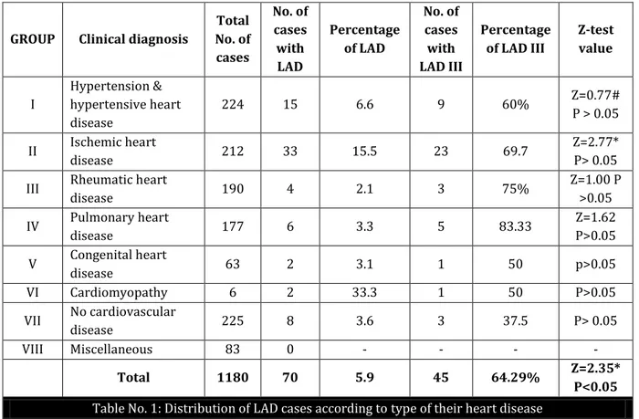
GROUP Clinical diagnosis
Total No. of cases No. of cases with LAD Percentage of LAD No. of cases with LAD III Percentage of LAD III
Z-test value I Hypertension & hypertensive heart disease
224 15 6.6 9 60% Z=0.77# P > 0.05
II Ischemic heart
disease 212 33 15.5 23 69.7
Z=2.77* P> 0.05 III Rheumatic heart
disease 190 4 2.1 3 75%
Z=1.00 P >0.05 IV Pulmonary heart
disease 177 6 3.3 5 83.33
Z=1.62 P>0.05 V Congenital heart
disease 63 2 3.1 1 50 p>0.05 VI Cardiomyopathy 6 2 33.3 1 50 P>0.05 VII No cardiovascular
disease 225 8 3.6 3 37.5 P> 0.05 VIII Miscellaneous 83 0 - - - -
Total 1180 70 5.9 45 64.29% Z=2.35*
P<0.05
Table No. 1: Distribution of LAD cases according to type of their heart disease
# 95% CL 71%, -31%
* Statistically significant; P < 0.05
4 cases of LAD were in 31 to 50 years of age, while 2 out of 3 cases of true LAD were above 51 years age. The mean age for LAD cases was 46.36 + 12.77 years and 58.6% cases were between 5th and 6th decades. The ratio of male to female was 7.3 irrespective of etiology. Male to female ratio increased from 1.5:1 to 3:1 from LAD I to LAD III and Males were more prone to have higher chances of LAD (P<0.05).
GROUP Clinical diagnosis No. of cases Percentage
I Hypertension and Hypertensive
heart disease 15 21.4
II Ischemic heart disease 33 47.1 III Rheumatic heart disease 4 5.7
IV Pulmonary heart disease
(Emphysema) 6 8.6
V Congenital heart disease 2 2.9
VI Cardiomyopathy 2 2.9
VII No cardiovascular disease 8 11.4
Total 70
J of Evolution of Med and Dent Sci/ eISSN- 2278-4802, pISSN- 2278-4748/ Vol. 3/ Issue 16/Apr 21, 2014 Page 4320
It could be observed from table 2 that ischemic heart disease with or without hypertension was responsible for about half of the cases. 11.4% of the cases having LAD had a clinical diagnosis of
No Organic heart disease .
In hypertension alone the incidence of LAD III was 25%, which increased to 72.7% with the development of hypertensive heart disease.
It was observed that patients with CCF had more evidence of true LAD than without CCF. Of the cases labeled as No organic heart disease, 3.6 % had LAD and 1.3% true LAD. Conversely among LAD cases No organic heart disease group was responsible for 11.4 % cases including 4.2% of true LAD
Emphysema was responsible for 3.3% of the cases of this series including 7.1% as true LAD. Of the two cases of cardiomyopathy with LAD, one case had true LAD. SIVB was responsible for 35% cases of ischemic heart disease, while PIB for 23.1%. SPIB was more common with anterolateral and IPIB with diaphragmatic infarct.
In congenital heart disease the average age was 18.5 years with range of 12 to 25 years, of these one case of tricuspid atresia had true LAD and 1 case of ventricular septal defect had LAD II.
Electrocardiographic Diagnosis No. of cases Percentage
Myocardial infarction 21 30
Only left axis deviation 18 25.71 Primary T and/ or ST changes 10 14.29 Left ventricular hypertrophy 8 11.43 Suggestive of emphysema 6 8.57 Bundle branch block:
Left bundle branch block (LBBB) 4 5.71 Right bundle branch block
(RBBB)
Without infarction With infarction
3
2 1
4.29
Total 70
Table No. 3: Electrocardiographic findings of LAD
Table 3 shows the electrocardiographic findings of 70 LAD cases. Myocardial infarction was most common and only LAD was present in 18 cases. Incidence of LAD III was highest in hypertensive ischemic heart disease, next being in hypertensive heart disease.
It was noticed that one LBBB case of hypertensive ischemic heart disease had true LAD similarly one case of rheumatic heart disease had true LAD.
All the cases of RBBB irrespective of etiology had true LAD and were of coronary thrombosis.
Phases of Breathing did make a Difference in Axis:
a) Normal axis remained normal, though it made an average difference of +23.8 degrees (Range +2 to + 66 degrees);
J of Evolution of Med and Dent Sci/ eISSN- 2278-4802, pISSN- 2278-4748/ Vol. 3/ Issue 16/Apr 21, 2014 Page 4321
c) True LAD with SIVB and SPIB did not make significant difference (Average +4.7 and Range +0 to +22 degrees).
DISCUSSION: Soon after the advent of electrocardiography it was recognized by Einthoven and others that characteristic deviation occurs in association with hypertrophy of cardiac chambers (Gubner et al, 1943).6
The definition of LAD is also under dispute Luisada (1965) would consider LAD between -30 to -90 degrees. While most of the workers (Friedberg7, 1966; Paul-wood 1968) would consider LAD from + 0 to –90 degrees.
Furthermore, marked (Curd et al8, 1961; Eliot et al9 1966) or true (Pryor et al, 1966)LAD would be considered when axis is superior to –30 degrees. The problem is further complicated by rotational effect of heart, for example, RBBB with horizontal heart often may give rise to LAD, while vertical heart in presence of or left bundle branch block may not be associated with LAD (McMullen, 1996)10.
The confusion worsens if respiration is taken into consideration. With deep inspiration position of diaphragm will change the anatomical axis of heart, downwards and so also of electrical axis. This results in reduction of height of R in lead I and depth of S in lead III and vice versa. Most of the authors are of opinion that axis should be calculated with normal breathing. While in cases with true LAD due to necrosis or bundle branch block, change in axis due to respiration is usually not more than 5 degrees. And hence often this is unlikely to alter the true LAD to minor LAD.
However, in true LAD associated with intraventricular or peri-infarction block respiratory variation will not make significant change. But in cases associated with LVH it may change to minor LAD or occasionally to true LAD.
On Studying LAD in different cardiovascular diseases in 3 grades, the observations show that LAD I and LAD II really do not reflect any quantitative correlation. On the contrary LAD III (-30 to –90 degrees) does reflect a definite pathological state.
Various workers have correctly called LAD III as marked or true LAD (Curd et al, 1961; Eliot et al, 1966 and Pryor et el, 1966). The proportion for true LAD were significant in IHD cases (Z = 2.29; P<0.05) and probability of true LAD was high in HHD and HTN cases. Significance of LAD is still debatable, in a healthy person. Incidences are reported to be varying from 0.2 to 10%.11
Whether the association of LAD in cases with LVH and LBBB is co-incidental or accidental? LAD is uncommon and was found in only 5.9% of the cases in this series. Various phases of breathing i.e., deep inspiration and expiration does change axis, some-times from –40 to+66 degrees. But observations of present Study showed breathing into abnormal axis either right or left does not change that normal axis.
J of Evolution of Med and Dent Sci/ eISSN- 2278-4802, pISSN- 2278-4748/ Vol. 3/ Issue 16/Apr 21, 2014 Page 4322
This fibrosis though in large majority of cases is of vascular origin associated with coronary sclerosis, and this fibrosis may give rise to SIVB. To name a few inflammation associated with myocarditis and cardiomyopathy, collagen disorders like rheumatic heart disease; rheumatoid lupus, scleroderma etc. Pryor et al (1966) has described hyperkalemia as an important cause of temporary LAD.
Other causes of LAD as mentioned by Banta et al include myotonic dystrophy, progressive muscular dystrophy, hemochromatosis and alcoholic cardiomyopathy. The list of non-cardiac causes which may be associated with LAD as Moller et al (1968)12 has found comprises of Henoch-Schoenlein purpura, myotonic dystrophy, plasma cell hepatitis, extra-hepatic billiary atresia rheumatoid arthritis is in association with incompletely diagnosed collagen disease, chronic lung disease like cystic fibrosis asthma, and bullous emphysema. However, in present series in the absence of autopsy studies it was not possible to pin point the etiology of fibrosis.
As the peak incidence was in elderly group, associated with ischemic heart disease it was justified to presume the lesion to be secondary to coronary atherosclerosis. The second common cause was peri-infarction block which was present in 33% of myocardial infarction cases. The SPIB being commonly associated with antero-lateral infarction and IPIB with diaphragmatic.13 This some time may be transient while at other times permanent depending upon the persistence of damage to superior radiation.
Of the 70 cases ischemic heart disease was responsible for 47.1% of the cases in this series. LAD was more found in cases of angina with hypertension than without it. However, in myocardial infarction the incidence was 3:2 in favor of normotensives as compared with hypertensive. The incidence of LAD progressively increased from hypertension to hypertensive heart disease to hypertensive ischemic heart disease to hypertensive coronary thrombosis. Considering that coronary artery disease without hypertension forms the largest group in any series.
It was also found to be responsible for the peak incidence of LAD in this series. Among various cardiovascular disorders the highest incidence of LAD was in cardiomyopathy (33.3%). Next being ischemic heart disease (15.5%). No organic heart disease cases showed 3.6% congenital heart disease2.9% and rheumatic heart disease (2.1%) has contributed for a very small percentage while in pulmonary heart disease it was 3.3%.
The incidence of LAD in hypertension and hypertensive heart disease was 6.6% while among LAD they formed 21.4%. 11.4% cases of LAD were associated with LVH, irrespective of latter s etiology, and of these 50% had true LAD. Hypertensive heart disease was responsible for only 25% LVH, while hypertensive ischemic heart disease for 37.5%.
The remaining were rheumatic heart disease and cardiomyopathy. This will justify the thinking that it is the ischemic factor, which is more responsible for production of LAD rather than hypertrophy itself alone.
Two of 4 cases of LBBB had only minor LAD. Three cases of RBBB had true LAD. LAD with RBBB usually suggests organic myocardial damage. Only a follow up study will justify the label No organic heart disease . The pathogenesis has been suggested to be due to either myocardial fibrosis or a congenital defect in the conduction system analogous to congenital RBBB.14
J of Evolution of Med and Dent Sci/ eISSN- 2278-4802, pISSN- 2278-4748/ Vol. 3/ Issue 16/Apr 21, 2014 Page 4323
various cardiovascular disorders the highest incidence of LAD was in cardiomyopathy (33.3%). Next being ischemic heart disease (15.5%) followed by hypertension and hypertensive heart disease (6.6%). No organic heart disease (3.6%), pulmonary heart disease (3.3%), congenital heart disease (3.1%), and rheumatic heart disease (2.1%), have contributed for a very small percentage.
Among the LAD cases Myocardial infarction was most common seen in 21 cases (30%). The ischemic factor was found to be more responsible for production of LAD rather than hypertrophy itself alone.
BIBLIOGRAPHY:
1. Okajima M, Simonson E. Electrical axis, measurement and definition in historical prospective. Am Heart J. 1961; 61:421.
2. Paul-Wood. Diseases of the heart and circulation, 1968; Ed 3:111.
3. Pryor R, Blount S.G. Jr. The clinical significance of true left axis deviation. Left intra- ventricular blocks. Am, Heart J., 1966; 72:391.
4. Yano K, Peskoe SM, Rhoads GG, Moore JO. Left axis deviation and left anterior hemiblock among 8, 000 Japanese- American men. Am J Cardiol, 1975; 35: 809-815.
5. Grant RP. Left axis deviation: An electrocardiographic pathologic correlation study. Circulation, 1956; 14:233.
6. Gubner R, Ungerleider HE. Electrocardiographic criteria of left ventricular hypertrophy. Arch Int Med. 1943; 72:196.
7. Friedberg, Diseases of the heart, W.B. Sunders Company, Philadelphia and London, Ed. 1966; 50-52, 172-173.
8. Curd GW, Hicks WM, Gyorkey F. Marked Left axis deviation. Indication of cardiac abnormality, Am Heart J. 1961; 62:462.
9. Eliot RS. Clinical significance of left axis deviation, P.G. Med., 1966; 39:349.
10.McMullen Michael 1996 Electrocardiographic significance of left axis deviation Ed. 1996.
11.Ostrander LD. Left axis deviation: prevalence, associated conditions and prognosis. Ann Intern Med. 1971; 75:23-28.
12.Moller J, Cartson E, Eliot RS. Left axis deviation in children. Dis Chest, 1968; 53:453.
13.Castle UT, Keane WM. Electrocardiographic "Periinfarction block"- A clinical correlation. Circulation, 1980; 31:403.
J of Evolution of Med and Dent Sci/ eISSN- 2278-4802, pISSN- 2278-4748/ Vol. 3/ Issue 16/Apr 21, 2014 Page 4324
AUTHORS:
1. S. S. Nelson 2. V. K. Mehta 3. A. C. Nagpal
PARTICULARS OF CONTRIBUTORS:
1. Assistant Professor, Department of Medicine, N.S.C.B. Medical College, Jabalpur.
2. Professor, Department of Medicine, N.S.C.B. Medical College, Jabalpur.
3. Professor, Department of Medicine, N.S.C.B. Medical College, Jabalpur.
NAME ADDRESS EMAIL ID OF THE CORRESPONDING AUTHOR:
Dr. S. S. Nelson, Silver Oak Compound,
Napier Town, Jabalpur-482001, M.P. E-mail: [email protected]