Image
Progression of Ventricular Repolarization in Left Bundle Branch
Block in Non-Compaction of the Myocardium
Lurildo Ribeiro Saraiva, Giordano Bruno Parente, Ricardo Loureiro, Thiago B. Saraiva Leão
Universidade Federal de Pernambuco, Recife, PE, BrazilMailing Address: lurildo r. saraiva •
Av. Prof. Moraes Rego, SN, Hospital das Clínicas UFPE,
Disciplina de Cardiologia, Cidade Universitária - 52070-420 - Recife, PE, Brazil E-mail: [email protected], [email protected]
Manuscript received November 15, 2005; revised manuscript received November 21, 2005; accepted November 21, 2005.
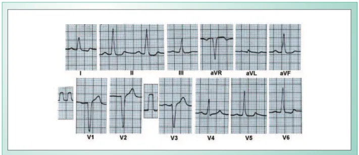
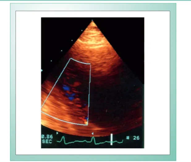
Male, 34-years-old, developed a rapidly progressive heart failure four years ago, resulting from a left ventricular (LV) noncompaction cardiomyopathy according to findings from magnetic resonance angiography. The initial electrocardiogram (Fig. 1) showed a first-degree AV block, left ventricular and left atrial overload and left bundle branch block, with SÂT at +30°, and biphasic T waves in D1 and V6. Recently, after a viral infection of the respiratory tract he presented with a severe exertional limitation. Doppler echocardiogram (Fig. 2) demonstrated diffuse hypokinesia and a significant decrease in LV ejection fraction (0.38) with mild mitral regurgitation, final LV diastolic diameter of 6.3cm and final systolic diameter of 5.1 cm, in addition to detecting the characteristic trabeculations in the internal face of the septal-apical myocardium with
Key words
Left bundle branch block, circumferential subendocardial ischemia, non-compacted myocardium.
intratrabecular blood flow. ECG (Figure 3) showed a higher degree of aberrant ventricular activation and significant T wave inversion in anterolateral and inferior walls, with a diametrically opposed SÂT at -145°.
Comments
The electrocardiographic concept of circumferential subendocardial ischemia because of SÂT displacement to the right upper quadrant in the three-vessel coronary artery disease or the like, indicative of a sudden increase in LV end-diastolic pressure, may be pertinent to this form of cardiomyopathy, for which information on ECG alterations is scarce.
Fig. 1 - Left ventricular and atrial overload and left bundle branch block. Observe biphasic T waves in D1 and V6, with straightening of the ST segment in the inferior wall. SÂT at +30º.
RGO, 34y, male
Image
saraiva e cols. prOGressIOn Of VentrICulAr repOlArIzAtIOn In left bundle brAnCh blOCK In nOn-COMpACted Of the MyOCArdIuM
Fig. 2 - Doppler echocardiogram demonstrating myocardial trabeculations in the septal-apical region of the left ventricle.
Fig. 3 - A higher degree of aberrant QRS complexes with SÂT at -145º is observed.
RGO, 34y, male