1. Department of Pathophysiology, National and Kapodistrian University of Athens, Medical School, “Laiko” General Hospital, Athens, Greece
2. Department of Pathology, “Evangelismos” General Hospital, Athens, Greece
3. Department of Thoracic Surgery, “Evangelismos” General Hospital, Athens, Greece
4. Tuberculosis Unit, “Sotiria” General Hospital of Chest Diseases, Athens, Greece
IntroductIon
Originally described by Liebow in 1973, bronchocentric granulomatosis (BcG) is an unusual form of gra -nulomatosis characterized by granulomatous destruc-tion of bronchial or bronchiolar walls and adjacent parenchyma, with debris and exudates filling the
air-way lumen1,2. Overall, the pathogenesis of BcG remains
uncertain3. It is suggested that BcG is one of the limi
-ted ways in which bronchi and bronchioles respond to
a variety of different types of stimuli4. In asthmatic
pa-tients, BcG probably results from a hypersensitivity re-action to inhaled fungi and has been associated with
allergic bronchopulmonary aspergillosis (ABPA)5. The
causative agent of hypersensitivity response in
non-asthmatic patients with BcG is obscure6. The histologi
-cal appearance differs between asthmatic and non-asth-matic patients: in the former, it is characterized by abundant tissue eosinophilia, while, in the latter,
tis-sue infiltration is primarily neutrophilic2,5. We herein
report the case of a middle-aged woman with rheuma-toid arthritis (RA) and BcG presenting as a sizeable pul-monary cavity, and subsequently, review all relevant cases previously reported in the English literature.
cAse presentAtIon
A 69-year-old female patient, non-smoker, with a 16--year history of RA treated with leflunomide and low dose methylprednisolone (4 mg/day), presented with low-grade fever, non-productive cough, excessive sweating, fatigue and malaise during the last two months, with no other accompanying symptoms, namely dyspnea or chest pain. She had no active arthri-tis and the Clinical Disease Activity Index (CDAI) score was 2 (inactive RA). She had received empirical antibi-otic therapy (clarithromycin and amoxicillin) without improvement of her symptoms. On admission, she had fever (38.3 C) and mid and end-inspiratory crackles at
Bronchocentric granulomatosis in rheumatoid
arthritis: case report and literature review
Pitsilka DA1, Kampolis CF1, Rontogianni D2, Zisis C3, Loukeri AA4, Vlachoyiannopoulos PG1 ACTA REUMATOL PORT. 2020;45:214-219
AbstrAct
Bronchocentric granulomatosis (BcG) is characterized by granulomatous destruction of bronchial or bron-chiolar walls and adjacent parenchyma, with debris and exudates filling the airway lumen. Approximately 50% of total cases have been associated with asthma and al-lergic bronchopulmonary aspergillosis, while it has been rarely reported in the context of rheumatoid arthritis (RA). We describe the case of a 69-year-old female RA patient with BcG presenting as a solitary cavi tary pulmonary mass. In addition, we conducted a li -terature review about the clinical and imaging features of BcG in RA patients.
A chronically immunosuppressed 69-year-old fe-male patient with a 16-year history of RA presented with constitutional symptoms (low-grade fever, exces-sive sweating and malaise) and a sizeable cavitary lung lesion. Open lung biopsy was performed and histopathological findings were consistent with the dia -gnosis of BcG. Other seven cases of BcG have been pre-viously reported in the context of RA, with clinical and laboratory characteristics described in five of them. Overall, pulmonary nodules or masses were the most frequent imaging finding of BcG, while no clear rela-tionship with disease activity or previous treatment modalities could be established. Surgical resection fol-lowed by administration of oral steroids was effective for achieving complete remission of symptoms and ra-diological stability in most cases.
Keywords: Pulmonary cavity; Rheumatoid arthritis;
the right lower lung field. Physical examination of the heart, abdomen, joints and nervous system was unre-markable.
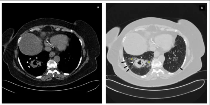
A chest CT scan was performed and revealed mild pulmonary fibrosis in the upper, mid and lower zones of bilateral lung fields and a 5-cm thick-walled pul-monary cavity in the right lower lobe (Figure 1, Pa nels a and b). In addition, the patient had mild normochromic normocytic anemia (Hb: 11.3 g/dl), signi -ficantly raised serum levels of inflammatory markers (Creactive protein was 120 mg/L and erythrocyte sedi -mentation rate was 100 mm/hour) and positive rheumatoid factor (RF) and anticitrullinated peptide antibodies (anti-CCPs). On the other hand, antinucle-ar and antineutrophil cytoplasmic antibodies, assays for the detection of human immunodeficiency virus infection, Mantoux tuberculin skin test and interferon--gamma release assays (IGRA) were all negative. Bron-choscopy with bronchoalveolar lavage (BAL) was sub-sequently performed. Bronchial mucosa had a normal macroscopic appearance. BAL fluid staining and cul-tures for bacteria, mycobacteria or fungi, and nuclear amplification assays for M. tuberculosis were also nega
-tive. No malignant cells were found in the cytological analysis. Neutrophils were moderately increased (40%) and lymphocytes were mildly elevated (13%) on BAL differential cell count.
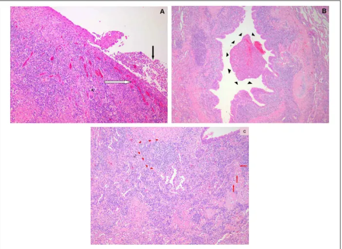
In order to establish a definite diagnosis, on the basis of a high pre-operative clinical suspicion of broncho-centric carcinoma, an open-lung procedure with right lower lobectomy was performed. Histological exa -mination was consistent with a diagnosis of BcG (Fi gure 2, Panels a to c). In particular, the epithelium of large-sized bronchi was focally replaced by an inflammatory granulomatous tissue and bronchiolar lumen was filled with fibrinopurulent exudates and necrotic debris. Chronic inflammatory infiltration, mainly consisting of lymphocytes, plasma cells, polymorphonuclear leuko-cytes, “foamy” cells and fibroblasts, also extended into in-terstitial space and alveolar lumens, thus forming foci of organizing pneumonia. A thorough examination of the resected lobe excluded the presence of pulmonary vas-culitis, fungal hyphae, malignancy or tissue infiltration by eosinophils. Besides surgical excision, no additional treatment was given, including corticosteroids.
Two years after surgical excision of the cavitary
le-FIGure 1. Bronchocentric granulomatosis presenting as a thick-walled cavity.
The major finding of chest computed tomography (CT) scan was a 5-cm thick walled cavity in the right lower lobe (Panels a + b). The lesion had well-defined margins with areas of spiculation (see white arrowheads, Panels a + b) , was adjacent to subsegmental bronchi and was surrounded by patchy ground glass opacities (see yellow asterisks) (Panel b). Small areas of bilateral septal thickening and/or ground glass attenuation restricted to the subpleural regions of the lower lung fields (see black arrows) were also visible on lung window and were indicative of mild interstitial lung disease (Panel b).
sion, the patient remained asymptomatic, complete blood count and inflammatory markers had norma lized and followup CT scan showed stable postope -rative fibrotic changes.
dIscussIon
Pulmonary involvement is among the most common extra-articular manifestations in RA and, along with cardiovascular disease and infections, is one of the lea -ding causes of morbidity and mortality, affecting the
course of the disease7. The wide spectrum of
pul-monary diseases in patients with RA includes
condi-tions that affect the parenchyma (infeccondi-tions, intersti-tial lung disease, rheumatoid nodules, drug toxicity, malignancy), pleura (pleural thickening and effusions), airways (bronchiectasis, bronchiolitis) or vasculature (rheumatoid vasculitis). BcG is a granulomatous lung disease of unknown cause, usually associated with al-lergic bronchopulmonary aspergillosis (ABPA) and asthma, but its occurrence in the context of RA has
been rarely reported in the literature3,5,8–11.
BcG should be considered not as a disease, but as a
de-scriptive pathological diagnosis12, which has been
as-sociated with two different clinical forms. About 50% of all reported cases of BcG are associated with asthma and ABPA (asthmatic form). The non-asthmatic form of FIGure 2. Major histopathological findings in bronchocentric granulomatosis (BcG).
The epithelium of medium- and large-sized bronchi has been focally replaced by granulomatous tissue (white arrow) with scarce multinucleated giant cells (asterisc). In addition, the bronchial lumen is filled with necrotic debris (black arrow) [Panel a, Hematoxylin & Eosin (H-E) stain (x100)]. Granulation tissue apparently arising from respiratory epithelium damage of central airways extends to the lumen of more peripheral ones (black arrowheads) [Panel b, H-E stain (x50)]. Chronic inflammatory infiltration by lymphocytes (red arrowheads), plasma cells, polymorphonuclear leukocytes, “foamy” cells (red arrows) and fibroblasts also extends into surrounding lung parenchyma (interstitial space and alveoli) [Panel c, H-E stain (x10)].
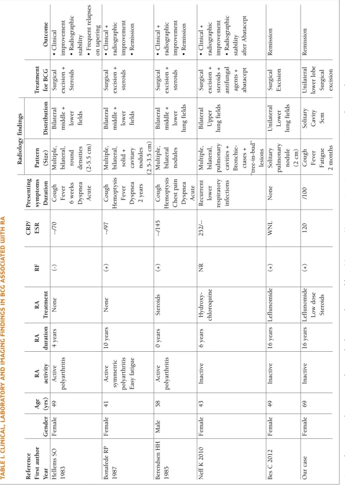
tA b Le I. c LI n Ic A L, L A b o r At o ry A n d Im A G In G F In d In G s In b cG A ss o cI At ed w It h r A R ad io lo g y f in d in g s R ef er en ce C R P / P re se n ti n g F ir st a u th o r A g e R A R A R A R F E S R sy m p to m s P at te rn T re at m en t Y ea r G en d er (y rs ) ac ti v it y d u ra ti o n T re at m en t D u ra ti o n (S iz e) D is tr ib u ti o n fo r B C G O u tc o m e H el le m s S O F em al e 4 9 A ct iv e 4 y ea rs N o n e (-) --/7 0 C o u g h M u lt ip le , B il at er al S u rg ic al • C li n ic al 1 9 8 3 p o ly ar th ri ti s F ev er b il at er al , m id d le + ex ci si o n + im p ro v em en t 6 w ee k s ro u n d lo w er S te ro id s • R ad io g ra p h ic D y sp n ea d en si ti es fi el d s st ab il it y A cu te (2 -5 .5 c m ) • F re q u en t re la p se o n t ap er in g B o n af ed e R P F em al e 4 1 A ct iv e 1 0 y ea rs N o n e (+ ) --/9 7 C o u g h M u lt ip le , B il at er al S u rg ic al • C li n ic al + 1 9 8 7 sy m m et ri c H em o p ty si s b il at er al , m id d le + ex ci si o n + ra d io g ra p h ic p o ly ar th ri ti s F ev er so li d + lo w er st er o id s im p ro v em en t E as y f at ig u e D y sp n ea ca v it ar y fi el d s • R em is si o n 2 y ea rs n o d u le s (2 .5 -3 .5 c m ) B er en d se n H H M al e 5 8 A ct iv e 0 y ea rs S te ro id s (+ ) --/1 4 5 C o u g h M u lt ip le , B il at er al S u rg ic al • C li n ic al + 1 9 8 5 p o ly ar th ri ti s H em o p ty si s b il at er al m id d le + ex ci si o n + ra d io g ra p h ic C h es t p ai n n o d u le s lo w er st er o id s im p ro v em en t D y sp n ea lu n g f ie ld s • R em is si o n A cu te N ef f K 2 0 1 0 F em al e 4 3 In ac ti v e 6 y ea rs H y d ro x y -N R 2 5 2 / -R ec u rr en t M u lt ip le , B il at er al S u rg ic al • C li n ic al + ch lo ro q u in e lo w er b il at er al , U p p er ex ci si o n + ra d io g ra p h ic re sp ir at o ry p u lm o n ar y lu n g f ie ld s st er o id s + im p ro v em en t in fe ct io n s ca v it ie s + an ti fu n g al • R ad io g ra p h ic B ro n ch ie -ag en ts + st ab il it y ct as es + ab at ac ep t af te r A b at ac ep t “t re e-in -b u d ” le si o n s B es C 2 0 1 2 F em al e 4 9 In ac ti v e 1 6 y ea rs L ef lu n o m id e (+ ) W N L N o n e S o li ta ry U n il at er al S u rg ic al R em is si o n p u lm o n ar y L o w er E x ci si o n n o d u le lu n g f ie ld s (2 c m ) O u r ca se F em al e 6 9 In ac ti v e 1 6 y ea rs L ef lu n o m id e (+ ) 1 2 0 /1 0 0 C o u g h S o li ta ry U n il at er al R em is si o n L o w d o se F ev er C av it y lo w er l o b e S te ro id s F at ig u e 5 cm S u rg ic al 2 m o n th s ex ci si o n B cG : B ro n ch o ce n tr ic g ra n u lo m at o si s, R A : R h eu m at o id a rt h ri ti s, R F : R h eu m at o id f ac to r, (+ ): p o si ti v e, ( -) : n eg at iv e, C R P : C -r ea ct iv e p ro te in , E S R : er y th ro cy te s ed im en ta ti o n r at e, N R : N o t re p o rt ed , W N L : W it h in n o rm al l im it s
BcG is usually idiopathic, but associations with pul-monary infections (mycobacterial, fungal,
echinococ-cal, viral)13–15, autoimmune diseases (RA, Wegener’ s
granulomatosis, ankylosing spondylitis)3,5,8–11,16,17,
chronic granulomatous disease, bronchogenic carci-noma, glomerulonephritis, red cell aplasia, diabetes
in-sipidus and scleritis6,18–22have been reported. In our
patient, RA was not complicated by scleritis. In addi-tion, although Mantoux tuberculin skin test and IGRA may be false negative in immunocompromised pa-tients, BAL fluid staining and cultures for M. tubercu-losis were also negative in our patient.
Patients with the asthmatic form, which is associa ted with eosinophilia, tend to be younger (9 to 50 years old) and mainly complain of respiratory symptoms such as cough, dyspnea, haemoptysis, wheeze and pleuritic pain. In the non-asthmatic form, patients are relatively older (with a range of 32 to 76 years) and pre-vailing symptoms are usually non-specific (malaise,
fa-tigue, fever)4,5,17. BcG radiographic findings are similar
for asthmatic and non-asthmatic patients. A wide va riety of radiographic appearances have been described, but they have consistently been divided into two main pat-terns: mass-like lesions/ nodules or focal areas of con-solidation which are usually confined to a single lobe
with a predilection for the upper lobes23,24. Cavitation
of solid lesions is less frequent25. BcG diagnosis cannot
be established solely on the basis of radiographic
fea-tures and open-lung biopsy is usually required23.
Smoking, advanced age, titer anti-CCPs, high-titer RF and family history of RA are among the most widely recognized risk factors for RA-associated inter-stitial lung disease, while data regarding the role of male
gender are inconsistent7. Our patient was elderly, but,
although RF and anti-CCPs were positive, their titers were not high (RF: 37 IU/ml, normal value <20, and anti-CCPs: 31 U/ml, normal value <17). Thus, the lim-ited radiological extent and the clinically insignificant manifestations of interstitial lung disease in our elder-ly female patient could partialelder-ly be attributed to the presence of low-titers of RF and anti-CCPs.
Treatment of underlying or associated condition is effective in most patients with BcG. In cases without an obvious cause, many patients improve without
medi-cal treatment2,5. Glucocorticoids are a mainstay
treat-ment for asthmatic patients, and, in a small number of BcG with recurrent or persistent disease, long-term
therapy may be required2,5,8,19. Although surgical
re-section as monotherapy may be curative in some
pa-tients2,11, there are no definite predictive factors of a
successful outcome.
Other seven cases of RA-associated BcG have been reported so far in the English literature, with clinical and laboratory characteristics described in five of them, as presented in Table I. The majority of patients were middle-aged (range: 41-69 years) women with a rela-tively long history of RA (at least 4 years), and 50% (n=3) had active polyarthritis on the diagnosis of BcG3,8,9. Recurrent fever, chronic cough with or
with-out hemoptysis and acute or chronic dyspnea were the main presenting respiratory symptoms. Solid or cavi-tated pulmonary nodules or masses (2-5.5 cm in size) was the usual radiographic pattern of BcG in RA pa-tients. In most cases, they were multiple and bilater-al3,8–10with a predilection for middle and/or lower lung
fields3,8,9,11. After surgical resection, two-thirds of
pa-tients (n=4) received systemic steroids3,8–10, with an
ini-tial daily dosage of prednisone ranging between 40 and 100 mg/day. One of them had frequent recurrences on
steroid tapering8, while, in another case, abatacept was
successfully administered after the failure of
conven-tional medical or surgical treatment10.
In conclusion, BcG seems to be a rare pulmonary manifestation among patients with RA, whose differ-ential diagnosis from cavitary lesions of another origin may be challenging. The exact pathogenetic mecha-nism remains elusive, since a causal relationship with a particular antigen, RA disease activity or the use of specific disease-modifying agents could not be sub-stantiated in the few cases published. The radiologic and clinical manifestations of BcG are non-specific and the diagnosis is almost impossible to be established without surgical lung biopsy. Most patients usually have a favorable prognosis after surgical excision of the pulmonary lesion.
correspondence to
Christos F Kampolis
Department of Pathophysiology
Athens University Medical School and “Laiko” General Hospital 75 M. Asias str.
1527, Athens, Greece E-mail: chkamp77@gmail.com
reFerences
1. Liebow AA. The J. Burns Amberson lecture--pulmonary angi-itis and granulomatosis. Am Rev Respir Dis 1973; 108: 1–18. 2. Koss MN, Robinson RG, Hochholzer L. Bronchocentric
granu-lomatosis. Hum Pathol 1981; 12: 632–638.
3. Berendsen HH, Hofstee N, Kapsenberg PD, van Reesema DR, Klein JJ. Bronchocentric granulomatosis associated with seropositive polyarthritis. Thorax 1985; 40: 396–397. 4. Clee MD, Lamb D, Clark RA. Bronchocentric granulomatosis:
a review and thoughts on pathogenesis. Br J Dis Chest 1983; 77: 227–234.
5. Katzenstein AL, Liebow AA, Friedman PJ. Bronchocentric gran-ulomatosis, mucoid impaction, and hypersensitivity reactions to fungi. Am Rev Respir Dis 1975; 111: 497–537.
6. Martínez-López MA, Peña JM, Quiralte J, Fernández MC, González JJ, Patrón M, et al. Bronchocentric granulomatosis as-sociated with pure red cell aplasia and lymphadenopathy. Tho-rax 1992; 47: 131–133.
7. Yunt ZX, Solomon JJ. Lung disease in rheumatoid arthritis. Rheum Dis Clin North Am 2015;41: 225–236.
8. Hellems SO, Kanner RE, Renzetti AD. Bronchocentric granulo-matosis associated with rheumatoid arthritis. Chest 1983; 83: 831–832.
9. Bonafede RP, Benatar SR. Bronchocentric granulomatosis and rheumatoid arthritis. Br J Dis Chest 1987; 81: 197–201. 10. Neff K, Stack J, Harney S, Henry M. The use of abatacept in
de-bilitating cavitating lung disease associated with rheumatoid arthritis, bronchocentric granulomatosis and aspergillosis. Tho-rax 2010; 65: 545–546.
11. Bes C, Kılıçgün A, Talay F, Yılmaz F, Soy M. Bronchocentric gran-ulomatosis in a patient with rheumatoid arthritis. Rheumatol Int 2012; 32: 3261–3263.
12. Myers JL. Bronchocentric granulomatosis. Disease or diagno-sis? Chest 1989; 96: 3–4.
13. Tazelaar HD, Baird AM, Mill M, Grimes MM, Schulman LL, Smith CR. Bronchocentric mycosis occurring in transplant re-cipients. Chest 1989; 96: 92–95.
14. Myers JL, Katzenstein AL. Granulomatous infection mimicking bronchocentric granulomatosis. Am J Surg Pathol 1986; 10: 317–322.
15. Den Hertog RW, Wagenaar SS, Wastermann CJ. Bronchocentric granulomatosis and pulmonary echinococcosis. Am Rev Respir Dis 1982; 126: 344–347.
16. Yousem SA. Bronchocentric injury in Wegener’s granulomato-sis: a report of five cases. Hum Pathol 1991; 22: 535–540. 17. Rohatgi PK, Turrisi BC. Bronchocentric granulomatosis and
ankylosing spondylitis. Thorax 1984; 39: 317–318.
18. Moltyaner Y, Geerts WH, Chamberlain DW, Heyworth PG, Noack D, Rae J, et al. Underlying chronic granulomatous dis-ease in a patient with bronchocentric granulomatosis. Thorax 2003; 58: 1096–1098.
19. Rossi GP, Pavan E, Chiesura-Corona M, Rea F, Poletti A, Pessi-na AC. Bronchocentric granulomatosis and central diabetes in-sipidus successfully treated with corticosteroids. Eur Respir J 1994; 7: 1893–8.
20. Wiedemann HP, Bensinger RE, Hudson LD. Bronchocentric granulomatosis with eye involvement. Am Rev Respir Dis 1982; 126: 347–50.
21. Warren J, Pitchenik AE, Saldana MJ. Bronchocentric granulo-matosis with glomerulonephritis. Chest 1985; 87: 832–4. 22. Houser SL, Mark EJ. Bronchocentric granulomatosis with
mu-cus impaction due to bronchogenic carcinoma. An association with clinical relevance. Arch Pathol Lab Med 2000; 124: 1168–71.
23. Bain GA, Flower CD. Pulmonary eosinophilia. Eur J Radiol 1996; 23: 3–8.
24. Ward S, Heyneman LE, Flint JD, Leung AN, Kazerooni EA, Müller NL. Bronchocentric granulomatosis: computed tomo-graphic findings in five patients. Clin Radiol 2000; 55: 296–300. 25. Robinson RG, Wehunt WD, Tsou E, Koss MN, Hochholzer L.
Bronchocentric granulomatosis: roentgenographic manifesta-tions. Am Rev Respir Dis 1982; 125: 751–756.