Anticancer activity of lichen substances:
a systematic review
Joana Maria Mendonça Marques
Faculdade de Ciências da Saúde Universidade Fernando Pessoa
Joana Maria Mendonça Marques
Anticancer activity of lichen substances: a systematic review
Faculdade de Ciências da Saúde Universidade Fernando Pessoa
________________________________________________________ Joana Maria Mendonça Marques
Trabalho apresentado à Universidade Fernando Pessoa como parte dos requisitos para obtenção de grau de Mestre em Ciências Farnacêuticas
i
Sumário
Os líquenes são fonte de uma grande variedade de metabolitos secundários, frequentemente exclusivos desta associação, com actividades biológicas importantes como a antibiótica, anti-inflamatória, antioxidante e anticancerígena, entre outras. A evidência científica demonstra que os efeitos anticancerígenos dos líquenes se produzem através da inibição da iniciação, crescimento e invasão de vários tipos de células cancerígenas in vitro e in vivo. Foi efectuada uma revisão sistemática da actividade anticancerígena e dos mecanismos de acção de extractos e respectivas substâncias isoladas de líquenes. Os termos “lichen”, “tumo(u)r” e “cancer” foram utilizados para pesquisar artigos científicos no PubMed e Web of Science publicados até dezembro de 2018. De um total de 368 artigos pesquisados, 142 cumpriram os critérios de inclusão e exclusão estabelecidos. O ácido úsnico foi a substância liquénica isolada mais vezes e citada em 40 artigos. A espécie de líquene mais utilizada, citada em 9 artigos, foi Evernia
prunastri (L.) Nyl. Os extractos e substâncias liquénicas têm a capacidade de interferir
com todas as propriedades biológicas das células cancerígenas que conduzem ao desenvolvimento de tumores, da seguinte forma: indução da inibição do ciclo celular, inibição das vias de sinalização do crescimento celular, activação da imunidade antitumoral, bloqueio da imortalidade replicativa por inibição da actividade da telomerase, inibição da inflamação, inibição da invasão e metástase, bloqueio da angiogénese, supressão da instabilidade do genoma, induzindo morte celular por apoptose, autofagia e necrose, e modulação do metabolismo energético. Muitas substâncias liquénicas revelaram-se eficazes contra muitos tipos de linhas celulares cancerígenas, quer isoladas quer em combinação com outros agentes anticancerígenos, e constituem por isso bons candidatos ao desenvolvimento de fármacos anticancerígenos.
Palavras-chave
Tumor, Farmacognosia, Fitoterapia, Angiogénese, Apoptose, Autofagia, Migração celular, Invasão celular, Viabilidade celular, Proliferação celular, Citotoxicidade, Senescência celular, Stress oxidativo.
ii
Abstract
Lichens are a source of a great variety of unique secondary metabolites with important biological activities, including antibiotic, anti-inflammatory, antioxidant and anticancer, among others. A large body of research has demonstrated anticancer effects of lichens by inhibition of initiation, growth and invasion of several cancer cell types in vitro and in
vivo. We performed a systematic review of the anticancer activity and mechanisms of
action of lichen extracts and substances isolated from lichens. The search terms “lichen”, “tumo(u)r” and “cancer” were used to retrieve articles in PubMed and Web of Science published until December 2018. From a total of 368 articles surveyed, 142 met the established inclusion and exclusion criteria. The most commonly isolated lichen substance was usnic acid, cited in 40 research articles. The species more frequently used was Evernia prunastri (L.) Nyl., cited in 9 research articles. Lichen extracts and isolated lichen substances are able to interfere with all currently recognized biological capabilities necessary for tumour growth and progression. They do so by inducing cell cycle arrest, inhibiting growth factor signalling, activating anti-tumour immunity, disabling replicative immortality by inhibiting telomerase activity, inhibiting tumour-promoting inflammation, inhibiting invasion and metastasis, blocking angiogenesis, supressing genome instability, inducing apoptotic, autophagic and necrotic cell death, and modulating energy metabolism. Many lichen substances have proved effective against many types of cancer cell lines, either isolated or in combination with other anticancer agents, and are therefore suitable candidates for anticancer drug development.
Keywords
Tumour, Pharmacognosy, Phytotherapy, Angiogenesis, Apoptosis, Autophagy, Cell migration, Cell invasion, Cell viability, Cell proliferation, Citotoxicity, Cellular senescence, Oxidative stress.
iii
Acknowledgements
I would like to thank my supervisor Prof. Pedro Barata for his encouragement and patience during the running of this project. I would also like to acknowledge my colleague Marina Swert for her valuable support.
iv Index
1. Introduction ... 1
1.1. Relevant aspects of lichen biology for cancer research ... 1
1.1.1. Biochemistry and secondary metabolites ... 2
2. Methods ... 4
2.1. Search strategy ... 4
2.2. Study selection ... 4
2.3. Data extraction ... 4
3. Results and discussion ... 6
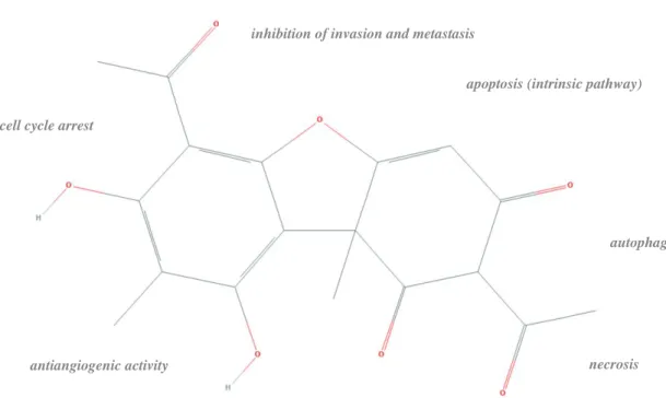
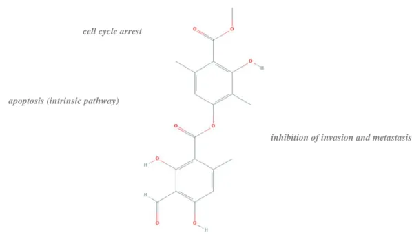
3.1. Usnic acid ... 31 3.2. Atranorin ... 33 3.3. Glucans ... 34 3.4. Protolichesterinic acid ... 35 3.5. Diffractaic acid ... 36 3.6. Physodic acid ... 37 3.7. Lobaric acid ... 38 3.8. Vulpinic acid ... 39 3.9. Salazinic acid ... 40 3.10. Lecanoric acid ... 41
3.11. Lichen extracts and other substances ... 42
4. Conclusions ... 47
v
List of figures
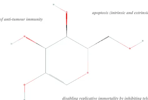
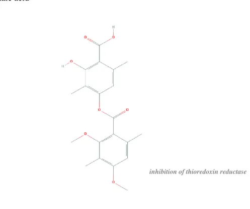
Figure 1. Flowchart of included research articles... 6 Figure 2. Number and cumulative number of research articles respecting the inclusion criteria published since 1959. ... 6 Figure 3. Geographical origin of the included research articles, mentioned lichen species, and lichen substances... 8 Figure 4. Most cited lichen species in the included research articles. ... 10 Figure 5. Structure of usnic acid and respective mechanisms of anticancer activity. .... 31 Figure 6. Structure of atranorin and respective mechanisms of anticancer activity. ... 33 Figure 7. Structure of a glucan (lichenin) and respective mechanisms of anticancer activity. ... 34 Figure 8. Structure of protolichesterinic acid and respective mechanisms of anticancer activity. ... 35 Figure 9. Structure of diffractaic acid and respective mechanisms of anticancer activity. ... 36 Figure 10. Structure of physodic acid and respective mechanisms of anticancer activity. ... 37 Figure 11. Structure of lobaric acid and respective mechanisms of anticancer activity. 38 Figure 12. Structure of vulpinic acid and respective mechanisms of anticancer activity. ... 39 Figure 13. Structure of salazinic acid and respective mechanisms of anticancer activity. ... 40 Figure 14. Structure of lecanoric acid and respective mechanisms of anticancer activity. ... 41 Figure 15. Schematic illustration of reported anticancer mechanisms of lichen substances. ... 43
vi
List of tables
Table 1. Number of research articles testing anticancer effects of isolated lichen substances or lichen extracts per type of cancer based on organ location. ... 10 Table 2. In vitro assays and techniques used in the included research articles for detection of anticancer activity of lichen substances or lichen extracts. ... 14 Table 3. Summary of the general mechanisms associated with anticancer activity of lichen extracts mentioned in the included research articles. ... 16 Table 4. Summary of the general mechanisms associated with anticancer activity of lichen substances mentioned in the included research articles... 26 Table 5. Summary of the molecular mechanisms associated with anticancer activity of lichen extracts mentioned in the included research articles. ... 43 Table 6. Summary of the molecular mechanisms associated with anticancer activity of lichen substances mentioned in the included research articles. ... 45
1
1. Introduction
The World Health Organization defines cancer as a generic term for a large group of diseases characterized by the uncontrolled proliferation of abnormal cells that are capable of invading adjoining parts of the body and/or spread to other organs. According to the global cancer statistics presented by the Global Cancer Observatory (GCO) (https://gco.iarc.fr/), cancer is the second leading cause of death globally, and is estimated to have accounted for 9,6 million deaths in 2018. Lung and breast cancers were the most common cancers worldwide, each contributing to approximately 12% of the total number of new cases diagnosed in 2018. Colorectal cancer was the third most common cancer, with 1,8 million new cases in 2018. Lung, prostate, colorectal, stomach and liver cancer are the most common types of cancer in men, while breast, colorectal, lung, cervical and thyroid cancer are the most common among women. Regardless of developments in the tools of disease diagnosis, treatment, and prevention measures, the number of cases is constantly increasing and estimated to reach an incidence of 29,5 million by 2040. Anticancer drugs currently approved by the Food and Drug Administration can be classified in four major groups: a) cytotoxic drugs; b) targeted-based agents; c) hormones and hormone antagonists; and d) immunomodulators (Liu et al., 2017). More than 70% of these drugs can be traced to natural products or synthetic to semi-synthetic substances derived from natural products (Katz and Baltz, 2016). Despite current progress in anticancer drug development, shifting from conventional nonspecific cytotoxic agents to specific target-based therapies and immune-related modulators (Liu et al., 2017), the discovery of new anticancer drugs from nature continues to be important for modern cancer research, as many potential sources of natural products remain largely unexplored (Stanojković, 2019).
1.1. Relevant aspects of lichen biology for cancer research
Lichens are symbiotic associations formed between at least one fungus, acting as host, and a mutualistic photosynthetic partner that is usually an alga and/or a cyanobacteria (Lumbsch and Rikkinen, 2017). The lichen fossil record is particularly poor but the probable origin of this association is currently set around 400–600 million years ago (Honegger et al., 2013; Yuan et al., 2005). The association has been such an evolutionary success that lichens can be found in most terrestrial ecosystems of the world and in a wide
2
variety of natural habitats, from sea level to high mountain peaks and from the hot deserts to the cold Arctic and Antarctic (Galloway, 1996). All over the world, they have been used in folk medicine, as a source of food, and also in the preparation of raw materials for dyes and perfumes (Crawford, 2019; Yamamoto et al., 2015). Almost 20 000 different lichen species have been described to date (Lücking et al., 2017), but since many regions of the world have been poorly collected, an even higher estimate may well be more realistic.
1.1.1. Biochemistry and secondary metabolites
Lichens produce a wide array of characteristic and unique primary (intracellular) and secondary (extracellular) metabolites (Elix, 1996). Primary metabolites include proteins, amino acids, polyols, carotenoids, polysaccharides and vitamins. Lichen secondary metabolites, often called lichen acids, are small but chemically complex substances grouped according to their chemical structure into the following classes: a) N-containing compounds; b) P-containing compounds; c) S-containing compounds; d) aliphatic and cycloaliphatic compounds; e) aromatic compounds; f) quinones; g) chromanes and chromones; h) xanthones; i) dibenzofurans; j) diphenylethers; k) biphenyls; l) diphenylmethanes; m) nostoclines; n) depsides; o) depsidones; p) depsones; q) naphthopyranes; r) terpenoids; s) pulvinic acid derivatives; and t) cleavage products of depsides and depsidones (Elix, 1996; Huneck and Yoshimura, 1996).
Research shows that both primary and secondary metabolites exert a wide variety of biological activities that include antibacterial, antifungal, antiviral, anti-inflammatory, antioxidant, anticancer, analgesic, antipyretic, enzyme inhibitory, anti-insecticidal and plant growth inhibitory (Calcott et al., 2018; Crawford, 2019; Molnár Katalin and Farkas Edit, 2014; Yamamoto et al., 2015; Zambare and Christopher, 2012). About 700 lichen substances were known by the time Huneck and Yoshimura (1996) published their compendium of lichen metabolites, and much more have been described since, with unique biological properties (e.g. Duong et al., 2017; Huneck, 2001; Le Pogam and Boustie, 2016; Nguyen et al., 2019). There are recent literature reviews addressing the anticancer activity of natural compounds isolated from lichens (Boustie and Lohézic-Le Dévéhat, 2008; Kim et al., 2015; Shrestha and Clair, 2013; Stanojković, 2019) but a detailed overview of all research available on their anticancer properties was still lacking. We have therefore performed, for the first time, a systematic review of all high-quality
3
research evidence of the anticancer activity of lichens and possible anticancer mechanisms of lichen extracts and isolated lichen substances.
4
2. Methods
This systematic review was conducted in accordance with the guidelines of Transparent Reporting of Systematic Reviews and Meta-Analyses (PRISMA Statement in Moher et
al., 2009 and PRISMA Elaboration and Explanation in Liberati et al., 2009).
2.1. Search strategy
Two internet sources were used to search for research articles that met the purpose of this review - PubMed (U. S. National Library of Medicine) and Web of Science (Clarivate Analytics) - using different combinations of the following search terms: “lichen”, “tumo(u)r” and “cancer”. In order to avoid confusion with the array of skin diseases named under the term “lichen” (ex: “lichen planus”) these were excluded as follows: aureus”, amyloidosis”, myxedematosus”, nitidus”, planopilaris”, planus”, “-purpuricus”, “-sclerosus”, “-simplex”, “-spinulosus”, “-striatus”.
The databases were searched for research articles published in the period up to and including December, 2018. Eligibility criteria were set to include any published research article that evaluated the anticancer activity of natural compounds obtained from lichens.
2.2. Study selection
All electronic search titles, selected abstracts, and full-text articles were independently reviewed by two reviewers (Joana Marques and Marina Swerts). The following exclusion criteria were defined: research articles focusing exclusively on endolichenic fungi, lichenicolous fungi, antimutagenic effects, normal cell lines or drug delivery systems; research articles not specifying the lichen species or lichen substance; review articles; meta-analyses; abstracts; conference proceedings; editorials/letters; and case reports (Figure 1).
2.3. Data extraction
Data were extracted by one reviewer (Joana Marques) using a predefined standardized form, and checked for completeness and accuracy by a second reviewer (Marina Swerts). Extracted information included data on the lichen substance (if specified), its origin
5
(natural, synthetic or commercial), chemical class (according to Huneck and Yoshimura, 1996), source (lichen species, if specified, and respective geographical origin), type of cancer addressed, applied assays and respective result, and suggested mechanism of action. Data on the experimental design was also extracted (use of controls and replicates).
6
3. Results and discussion
The primary search identified 368 research articles for preliminary review from electronic and manual searches, with 221 from PubMed, 101 from Web of Science and 46 from manual selection. After the removal of duplicates (64) and screening for relevant titles and abstracts, a total of 154 research articles was submitted for a full-text review. A total of 142 research articles met the inclusion and exclusion criteria established. A flowchart illustrating the selection process and number of research articles at each stage was performed as suggested in Liberati et al., 2009 (Figure 1).
Figure 2. Number and cumulative number of research articles respecting the inclusion criteria published since 1959.
0 25 50 75 100 125 150 0 2 4 6 8 10 12 14 16 1 9 5 9 1 9 6 1 1 9 6 3 1 9 6 5 1 9 6 7 1 9 6 9 1 9 7 1 1 9 7 3 1 9 7 5 1 9 7 7 1 9 7 9 1 9 8 1 1 9 8 3 1 9 8 5 1 9 8 7 1 9 8 9 1 9 9 1 1 9 9 3 1 9 9 5 1 9 9 7 1 9 9 9 2 0 0 1 2 0 0 3 2 0 0 5 2 0 0 7 2 0 0 9 2 0 1 1 2 0 1 3 2 0 1 5 2 0 1 7
Number of research articles Cumulative number of research articles
7
The 142 research articles here reviewed (Figure 2) mention 296 lichen species collected in 31 countries (Figure 3). Only 70% of the mentioned lichen names are accompanied by the name of their author(s). A species name is only complete if including author citation, i.e. the name(s) of the author(s) responsible for the establishment or introduction of a species name (Turland et al., 2018). Author citation is essential to track down the original species description and guarantee proper linking to species identity - quite frequently there are cases where at least two different author names are given after the same scientific species name; if after a taxonomic revision these are found to be different species, with different chemistry and medicinal/pharmaceutical usage, author citation is the only means to distinguish between the two. The most cited lichen species include: Evernia prunastri (L.) Ach. (n= 9), Cetraria islandica (L.) Ach. (n= 9), Hypogymnia physodes (L.) Nyl. (n= 8), Cladonia furcata (Huds.) Schrad. (n= 7), Stereocaulon alpinum Laur. (n= 7),
Gyrophora esculenta Miyoshi (n= 7), Parmelia caperata (L.) Ach. (n= 6), Thamnolia vermicularis (dubious author citation) (n= 5), Lasallia pustulata (L.) Mérat (n = 5), Parmelia sulcata Taylor (n= 5) and Xanthoria parietina (L.) Th. Fr. (n= 5) (Figure 4).
Among the reviewed research articles, 50 use lichen extracts and 114 use isolated lichen substances. A total of 137 isolated substances have been tested, belonging to 18 classes of primary and secondary metabolites (adapted from Huneck and Yoshimura, 1996): 1) depsidones (represented by physodic acid, lobaric acid, salazinic acid and 23 other substances); 2) depsides (represented by atranorin, diffractaic acid, lecanoric acid and 20 other substances); 3) aliphatic and cycloaliphatic compounds (represented by protolichesterinic acid and 17 other substances); 4) aromatic compounds (represented by 18 substances); 5) quinones; 6) dibenzofurans (represented by usnic, usnetic and usnolic acids), 7) polysaccharides (mainly glucans like lichenin and isolichenin); 8) N-containing compounds; 9) diphenylethers; 10) xanthones; 11) cleavage products of depsides and depsidones; 12) terpenoids; 13) peptides; 14) glutamic acid derivatives; 15) alkaloids; 16) pulvinic acid derivatives; 17) flavanols; and 18) tannins. Top ten cited substances are usnic acid (n= 40) - including its enantiomers (+)-usnic acid (n= 14) and (-)-usnic acid (n= 8), and salt forms (n= 1) - atranorin (n= 13), glucans (n= 10), protolichesterinic acid (n= 8), diffractaic acid (n= 7), physodic acid (n= 7), lobaric acid (n= 7), vulpinic acid (n= 6), salazinic acid (n= 5) and lecanoric acid (n= 4).
8
Figure 3. Geographical origin of the included research articles (A), mentioned lichen species (B), and lichen substances (C).
A
B
9
A B
C D
E F
10
Figure 4. Most cited lichen species in the included research articles. (A) Evernia
prunastri, (B) Cetraria islandica, (C) Hypogymnia physodes, (D) Cladonia furcata, (E) Stereocaulon alpinum, Gyrophora esculenta (no image available), (F) Parmelia caperata, (G) Thamnolia vermicularis, (H) Lasallia pustulata, (I) Parmelia sulcata, (J)
Xanthoria paritina.
Lichen substances have been tested against breast cancer (31% of the included research articles), colorectal cancer (22%), leukaemia (18%), lung cancer (16%), melanoma (13%), sarcoma (13%), cervical cancer (9%), brain cancer (8%), liver cancer (8%), prostate cancer (7%), pancreas cancer (4%), ovary cancer (4%), kidney cancer (4%), stomach cancer (4%), larynx cancer (2%), myeloma (2%), endometrial cancer (1%), oral cavity and pharynx cancer (1%), lymphoma (1%), bone and joint cancer (1%), head and neck cancer (1%), uterus cancer (1%), bladder cancer (1%), vulvar cancer (1%) and mastocytoma (1%) cell lines (Table 1).
Table 1. Number of research articles testing anticancer effects of isolated lichen substances or lichen extracts per type of cancer based on organ location.
Substance blad d er b on e an d j oin t b rain b re as t ce rvical colon an d r ec tu m en d om et rial h ead an d n ec k K id n ey larynx leuk em ia li ve r lu n g lym p h om a m as toc yt om a m elan om a m ye lom a or al cavit y an d p h ar yn x ovar y p an cr eas p ros tat e sar com a st om ac h u te ru s vu lvar Num b er of r es ear ch ar ticle s usnic acid 1 2 5 4 5 3 2 4 4 1 1 1 1 17 (+)-usnic acid 2 4 2 1 2 1 3 1 1 1 1 1 14 atranorin 1 1 2 1 4 2 1 5 2 1 13 glucan 9 10 (-)-usnic acid 2 3 1 3 1 5 1 1 8 protolichesterinic acid 4 2 2 1 3 2 1 1 2 1 8 diffractaic acid 1 1 1 1 1 1 7 lobaric acid 1 3 2 3 2 1 1 1 1 1 1 7 physodic acid 1 1 1 1 3 1 2 1 7 vulpinic acid 1 1 1 1 2 2 1 6 salazinic acid 1 1 3 1 1 1 5 I J
11 Substance blad d er b on e an d j oin t b rain b re as t ce rvical colon an d r ec tu m en d om et rial h ead an d n ec k K id n ey larynx leuk em ia li ve r lu n g lym p h om a m as toc yt om a m elan om a m ye lom a or al cavit y an d p h ar yn x ovar y p an cr eas p ros tat e sar com a st om ac h u te ru s vu lvar Num b er of r es ear ch ar ticle s evernic acid 1 1 1 1 4 glucan derivative 3 4 gyrophoric acid 1 1 2 1 1 2 1 1 4 lecanoric acid 1 1 1 1 1 1 1 4 lichenin 1 3 4 polysaccharide 1 1 3 4 norstictic acid 1 1 2 3 parietin 2 1 2 1 2 3
polyporic acid derivative 3 3
protocetraric acid 1 2 3 baeomycesic acid 1 1 1 1 1 1 2 1 2 barbatic acid 1 1 1 1 2 divaricatic acid 1 1 2 ethyl orsellinate 1 1 1 1 1 2 everninic acid 1 1 1 2 fumarprotocetraric acid 1 1 1 1 1 1 1 2 lichesterinic acid 1 1 1 1 2 lichexanthone 1 1 1 1 2 lobarstin 1 1 1 2 methyl orsellinate 2 1 1 1 1 1 2 olivetoric acid 1 2 pannarin 1 1 2 polyporic acid 1 2 1 2 psoromic acid 1 1 2 ramalin 1 1 2 rangiformic acid 1 2 sphaerophorin 1 1 2 squamatic acid 1 1 2 stictic acid 1 1 1 2
usnic acid derivative 1 1 2 2 2
variolaric acid 1 1 1 2 1 2
vicanicin 1 1 1 1 2
(+)-D-montagnetol 1 1 1 1
(+)-protolichesterinic acid 1 1 1
(+)-usnic acid analogue 1 1
16-O-acetyl-leucotylic acid 1 1
3-hydroxy physodic acid 1 1
6-methyl praesorediosate A 1 1 1 1 6-methyl vinapraesorediosate A 1 1 1 1 9'-(O-methyl)protocetratic acid 1 1 1 1 1 1 ambewelamide A 1 1 barbatolic acid 1 1 caperatic acid 1 1 catechin 1 1 1 D-(+)-erythrin 1 1 1 1 epiphorellic acid-1 1 1 hydrofurylmontagnetol 1 1 1 1 isolichenin 1 1 isopropyl orsellinate 1 1 1 1 1 leucotylic acid 1 1 lobarin 1 1 methyl-β-orcinol carboxylate 1 1 n-butyl orsellinate 1 1 1 1 1 norlichexanthone 1 1 1 1 norlichexanthone derivative 1 1 1 1 norlobaridone 1 1 1 1 1 n-pentyl orsellinate 1 1 1 1 1 n-propyl orsellinate 1 1 1 1 1 orcinol 1 1 1
orsellinic acid methyl ester 1 1 1
orsellinylmontagnetol A 1 1 1 orsellinylmontagnetol C 1 1 1 parmoether A 1 1 1 parmoether B 1 1 1 1 parmosidone A 1 1 1 1 parmosidone C 1 1 1 1 parmosidone D 1 1 perlatolic acid 1 1 physciosporin 1 1 physodalic acid 1 1 praesorediosic acid 1 1 1 1 purpurin 1 1 1 pygmeine 1 1
12 Substance blad d er b on e an d j oin t b rain b re as t ce rvical colon an d r ec tu m en d om et rial h ead an d n ec k K id n ey larynx leuk em ia li ve r lu n g lym p h om a m as toc yt om a m elan om a m ye lom a or al cavit y an d p h ar yn x ovar y p an cr eas p ros tat e sar com a st om ac h u te ru s vu lvar Num b er of r es ear ch ar ticle s pygmeine derivative 1 1 reserpin 1 1 1 roccellic acid 1 1 1
salazinic acid derivative 1 1 1 1 scabrosin acetate butyrate 1 1 1 scabrosin diacetate 1 1 scabrosin dibutyrate 1 1 scabrosin ester 1 1 sec-butyl orsellinate 1 1 1 1 1 stereocalpin A 1 1 1 1 stictamide A 1 1 tannic acid 1 1 1 tenuiorin 1 1 1 1 tert-butyl orsellinate 1 1 1 1 1 thamnoliadepside A 1 1 thamnoliadepside B 1 1 thamnolic acid A 1 1 tumidulin 1 1 usnolic acid 1 1 UTP-1 1 1 1 1 1 1 1 UTP-2 1 1 1 1 1 1 1 vermicularin 1 1 vinapraesorediosic acid A 1 1 1 1 vinapraesorediosic acid B 1 1 1 1 vinapraesorediosic acid C 1 1 1 1 β-orcinol 1 1 β-resorcylic acid 1 1 ω-aminoalkoxylxanthone derivative 1 1 1 1 1 1 ω-bromoalkoxylxanthone derivative 1 1 1 1 1 1
extract (substance not specified) 4 17 3 14 1 1 1 2 8 3 10 1 9 2 2 1 5 4 3 1 51
About 77% of the included studies are based on in vitro assays (Table 2) addressing cell viability/proliferation or cytotoxicity (92%), apoptosis (44%), gene and protein expression (16%), cell migration and invasion (12%), autophagy (4%), angiogenesis (4%) and cellular senescence (3%). A good number of studies (12%) address oxidative stress induced by lichen substances since oxidative stress is known to activate inflammatory pathways leading to transformation of normal cells into tumour cells, and influence tumour cell survival, proliferation, resistance to anticancer therapies, invasion and angiogenesis. Antioxidant activity of lichen substances is also addressed in 13% of the included studies (data not shown) as it is well documented that antioxidants help reducing the risk of cancer development by means of chemically induced apoptosis (White et al., 2014). In vivo assays are used in 23% of the included research articles, achieved by injection of tumour-bearing mice with lichen substances and measurement of variation in tumour volume or weight, after a certain period of time.
Methylthiazolyldiphenyl-tetrazolium bromide (MTT) assay, Propidium Iodine (PI) assay and Trypan Blue Dye assay are among the most common assays used for the determination of cell viability. Apoptosis is more commonly addressed by the detection
13
of the expression of apoptosis-related genes and proteins through western blotting, followed by the caspase activity assay and Annexin-V staining assays. Cell migration and cell invasion processes were mainly assessed by Boyden chamber/transwell migration assays and scratch/wound-healing assays, respectively.
Lichen substances, either isolated or in extracts, have been found to act on the three classical forms of cell death (Chen et al., 2018): apoptosis, autophagy and necrosis - often with low cytotoxicity to normal cells (78% of the tested cases). The general mechanisms of anticancer activity of isolated lichen substances and lichen extracts are summarized in Tables 3-4. The following sections will detail current knowledge about the anticancer activity of the most promising and frequently reported lichen substances.
14
Table 2. In vitro assays and techniques used in the included research articles for detection of anticancer activity of lichen substances or lichen extracts, following Ediriweera et al. (2019), with adaptations.
Angiogenesis Apoptosis Autophagy Cell migration/invasion Cell viability/proliferation Cellular senescence Gene/protein expression Oxidative stress
CAM assay Light microscopy Fluorescence microscopy BME (Basement Membrane Chamber)
invasion assay Based on cellular enzymes and proteins TRAP assay Immunoassays Enzymes
Tube formation assay Confocal microscopy Muse autophagy LC3-antibody assay Boyden chamber/transwell migration
assay Acid phosphatase assay immunocytochemistry assay CAT activity
Acridine orange staining Lysotracker assay Invasion assay Arachidonic acid release p53/p21 induction Thioredoxin reductase inhibition assay Fluorescence microscopy
Detection of the expression of autophagy related genes and proteins
Scratch/wound-healing assay ATP assay Plk1 enzyme assay Markers
Acridine orange/EB staining assay Immunocytochemistry assay Spheroid‐based migration assay ATPase activity assay ser15 phosphorylation 8‐OH‐dG assay
Annexin-V/FITC staining assay Reporter assay
Detection of the expression of migration/invasion related genes and proteins
CCK-8 assay AIF translocation GSH levels
DAPI staining assay Western blotting Western blotting Cytochrome P450 activity assay qPCR LPO levels FITC assay Real‐time cell monitoring of cell
proliferation and imaging DNA ligase adenylation assay Reporter assay RNS production assay
FRAP assay DNA ligase ligation UPLC-MS/MS ROS production assay
Hoechst dye staining assay GTPases precipitation Western blotting TOS assay
PI assay LDH leakage assay DCF assay
Electron microscopy MTS assay
Coulter counter MTT assay
Flow cytometry Resazurin assay
Acridine orange staining SRB assay
Annexin-V staining assay Taurine release
Annexin-V/7-AAD staining assay WST assay
Annexin-V/FITC staining assay Based on DNA synthesis
Annexin-V/FITC/PI staining assay BrdU assay
Annexin-V/PI double staining assay Mitotic index assay
PI assay PCNA assay
15
Angiogenesis Apoptosis Autophagy Cell migration/invasion Cell viability/proliferation Cellular senescence Gene/protein expression Oxidative stress
acridine orange staining PicoGreen dsDNA quantitation assay
DNA fragmentation and damage Thymidine incorporation assay
COMET assay Colony formation assay
TUNEL assay Dye exclusion assays
Electroforesis Crystal violet assay
Mitochondial membrane potential NRU assay
rhodamine 123 dye assay (plate
reader) Trypan blue assay
mitochondrial membrane potential
assay Coulter counter
DiOC6 assay (flow cytometry) Flow cytometry
JC-1 assay (fluorescence microscopy) Fluorescence microscopy
Detection of caspases Cytoskeletal immunofluorescence
caspase activity assay Cytoskeleton staining
M30 assay Real‐time cell monitoring of cell
16
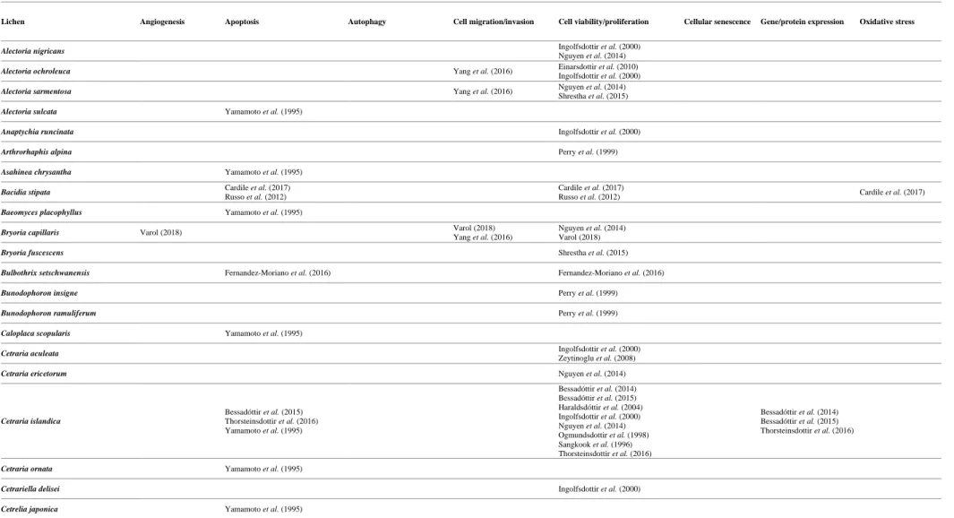
Table 3. Summary of the general mechanisms associated with anticancer activity of lichen extracts mentioned in the included research articles.
Lichen Angiogenesis Apoptosis Autophagy Cell migration/invasion Cell viability/proliferation Cellular senescence Gene/protein expression Oxidative stress
Alectoria nigricans Ingolfsdottir et al. (2000)
Nguyen et al. (2014)
Alectoria ochroleuca Yang et al. (2016) Einarsdottir et al. (2010) Ingolfsdottir et al. (2000)
Alectoria sarmentosa Yang et al. (2016) Nguyen et al. (2014) Shrestha et al. (2015)
Alectoria sulcata Yamamoto et al. (1995)
Anaptychia runcinata Ingolfsdottir et al. (2000)
Arthrorhaphis alpina Perry et al. (1999)
Asahinea chrysantha Yamamoto et al. (1995)
Bacidia stipata Cardile et al. (2017)
Russo et al. (2012)
Cardile et al. (2017)
Russo et al. (2012) Cardile et al. (2017)
Baeomyces placophyllus Yamamoto et al. (1995)
Bryoria capillaris Varol (2018) Varol (2018) Yang et al. (2016)
Nguyen et al. (2014) Varol (2018)
Bryoria fuscescens Shrestha et al. (2015)
Bulbothrix setschwanensis Fernandez-Moriano et al. (2016) Fernandez-Moriano et al. (2016)
Bunodophoron insigne Perry et al. (1999)
Bunodophoron ramuliferum Perry et al. (1999)
Caloplaca scopularis Yamamoto et al. (1995)
Cetraria aculeata Ingolfsdottir et al. (2000)
Zeytinoglu et al. (2008)
Cetraria ericetorum Nguyen et al. (2014)
Cetraria islandica Bessadóttir et al. (2015) Thorsteinsdottir et al. (2016) Yamamoto et al. (1995) Bessadóttir et al. (2014) Bessadóttir et al. (2015) Haraldsdóttir et al. (2004) Ingolfsdottir et al. (2000) Nguyen et al. (2014) Ogmundsdottir et al. (1998) Sangkook et al. (1996) Thorsteinsdottir et al. (2016) Bessadóttir et al. (2014) Bessadóttir et al. (2015) Thorsteinsdottir et al. (2016)
Cetraria ornata Yamamoto et al. (1995)
Cetrariella delisei Ingolfsdottir et al. (2000)
17
Lichen Angiogenesis Apoptosis Autophagy Cell migration/invasion Cell viability/proliferation Cellular senescence Gene/protein expression Oxidative stress
Chondropsis semiviridis Perry et al. (1999)
Cladia aggregata Yamamoto et al. (1995) Martins et al. (2016) Perry et al. (1999)
Cladia retipora Perry et al. (1999)
Cladia sullivanii Perry et al. (1999)
Cladina confusa Brandão et al. (2013)
Perry et al. (1999)
Cladina dendroides Nascimento et al. (1994)
Cladina mitis Perry et al. (1999)
Cladonia amaurocraea Yamamoto et al. (1995)
Cladonia arbuscula
Bessadottir et al. (2012) Einarsdottir et al. (2010) Galanty et al. (2017)
Bessadottir et al. (2012) Galanty et al. (2017)
Bessadottir et al. (2012) Einarsdottir et al. (2010) Galanty et al. (2017)
Cladonia coniocraea Delebassée et al. (2017)
Cladonia convoluta Bézivin et al. (2004)
Coskun et al. (2015)
Açıkgöz et al. (2014) Bézivin et al. (2003) Bézivin et al. (2004) Coskun et al. (2015)
Cladonia crispatula Nascimento et al. (1994)
Cladonia cristatella Yamamoto et al. (1995)
Cladonia ecmocyna Ingolfsdottir et al. (2000)
Cladonia fimbriata Perry et al. (1999)
Cladonia foliacea Koparal et al. (2015) Mitrovic et al. (2011)
Koparal et al. (2006) Koparal et al. (2015) Mitrovic et al. (2011) Cladonia furcata Lin et al. (2001) Lin et al. (2003) Yamamoto et al. (1995) Ingolfsdottir et al. (2000) Kosanić et al. (2014) Lin et al. (2001) Lin et al. (2003) Ranković et al. (2011) Shrestha et al. (2015) Lin et al. (2003)
Cladonia glauca Delebassée et al. (2017)
Cladonia gracilis Ingolfsdottir et al. (2000)
Cladonia gracilis tenerrima Perry et al. (1999)
Cladonia gracilis var. dilatata Yamamoto et al. (1995)
18
Lichen Angiogenesis Apoptosis Autophagy Cell migration/invasion Cell viability/proliferation Cellular senescence Gene/protein expression Oxidative stress
Cladonia macrophylla Nguyen et al. (2014)
Cladonia mitis Nguyen et al. (2014)
Cladonia nigripes Yamamoto et al. (1995)
Cladonia parasitica Delebassée et al. (2017)
Cladonia pleurota Yamamoto et al. (1995)
Cladonia pocillum Ersoz et al. (2017) Ersoz et al. (2017) Ersoz et al. (2017)
Cladonia pyxidata Kosanić et al. (2014)
Cladonia rangiferina Yamamoto et al. (1995) Ingolfsdottir et al. (2000) Kosanić et al. (2014)
Cladonia rangiformis Coskun et al. (2015)
Açıkgöz et al. (2014) Bézivin et al. (2003) Coskun et al. (2015) Delebassée et al. (2017)
Cladonia squamosa Delebassée et al. (2017)
Cladonia stricta Ingolfsdottir et al. (2000)
Cladonia substellata Nascimento et al. (1994)
Cladonia uncialis Paluszczak et al. (2018) Paluszczak et al. (2018) Paluszczak et al. (2018)
Cladonia vulcani Yamamoto et al. (1995)
Coccocarpia palmicola Perry et al. (1999)
Coeleocaulon aculeatum Perry et al. (1999)
Coenogonium implexum Perry et al. (1999)
Collema flaccidum Řezanka et al. (2006)
Cornicularia aculeata Brisdelli et al. (2013)
Brisdelli et al. (2016)
Brisdelli et al. (2013)
Brisdelli et al. (2016) Brisdelli et al. (2013)
Cornicularia epiphorella Russo et al. (2006) Russo et al. (2006) Russo et al. (2006)
Dirinaria aspera Brandão et al. (2013)
Evernia divaricata Yang et al. (2016) Nguyen et al. (2014)
Evernia prunastri Mitrovic et al. (2011)
Yamamoto et al. (1995) Bézivin et al. (2003) Burlando et al. (2009) Delebassée et al. (2017) Kosanić et al. (2013) Mitrovic et al. (2011) Shrestha et al. (2015) Triggiani et al. (2009)
19
Lichen Angiogenesis Apoptosis Autophagy Cell migration/invasion Cell viability/proliferation Cellular senescence Gene/protein expression Oxidative stress
Everniastrum catawbiense Shrestha et al. (2015)
Everniastrum vexans Zhou et al. (2017) Zhou et al. (2017) Zhou et al. (2017)
Everniopsis trulla Yang et al. (2018)
Flavocetraria cucullata Nguyen et al. (2014) Nguyen et al. (2014) Nguyen et al. (2014) Nguyen et al. (2014)
Flavocetraria nivalis Yang et al. (2016) Ingolfsdottir et al. (2000) Nguyen et al. (2014)
Flavoparmelia caperata Fernandez-Moriano et al. (2016)
Mitrovic et al. (2011) Yang et al. (2015)
Fernandez-Moriano et al. (2016) Mitrovic et al. (2011)
Flavoparmelia euplecta Fernandez-Moriano et al. (2016) Fernandez-Moriano et al. (2016)
Flavoparmelia haysomii Fernandez-Moriano et al. (2016) Fernandez-Moriano et al. (2016)
Fuscoderma applanatum Perry et al. (1999)
Gymnoderma coccocarpum Yamamoto et al. (1995)
Hypocenomyce scalaris Paluszczak et al. (2018) Paluszczak et al. (2018) Paluszczak et al. (2018)
Hypogymnia lugubris Cardile et al. (2017) Cardile et al. (2017)
Perry et al. (1999) Cardile et al. (2017)
Hypogymnia physodes Ari et al. (2014) Mitrovic et al. (2011) Paluszczak et al. (2018) Studzinska-Sroka et al. (2016) Yamamoto et al. (1995) Paluszczak et al. (2018) Yang et al. (2016) Ari et al. (2014) Mitrovic et al. (2011) Nguyen et al. (2014) Paluszczak et al. (2018) Stojanovic et al. (2014) Studzinska-Sroka et al. (2016) Paluszczak et al. (2018)
Hypotrachyna cirrhata Fernandez-Moriano et al. (2016) Fernandez-Moriano et al. (2016)
Hypotrachyna sinuosa Yang et al. (2015)
Lasallia pustulata Burlando et al. (2009)
Delebassée et al. (2017)
Lecanora atra Ranković et al. (2011)
Lecanora epibryon subsp. broccha Perry et al. (1999)
Lecanora muralis Ranković et al. (2011)
Leifidium tenerum Perry et al. (1999)
Leprocaulon microscopicum Delebassée et al. (2017)
Leproloma membranaceum Delebassée et al. (2017)
Leptogium cyanescens Perry et al. (1999)
Letharia vulpina Koparal et al. (2015) Yamamoto et al. (1995)
Burlando et al. (2009) Koparal et al. (2015) Shrestha et al. (2015)
20
Lichen Angiogenesis Apoptosis Autophagy Cell migration/invasion Cell viability/proliferation Cellular senescence Gene/protein expression Oxidative stress
Lethariella canariensis Fernandez-Moriano et al. (2016) Fernandez-Moriano et al. (2016)
Lethariella zahlbruckneri Lee et al. (2012)
Ren et al. (2009)
Lee et al. (2012) Ren et al. (2009)
Lee et al. (2012) Ren et al. (2009)
Lichina pygmaea Roullier et al. (2010)
Lobaria pulmonaria
Delebassée et al. (2017) Pejin et al. (2017) Shrestha et al. (2015)
Melanelia hepatizon Ingolfsdottir et al. (2000)
Micarea austroternaria Perry et al. (1999)
Myelochroa aurulenta Tokiwano et al. (2009)
Myelochroa irrugans Fernandez-Moriano et al. (2016) Fernandez-Moriano et al. (2016)
Neocatapyrenium sp. Yang et al. (2018)
Nephroma australe Perry et al. (1999)
Nephroma expallidum Ingolfsdottir et al. (2000)
Nephroma laevigatum Delebassée et al. (2017)
Nephroma parile Delebassée et al. (2017)
Nephroma plumbeum var. isidiatum Perry et al. (1999)
Nephroma sp. Yang et al. (2015)
Neuropogon acromelanus Perry et al. (1999)
Niebla sp. Yang et al. (2018) Yang et al. (2018) Yang et al. (2018)
Ochrolechia deceptionis Cardile et al. (2017) Brisdelli et al. (2013) Cardile et al. (2017)
Brisdelli et al. (2013) Cardile et al. (2017)
Pannaria hookeri Perry et al. (1999)
Parmelia caperata
Bézivin et al. (2003) Kosanić et al. (2012) Manojlovic et al. (2012)
Parmelia omphalodes Fernandez-Moriano et al. (2016) Fernandez-Moriano et al. (2016) Ingolfsdottir et al. (2000)
Parmelia perlata Bézivin et al. (2003)
Parmelia saxatilis
Delebassée et al. (2017) Ingolfsdottir et al. (2000) Kosanić et al. (2012) Manojlovic et al. (2012)
21
Lichen Angiogenesis Apoptosis Autophagy Cell migration/invasion Cell viability/proliferation Cellular senescence Gene/protein expression Oxidative stress
Parmelia subrudecta Ivanova et al. (2010)
Parmelia sulcata Ari et al. (2014) Ari et al. (2015) Mitrovic et al. (2011) Paluszczak et al. (2018) Ari et al. (2014) Ari et al. (2015) Kosanić et al. (2012) Mitrovic et al. (2011) Paluszczak et al. (2018) Paluszczak et al. (2018)
Parmelia tenuirima Perry et al. (1999)
Parmotrema dilatatum Brandão et al. (2013)
Parmotrema lichexanthonicum Brandão et al. (2013)
Micheletti et al. (2009)
Parmotrema praesorediosum Huynh et al. (2016)
Parmotrema reticulatum Ghate et al. (2013) Ghate et al. (2013) Shrestha et al. (2015)
Parmotrema sp. Williams et al. (2011)
Parmotrema tinctorum Bogo et al. (2010)
Parmotrema tsavoense Duong et al. (2015)
Peltigera aphtosa Shrestha et al. (2015)
Peltigera canina Ingolfsdottir et al. (2000)
Munzi et al. (2014)
Peltigera degenii Perry et al. (1999)
Peltigera dolichorhiza Perry et al. (1999)
Peltigera elisabethae Munzi et al. (2014)
Peltigera horizontalis Delebassée et al. (2017)
Peltigera leucophlebia Ingolfsdottir et al. (2000)
Ingolfsdottir et al. (2002)
Peltigera membranacea Perry et al. (1999)
Peltigera praetextata Munzi et al. (2014)
Peltigera venosa Nguyen et al. (2014)
Pertusaria oculata Ingolfsdottir et al. (2000)
Physcia sp. Yang et al. (2015)
Placopsis contortuplicata Cardile et al. (2017) Cardile et al. (2017) Cardile et al. (2017)
22
Lichen Angiogenesis Apoptosis Autophagy Cell migration/invasion Cell viability/proliferation Cellular senescence Gene/protein expression Oxidative stress
Platismatia glauca Paluszczak et al. (2018) Paluszczak et al. (2018)
Bézivin et al. (2003) Delebassée et al. (2017) Nguyen et al. (2014) Paluszczak et al. (2018)
Paluszczak et al. (2018)
Pleurosticta acetabulum Delebassée et al. (2017) Delebassée et al. (2017)
Protousnea magellanica Russo et al. (2012) Brisdelli et al. (2013)
Russo et al. (2012) Brisdelli et al. (2013)
Protousnea malacea Russo et al. (2012) Russo et al. (2012)
Protousnea sp. Yang et al. (2015)
Pseudevernia furfuracea Yamamoto et al. (1995)
Emsen et al. (2016) Kosanic et al. (2013) Nguyen et al. (2014)
Emsen et al. (2016)
Pseudevernia furfuracea var. ceratea Koparal et al. (2010) Koparal et al. (2010)
Pseudocyphellaria ardesiaca Perry et al. (1999)
Pseudocyphellaria argyracea Yang et al. (2015)
Pseudocyphellaria billardierei Perry et al. (1999)
Pseudocyphellaria carpoloma Perry et al. (1999)
Pseudocyphellaria cinnamomea Perry et al. (1999)
Pseudocyphellaria colensoi Perry et al. (1999)
Pseudocyphellaria coriacea Yang et al. (2015) Perry et al. (1999)
Yang et al. (2015) Yang et al. (2015)
Pseudocyphellaria coronata Perry et al. (1999)
Pseudocyphellaria degelii Perry et al. (1999)
Pseudocyphellaria dissimilis Perry et al. (1999)
Pseudocyphellaria faveolata Perry et al. (1999)
Pseudocyphellaria fimbriatoides Perry et al. (1999)
Pseudocyphellaria glabra Yang et al. (2015) Perry et al. (1999)
Pseudocyphellaria granulata Perry et al. (1999)
Pseudocyphellaria homoeophylla Perry et al. (1999)
Pseudocyphellaria maculata Perry et al. (1999)
Pseudocyphellaria multifida Perry et al. (1999)
23
Lichen Angiogenesis Apoptosis Autophagy Cell migration/invasion Cell viability/proliferation Cellular senescence Gene/protein expression Oxidative stress
Pseudocyphellaria pickeringii Perry et al. (1999)
Pseudocyphellaria rubella Perry et al. (1999)
Pseudocyphellaria rufovirescens Perry et al. (1999)
Pseudocyphellaria verrucosa Yang et al. (2015)
Psoroma buchananii Perry et al. (1999)
Psoroma dimorphum Russo et al. (2012) Russo et al. (2012) Russo et al. (2012)
Psoroma hirsutulum Perry et al. (1999)
Psoroma leprolomum Perry et al. (1999)
Psoroma microphyllizans Perry et al. (1999)
Psoroma pallidum Brisdelli et al. (2013)
Perry et al. (1999)
Psoroma spp. Russo et al. (2006)
Russo et al. (2008)
Russo et al. (2006) Russo et al. (2008)
Russo et al. (2006) Russo et al. (2008)
Ramalina celastri Leão et al. (1997)
Ramalina cuspidata Bézivin et al. (2003)
Ramalina farinacea Koparal et al. (2006)
Ramalina menziesii Shrestha et al. (2015)
Ramalina sp. Zhou et al. (2017) Brandão et al. (2013) Yang et al. (2018)
Ramalina terebrata Lee et al. (2016)
Suh et al. (2017) Lee et al. (2016) Suh et al. (2017)
Lee et al. (2016)
Suh et al. (2017) Suh et al. (2017)
Rhizoplaca chrysoleuca Zhou et al. (2017) Shrestha et al. (2015)
Rhizoplaca melanophthalma Russo et al. (2012) Yang et al. (2015) Emsen et al. (2016) Russo et al. (2012)
Emsen et al. (2016) Russo et al. (2012)
Rhizoplaca peltata Shrestha et al. (2015)
Roccella montagnei Duong et al. (2017)
Mishra et al. (2017)
Roccella sp. Yang et al. (2018)
Siphula dissoluta Perry et al. (1999)
Solorina crocea Yamamoto et al. (1995) Ingolfsdottir et al. (2000)
24
Lichen Angiogenesis Apoptosis Autophagy Cell migration/invasion Cell viability/proliferation Cellular senescence Gene/protein expression Oxidative stress
Sphaerophorus globosus Russo et al. (2006)
Russo et al. (2008) Ingolfsdottir et al. (2000) Russo et al. (2006) Russo et al. (2008) Russo et al. (2006) Russo et al. (2008)
Sphaerophorus stereocauloides Perry et al. (1999)
Stereocaulon alpinum Hong et al. (2018)
Brisdelli et al. (2013) Haraldsdóttir et al. (2004) Hong et al. (2018) Ingolfsdottir et al. (2000) Nguyen et al. (2014) Ogmundsdottir et al. (1998) Brisdelli et al. (2013)
Stereocaulon arcticum Ingolfsdottir et al. (2000)
Stereocaulon fronduliferum Perry et al. (1999)
Stereocaulon halei Ismed et al. (2012)
Stereocaulon intermedium Yamamoto et al. (1995)
Stereocaulon ramulosum Perry et al. (1999)
Stereocaulon spathuliferum Ingolfsdottir et al. (2000)
Sticta filix Perry et al. (1999)
Sticta latifrons Perry et al. (1999)
Sticta martinii Perry et al. (1999)
Sticta sp. Liang et al. (2011) Liang et al. (2011)
Teloschistes fasciculatus Perry et al. (1999)
Thamnolia subliformis Yamamoto et al. (1995)
Thamnolia vermicularis Zhou et al. (2017)
Guo et al. (2011) Manojlović et al. (2010) Nguyen et al. (2014) Perry et al. (1999)
Thamnolia vermicularis var.
subuliformis Haraldsdóttir et al. (2004) Toninia candida Ranković et al. (2012)
Trapeliopsis congregans Perry et al. (1999)
Tuckermannopsis ciliaris Shrestha et al. (2015) Shrestha et al. (2015) Shrestha et al. (2015)
Umbilicaria arctica Ingolfsdottir et al. (2000)
Umbilicaria crustulosa Kosanić et al. (2012)
Umbilicaria cylindrica Kosanić et al. (2012)
25
Lichen Angiogenesis Apoptosis Autophagy Cell migration/invasion Cell viability/proliferation Cellular senescence Gene/protein expression Oxidative stress
Umbilicaria esculenta Sun et al. (2018) Sun et al. (2018)
Xu et al. (2014) Xu et al. (2014) Sun et al. (2018)
Umbilicaria hirsuta Bačkorová et al. (2011) Bačkorová et al. (2012) Bačkorová et al. (2011) Bačkorová et al. (2012)
Umbilicaria kisovana Yamamoto et al. (1995)
Umbilicaria mammulata Shrestha et al. (2015)
Umbilicaria polyphylla Kosanić et al. (2012)
Umbilicaria proboscidea Ingolfsdottir et al. (2000)
Umbilicaria tornata Shang et al. (2018)
Usnea aurantiacoatra Fernandez-Moriano et al. (2016) Fernandez-Moriano et al. (2016)
Usnea barbata Zugic et al. (2016) Ranković et al. (2012) Zugic et al. (2016)
Usnea capillacea Perry et al. (1999)
Usnea cf. inermis Perry et al. (1999)
Usnea ciliifera Perry et al. (1999)
Usnea contexta Fernandez-Moriano et al. (2016) Fernandez-Moriano et al. (2016)
Usnea diffracta Dincsoy & Duman (2017)
Usnea fasciata Periera (1994)
Usnea filipendula Ari et al. (2014) Ari et al. (2014)
Usnea florida Yang et al. (2016) Nguyen et al. (2014)
Usnea longissima Yamamoto et al. (1995)
Usnea rubicunda Bézivin et al. (2003)
Usnea sp. Brandão et al. (2013)
Usnea strigosa Ebrahim et al. (2016) Ebrahim et al. (2016)
Shrestha et al. (2015) Ebrahim et al. (2016)
Usnea subcavata Brandão et al. (2013)
Vulpicida canadensis Shrestha et al. (2015)
Xanthoparmelia chlorochroa Shrestha et al. (2015) Shrestha et al. (2015) Shrestha et al. (2015)
Xanthoparmelia scabrosa Moerman et al. (2003) Ernst-Russell et al. (1999) Moerman et al. (2003)
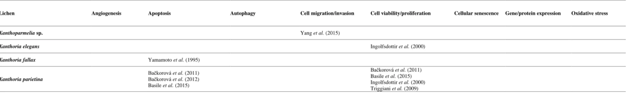
26
Lichen Angiogenesis Apoptosis Autophagy Cell migration/invasion Cell viability/proliferation Cellular senescence Gene/protein expression Oxidative stress
Xanthoparmelia sp. Yang et al. (2015)
Xanthoria elegans Ingolfsdottir et al. (2000)
Xanthoria fallax Yamamoto et al. (1995)
Xanthoria parietina Bačkorová et al. (2011) Bačkorová et al. (2012) Basile et al. (2015) Bačkorová et al. (2011) Basile et al. (2015) Ingolfsdottir et al. (2000) Triggiani et al. (2009)
Table 4. Summary of the general mechanisms associated with anticancer activity of lichen substances mentioned in the included research articles.
Substance Angiogenesis Apoptosis Autophagy Cell migration and invasion Cell viability/proliferation Cellular senescence Gene/protein expression Oxidative stress
(-)-usnic acid Koparal et al. (2015)
Bézivin et al. (2004) Takai et al. (1979) Yamamoto et al. (1995) Bazin et al. (2008) Bézivin et al. (2004) Einarsdottir et al. (2010) Koparal et al. (2006) Koparal et al. (2015)
(+)-protolichesterinic acid Bessadóttir et al. (2015) Bessadóttir et al. (2015) Bessadóttir et al. (2015)
(+)-usnic acid Bazin et al. (2008) Bačkorová et al. (2012) Bessadottir et al. (2012) Einarsdottir et al. (2010) Geng et al. (2018) Sahu et al. (2012) Yamamoto et al. (1995) Bessadottir et al. (2012) Ebrahim et al. (2017) Wu et al. (2018) Ebrahim et al. (2017) Wu et al. (2018) Yang et al. (2016) Bazin et al. (2008) Bessadottir et al. (2012) Burlando et al. (2009) Ebrahim et al. (2017) Einarsdottir et al. (2010) Emsen et al. (2018) Geng et al. (2018) Koparal et al. (2006) Sahu et al. (2012) Wu et al. (2018) Yang et al. (2016) Sahu et al. (2012) Wu et al. (2018) Yang et al. (2016) Bačkorová et al. (2012) Emsen et al. (2018) Sahu et al. (2012)
(+)-usnic acid analogue Ebrahim et al. (2017) Ebrahim et al. (2017) Ebrahim et al. (2017)
16-O-acetyl-leucotylic acid Tokiwano et al. (2009)
3-hydroxy physodic acid Stojanovic et al. (2014)
atranorin Bačkorová et al. (2011) Bačkorová et al. (2012) Cardile et al. (2017) Galanty et al. (2017) Russo et al. (2012) Solar et al. (2016) Galanty et al. (2017) Zhou et al. (2017) Bačkorová et al. (2011) Brandão et al. (2013) Cardile et al. (2017) Galanty et al. (2017) Kosanić et al. (2014) Paluszczak et al. (2018) Russo et al. (2012) Solar et al. (2016) Zhou et al. (2017) Paluszczak et al. (2018) Zhou et al. (2017) Bačkorová et al. (2012) Cardile et al. (2017)
27
Substance Angiogenesis Apoptosis Autophagy Cell migration and invasion Cell viability/proliferation Cellular senescence Gene/protein expression Oxidative stress
baeomycesic acid Haraldsdóttir et al. (2004)
barbatic acid Martins et al. (2016)
barbatolic acid Varol (2018) Varol (2018) Varol (2018)
caperatic acid Paluszczak et al. (2018) Paluszczak et al. (2018) Paluszczak et al. (2018) Paluszczak et al. (2018)
catechin Ghate et al. (2013)
colleflaccinoside A Řezanka et al. (2006)
colleflaccinoside B Řezanka et al. (2006)
diffractaic acid Russo et al. (2012)
Brandão et al. (2013) Brisdelli et al. (2013) Emsen et al. (2018) Russo et al. (2012) Emsen et al. (2018) Ozgencli et al. (2018)
divaricatic acid Russo et al. (2012) Brandão et al. (2013)
Russo et al. (2012)
epiphorellic acid-1 Russo et al. (2006) Russo et al. (2006) Russo et al. (2006)
ethyl orsellinate Bogo et al. (2010)
evernic acid Yamamoto et al. (1995) Kosanic et al. (2013) Ozgencli et al. (2018)
everninic acid Mishra et al. (2017)
fumarprotocetraric acid Kosanić et al. (2014)
glucan Leão et al. (1997)
glucan derivative Leão et al. (1997) Leão et al. (1997)
gyrophoric acid Bačkorová et al. (2011) Bačkorová et al. (2012) Cardile et al. (2017) Bačkorová et al. (2011) Cardile et al. (2017) Bačkorová et al. (2012) Cardile et al. (2017)
isopropyl orsellinate Bogo et al. (2010)
lecanoric acid
Bogo et al. (2010) Ivanova et al. (2010) Paluszczak et al. (2018)
Paluszczak et al. (2018) Ozgencli et al. (2018)
leucotylic acid Tokiwano et al. (2009)
lichenin Lin et al. (2003) Lin et al. (2003) Lin et al. (2003)
lichesterinic acid Yamamoto et al. (1995)
lobaric acid Hong et al. (2018)
Brisdelli et al. (2013) Emsen et al. (2018) Haraldsdóttir et al. (2004) Hong et al. (2018) Ogmundsdottir et al. (1998) Emsen et al. (2018) Ozgencli et al. (2018)
lobarstin Hong et al. (2018)
Kim et al. (2013)
Hong et al. (2018)
Kim et al. (2013) Kim et al. (2013)
methyl orsellinate Bogo et al. (2010)
Ingolfsdottir et al. (2002)
n-butyl orsellinate Bogo et al. (2010)
norlichexanthone Micheletti et al. (2009)
28
Substance Angiogenesis Apoptosis Autophagy Cell migration and invasion Cell viability/proliferation Cellular senescence Gene/protein expression Oxidative stress
norlobaridone Talapatra et al. (2016)
norstictic acid Ebrahim et al. (2016)
Brandão et al. (2013) Ebrahim et al. (2016) Ranković et al. (2012)
Ebrahim et al. (2016)
n-pentyl orsellinate Bogo et al. (2010)
n-propyl orsellinate Bogo et al. (2010)
olivetoric acid Koparal et al. (2010) Emsen et al. (2016)
Koparal et al. (2010) Emsen et al. (2016)
orcinol Ivanova et al. (2010)
orsellinic acid methyl ester Ivanova et al. (2010)
orsellinylmontagnetol A Duong et al. (2017)
pannarin Russo et al. (2006)
Russo et al. (2008)
Russo et al. (2006) Russo et al. (2008)
Russo et al. (2006) Russo et al. (2008)
parietin Bačkorová et al. (2012) Bačkorová et al. (2011)
parmoether A Duong et al. (2015)
parmoether B Duong et al. (2015)
parmosidone C Duong et al. (2015)
perlatolic acid Brandão et al. (2013)
physciosporin Yang et al. (2015) Yang et al. (2015) Yang et al. (2015)
physodalic acid Stojanovic et al. (2014)
physodic acid Cardile et al. (2017)
Paluszczak et al. (2018) Cardile et al. (2017) Emsen et al. (2016) Kosanic et al. (2013) Paluszczak et al. (2018) Stojanovic et al. (2014) Studzinska-Sroka et al. (2016) Talapatra et al. (2016)
Paluszczak et al. (2018) Cardile et al. (2017) Emsen et al. (2016)
protocetraric acid Brandão et al. (2013)
Manojlovic et al. (2012) protolichesterinic acid Brisdelli et al. (2013) Russo et al. (2012) Thorsteinsdottir et al. (2016) Bessadóttir et al. (2014) Brisdelli et al. (2013) Brisdelli et al. (2016) Haraldsdóttir et al. (2004) Ogmundsdottir et al. (1998) Russo et al. (2012) Sangkook et al. (1996) Thorsteinsdottir et al. (2016)
Bessadóttir et al. (2014) Russo et al. (2012)
psoromic acid Brandão et al. (2013)
Emsen et al. (2016) Emsen et al. (2016)
purpurin Ghate et al. (2013)
pygmeine Roullier et al. (2010)
pygmeine derivative Roullier et al. (2010)
ramalin Lee et al. (2016)
Suh et al. (2017) Lee et al. (2016) Suh et al. (2017)
Lee et al. (2016)