UNIVERSIDADE DA BEIRA INTERIOR
Ciências da Saúde
Estrogenic Regulation of the SCF/c-KIT System
in Prostate Cells:
a Relationship with Prostate Cancer?
Marília Isabel Neto Figueira
Dissertação para obtenção do Grau de Mestre em
Ciências Biomédicas
(2º ciclo de estudos)
Orientador: Prof.
aDoutora Sílvia Socorro
Co-orientador: Prof. Doutor Cláudio Maia
Dedico esta dissertação às pessoas mais importantes da minha vida, os meus pais.
“Vocês deixaram os vossos sonhos para que eu sonhasse. Derramaram lágrimas para que eu fosse feliz.
Perderam noites de sono para que eu dormisse tranquila. Acreditaram em mim, apesar dos meus erros.
Jamais esqueçam que eu levarei para sempre um pedaço do vosso ser dentro do meu próprio ser…”
É um privilégio, para mim, poder agradecer a todos aqueles que tornaram a realização deste projecto possível e bem sucedido.
Em primeiro lugar, quero agradecer à melhor orientadora com que eu e este projecto poderíamos contar, a Professora Doutora Sílvia Socorro. Estou grata por todo o empenho, dedicação e sugestões sábias durante o acompanhamento deste percurso. Foi incansável até ao último minuto, obrigada por acreditar no meu trabalho. Agradeço ao meu co-orientador, o Professor Cláudio Maia, pela disponibilidade e espírito positivo que sempre transmitiu.
Agradeço a todos os colegas de laboratório pelas sugestões valiosas, que fizeram a diferença, em particular à Sara Correia e à Cátia Vaz, pelo tempo que dedicaram a acompanhar-me no laboratório, e ao Carlos Gaspar pela ajuda na aquisição de imagens de microscopia de confocal. De um modo particular ainda, quero agradecer ao Henrique Cardoso por ter sido mais que um colega de laboratório, um amigo. Um amigo que aturou os meus momentos bons e menos bons, um amigo que festejou comigo um bom resultado e que chorou comigo um fracasso. Agradeço também à Mestre Catarina Ferreira pela ajuda na aquisição de imagens de microscopia de fluorescência.
Agradeço aos meus pais, por toda a força e sábios conselhos, pelos valores que me transmitiram e que fazem de mim aquilo que hoje sou, e por acreditarem em mim, às vezes até mais que eu própria. Agradeço aos meus irmãos, cunhadas e sobrinhos por estarem sempre do meu lado, com a palavra ou atitude certa em cada momento.
Agradeço aos meus amigos de agora e de sempre, pela paciência com que tantas vezes aceitaram a minha ausência e por continuarem lá sempre que eu precisava.
Agradeço também a todas as pessoas que disseram: “não vais conseguir”, “é impossível”, “não vale a pena”, porque me motivaram ainda mais para seguir em frente e nunca desistir.
Agradeço à Mia e ao Pepe pela calma que me transmitiram quando mais precisei.
E agradeço a Deus, porque acredito que há sempre uma força invisível aos olhos dos homens e da ciência que nos move e nos ajuda nas alturas mais difíceis.
“Algo só é impossível até que alguém decida provar o contrário.”
Albert Einstein
“Nunca tenha certeza de nada, porque a sabedoria começa com a dúvida.”
O cancro da próstata é o tipo de doença oncológica mais comum entre os homens, a qual tem apresentado uma incidência crescente ao longo dos anos. O desenvolvimento e a progressão do cancro da próstata têm vindo a ser relacionados com o ambiente hormonal intraprostático, assim como com os níveis séricos hormonais. Apesar do papel dos androgénios como agentes estimuladores do crescimento do cancro da próstata se encontrar bem descrito, também os estrogénios parecem estar envolvidos na carcinogénese da próstata. Contudo, a dualidade dos possíveis efeitos dos estrogénios na próstata é um assunto que tem vindo a ganhar consistência nos últimos anos. Se alguns estudos defendem que os estrogénios são agentes causadores do cancro da próstata, outras evidências, igualmente fortes, indicam que estas hormonas podem ser protectoras contra o cancro da próstata. O receptor tirosina cinase c-KIT e o seu ligando, o Stem Cell Factor (SCF), são fortes estimuladores da proliferação celular num vasto conjunto de tecidos, e a interacção ligando/receptor parece ter um papel fundamental na carcinogénese. Para além disso, tem sido demonstrado que os estrogénios regulam a expressão do sistema SCF/c-KIT em vários tecidos, desconhecendo-se o que acontece ao nível da próstata. O presente trabalho tem como objectivo avaliar o papel dos estrogénios na regulação do sistema SCF/c-KIT em linhas celulares de próstata humana e em próstata de rato in vivo. Pretende-se igualmente averiguar o consequente efeito dos estrogénios na proliferação e apoptose de células da próstata.
Para isso, linhas celulares de próstata humana neoplásicas (LNCaP, DU145 e PC3) e não-neoplásicas (PNT1A) foram mantidas em cultura na presença ou ausência de 100 nM de 17β-estradiol (E2) durante diferentes períodos de tempo. Por outro lado, ratos machos adultos
da estirpe Wistar foram injectados diariamente com veículo (controlo) ou com E2 (250
mg/dia/kg), durante 5 dias. Após o tratamento, os animais foram eutanasiados sob anestesia e removeram-se as próstatas, as quais foram pesadas e fixadas em paraformaldeído 4 % ou, alternativamente, congeladas em azoto líquido. A análise da expressão do SCF e do c-KIT em resposta ao tratamento com E2 foi efectuada através de PCR em tempo real, Western Blot e
imunocito/histoquímica. O índice de proliferação celular na próstata de rato foi estimado com base na marcação do Ki67 usando imunohistoquímica de fluorescência. O rácio proteico Bax (pro-apoptótica)/Bcl-2 (anti-apoptótica), a expressão da caspase-9, do Fas e do Fas-L, o ensaio enzimático da actividade da caspase-3 e o ensaio do TUNEL foram as metodologias usadas para avaliar a apoptose.
Os resultados obtidos revelaram uma diminuição da expressão do SCF e do c-KIT em resposta ao E2, quer em linhas celulares de próstata humana, quer na próstata de rato in vivo,
actividade enzimática da capase-3 encontrou-se aumentada em resposta ao E2, o que indica
que os estrogénios induziram a apoptose das células da próstata de rato. O aumento da expressão do sistema Fas nos animais tratados com E2 sugere o envolvimento da via extrínseca
na apoptose induzida pelos estrogénios.
Em conclusão, o presente trabalho demonstrou que os estrogénios diminuem a expressão do sistema SCF/c-KIT em células neoplásicas e não neoplásicas de próstata humana, assim como, na próstata de rato. Ficou ainda estabelecido que os estrogénios têm efeitos anti-proliferativos e pro-apoptóticos na próstata, os quais provavelmente dependem da diminuição da expressão do sistema SCF/c-KIT. Por fim, estes resultados constituem uma base molecular fundamental de suporte ao papel protector dos estrogénios no desenvolvimento do cancro da próstata.
Palavras-chave
Apoptose, cancro da próstata, DU145, estrogénios, c-KIT, LNCaP, PC3, PNT1A, proliferação, ratos, “Stem Cell Factor”.
O cancro da próstata é o tipo de doença oncológica mais comum entre os homens, a qual afecta principalmente os homens com idades acima dos 50 anos. Com o envelhecimento da população e o aumento da esperança média de vida, a incidência desta doença tem vindo a aumentar, pelo que se torna necessário continuar a melhorar as suas formas de diagnóstico e tratamento.
O desenvolvimento e a progressão do cancro da próstata têm vindo a ser relacionados com o efeito das hormonas presentes na circulação, assim como, com o ambiente hormonal no interior da próstata. Os androgénios desempenham um papel crucial no desenvolvimento da próstata com acções importantes desde o período embrionário. O papel dos androgénios como agentes estimuladores do aparecimento e desenvolvimento do cancro da próstata encontra-se bem descrito, e há várias décadas que as terapias de privação androgénica são a base dos tratamentos de neoplasias prostáticas. Embora, os estrogénios sejam essencialmente conhecidos como hormonas femininas, estes estão presentes no organismo masculino, ainda que em quantidades muito pequenas. Na verdade, também os estrogénios parecem estar envolvidos na carcinogénese prostática. Contudo, a dualidade dos possíveis efeitos dos estrogénios na próstata é um assunto que tem vindo a ganhar consistência nos últimos anos. Se por um lado alguns estudos defendem que os estrogénios são agentes causadores de cancro da próstata, por outro lado, há também fortes evidências experimentais que apontam estes esteróides como protectores contra o cancro da próstata.
O c-KIT é um receptor tirosina cinase cujo ligando específico é uma citocina, o “Stem Cell Factor” (SCF). O c-KIT apresenta diferentes isoformas, as quais diferem, entre outros aspectos, pela sua localização subcelular, na membrana ou no citoplasma da célula. A forma completa do c-KIT, e com efeitos melhor documentados, corresponde à forma membranar. No entanto, o c-KIT apresenta também uma forma truncada (tr-KIT), originada por mecanismos de uso alternativo do promotor, a qual se localiza no citoplasma e contém apenas com a parte do domínio cinase e o terminal-C, e uma forma solúvel, originada por clivagem proteolítica, contendo apenas o domínio extracelular. Tal como o c-KIT, também o SCF existe na sua forma membranar (mSCF) e na forma solúvel (sSCF) libertado para o meio extracelular.
A interacção do SCF com o c-KIT provoca a dimerização do receptor, activando a sua actividade de tirosina cinase e iniciando um conjunto de vias de transdução de sinal. O sistema SCF/c-KIT está envolvido na estimulação da proliferação num vasto conjunto de tecidos, e a sua interacção parece ter um papel fundamental na carcinogénese. Para além
O presente trabalho tem como objectivo avaliar o papel dos estrogénios na regulação do sistema SCF/c-KIT em linhas celulares de próstata humana e em próstata de rato in vivo. Pretende-se igualmente averiguar o consequente efeito dos estrogénios na proliferação e apoptose de células da próstata.
Para isso, foram efectuados estudos experimentais in vitro e in vivo. Na abordagem in
vitro, linhas celulares de próstata humana neoplásicas (LNCaP, DU145 e PC3) e não neoplásica
(PNT1A) foram mantidas em cultura na presença ou ausência de 100 nM de 17β-estradiol (E2)
durante 0, 12, 24 e 48 horas. No segundo caso, ratos machos adultos da estirpe Wistar foram injectados diariamente com veículo (controlo) ou com E2 (250 mg/dia/kg), durante 5 dias. Após o tratamento, os animais foram eutanasiados sob anestesia e removeram-se as próstatas, as quais foram pesadas e fixadas em paraformaldeído 4 % ou, alternativamente, congeladas em azoto líquido. Seguidamente, procedeu-se à extracção de RNA e proteína total das linhas celulares e dos tecidos prostáticos. Sintetizou-se DNA complementar (cDNA) a partir do RNA extraído. A análise da expressão do SCF e do c-KIT em resposta ao E2 foi
efectuada através de PCR em tempo real, Western Blot e imunocitoquímica (linhas celulares) ou imunohistoquímica (ratos). Relativamente à estimulação in vivo, procedeu-se ainda à avaliação do efeito dos estrogénios na proliferação e apoptose celular na próstata de rato. O índice de proliferação celular na próstata de rato foi estimado com base na marcação do Ki67 usando imunohistoquímica de fluorescência. O rácio proteico Bax (pro-apoptótica)/Bcl-2 (anti-apoptótica), a expressão da caspase-9, do Fas e do Fas-L, o ensaio enzimático da actividade da caspase-3 e o ensaio do TUNEL foram as metodologias usadas para avaliar a apoptose.
Como estratégia inicial confirmou-se expressão do c-KIT e do SCF em todas as linhas celulares de próstata humana e na próstata de rato, quer ao nível do mRNA quer da proteína. Os resultados obtidos confirmaram a expressão do sistema SCF/c-KIT, e das suas várias isoformas, em todas as linhas celulares estudadas e igualmente na próstata de rato, ainda que apresentando um expressão diferencial. Relativamente às linhas celulares, verificou-se uma diminuição da expressão proteica do SCF e c-KIT (incluindo as formas membranares e truncadas) em resposta ao E2 nas células PNT1A, LNCaP e DU145, ao contrário do que
aconteceu nas células PC3, nas quais o tratamento aumentou a expressão do sistema SCF/c-KIT. Estes resultados foram obtidos por Western Blot e confirmados por imunicitoquímica. A estimulação com E2 também diminuiu a expressão do SCF e do c-KIT na próstata de rato,
contudo, este efeito só foi observado para a forma membranar; as formas truncadas do c-KIT (30 and 50 kDa) apresentaram expressão aumentada em resposta ao E2. Ainda assim, a função
destas isoformas permanece desconhecida, sendo a forma membranar a que tem sido relacionada com a estimulação da proliferação celular.
confirmado in vivo, pela diminuição do peso da próstata e pela redução do índice de proliferação Ki67 observadas no grupo tratado. Para além disso, a actividade enzimática da capase-3 também se encontrou aumentada em resposta ao E2 o que indica que os estrogénios
induziram a apoptose das células da próstata de rato. Numa tentativa de clarificar quais os mecanismos moleculares envolvidos na apoptose induzida pelo E2 estudou-se a expressão de
elementos chave nas diferentes vias apoptóticas. O aumento da expressão do sistema Fas nos animais tratados com E2 e a ausência de alterações na expressão da caspase-9, sugerem o
envolvimento da via extrínseca da apoptose.
Em conclusão, o presente trabalho demonstrou que os estrogénios diminuem a expressão do sistema SCF/c-KIT em células neoplásicas e não neoplásicas de próstata humana, assim como, na próstata de rato. Ficou ainda estabelecido que os estrogénios têm efeitos anti-proliferativos e pro-apoptóticos na próstata de rato, os quais, provavelmente dependem da diminuição da expressão do sistema SCF/c-KIT. Por fim, estes resultados constituem uma base molecular fundamental de suporte ao seu papel protector no desenvolvimento do cancro da próstata.
Prostate cancer (PCa) is the most common type of oncological disorder in men, for which an increasing incidence has been reported. Development and progression of PCa have been highly related with the circulating and intraprostatic hormonal milieu. Despite the classical role of androgens as stimulating agents in PCa growth, currently, estrogens also have been implicated in the prostate carcinogenesis. However, a duality for the possible role of estrogens in prostate cells has been gaining consistency over the last years. If some studies defend that estrogens are potential causative agents of PCa, other strong evidences indicate that these steroids may be protective against PCa. The tyrosine kinase receptor c-KIT and its ligand, the Stem Cell Factor (SCF) are powerful agents stimulating cell proliferation in a broad range of tissues, and the SCF/c-KIT interaction seems to play a crucial role in carcinogenesis. Moreover, it has been shown that estrogens modulate the expression of SCF/c-KIT system in several tissues, except the prostate. The present work aims to evaluate the role of estrogens regulating the SCF/c-KIT expression in human prostate cell lines and in rat prostate in vivo. The consequent effects on proliferation and apoptosis of prostatic cells will also be determined.
Neoplastic (LNCaP, DU145 and PC3) and non-neoplastic (PNT1A) human prostate cell lines were cultured in presence or absence of 100 nM 17β-estradiol (E2) for different time
periods. Adult male Wistar rats were daily injected with vehicle (control) or E2 (250
mg/day/kg) for 5 days. After treatment, animals were euthanized under anesthesia and prostates were collected, weighted and either fixed in 4 % paraformaldehyde or snap frozen in liquid nitrogen. The expression analysis of SCF and c-KIT in response to E2 was performed
by means of real-time PCR, Western Blot and immunocito/histochemistry. The proliferation in rat prostate cells was estimated via fluorescent immunohistochemistry of Ki67. The protein ratio of Bax (pro-apoptotic)/Bcl-2 (anti-apoptotic), the expression of caspase-9, Fas and Fas-L, the enzymatic activity of caspase-3 and a TUNEL assay were used to evaluate apoptosis.
The results obtained showed a decreased expression of both SCF and c-KIT in response to E2-treatment either in human prostate cells or rat prostate in vivo, which suggested a
restricted proliferation of prostate cells in response to estrogens. This fact was confirmed in
vivo by the diminished prostate weight and reduced Ki67 proliferation index observed in the
E2-treated group. In addition, the enzymatic activity of caspase-3 was increased in response
to E2, which indicates that estrogens induced apoptosis in rat prostate. The enhanced
expression of the Fas system in the prostate of E2-treated animals suggests the involvement of
vivo. Moreover, estrogens have anti-proliferative and apoptosis-inducer effects in prostate exerted likely through the down-regulation of the SCF/c-KIT system. These findings also provided a body of evidence supporting the protective role of estrogens against development of PCa.
Keywords
Apoptosis, DU145, estrogens, c-KIT, LNCaP, PC3, PNT1A, PCa, proliferation, rat, Stem Cell Factor.
I. Introduction ... 1
1. Anatomy and Physiology of Prostate Gland: Brief Overview ... 3
2. Prostate Cancer ... 5
3. Hormonal Actions and Prostate Carcinogenesis ... 8
3.1. Androgens ... 8
3.2. Estrogens ... 11
4. The Stem Cell Factor (SCF)/c-KIT System ... 15
4.1. The c-KIT Receptor: Molecular Biology and Signaling ... 15
4.2. Molecular and Functional Aspects of SCF, the c-KIT Ligand ... 17
4.3. The Role of SCF/c-KIT System in Prostate and PCa ... 19
4.4. Hormonal Regulation of the SCF/c-KIT System ... 20
II. Objectives ... 23
III. Material and Methods ... 27
1. Cells Lines and Animals ... 28
1.1. Neoplastic and Non-neoplastic Human Prostate Cell Lines ... 28
1.2. Animals ... 28
2. Human Prostate Cell Culture and In Vitro Hormonal Treatment ... 28
3. In vivo Hormonal Treatment ... 29
4. RNA Extraction ... 29
5. cDNA Synthesis ... 30
6. Polymerase Chain Reaction (PCR) ... 30
7. Real-time Quantitative Polymerase Chain Reaction (qPCR) ... 31
8. Total Protein Extraction ... 32
9. Western Blot (WB) ... 33
10. Fluorescent Immunocitochemistry ... 33
11. Fluorescent Immunohistochemistry ... 34
12. Proliferation Index ... 34
13. Caspase-3 Activity Assay ... 35
IV. Results ... 37
1. Characterization of the SCF/c-KIT System Expression in Human Prostate Cell Lines and Rat
Prostate ... 39
1.1. SCF and c-KIT are Expressed in Both Neoplastic and Non-neoplastic Prostate Cell Lines
... 39
1.2. The SCF/c-KIT System is Expressed in Rat Prostate ... 41
2. Estrogens Regulate the Expression of SCF/c-KIT System in Neoplastic and Non-neoplastic
Prostate Cell Lines ... 43
3. Estrogenic Regulation of SCF/c-KIT System Expression in Rat Prostate ... 50
3.1. E
2Down-Regulates the Expression of SCF and c-KIT in vivo ... 50
3.2. Diminished Expression of c-KIT in Response to E
2is Concomitant with Decreased
Prostate Weight, Diminished Proliferation and Increased Apoptosis ... 52
V. Discussion ... 57
VI. Conclusion ... 65
VII. References ... 69
VIII. Publications and Communications ... 83
1. Articles in International Peer-Reviewd Journal ... 85
2. Oral Communications ... 85
Figure I-1. Localization of the prostate gland in the male reproductive system. ... 3
Figure I-2. The zonal anatomy of the prostate gland (Eylert and Persad, 2012)... 4
Figure I-3. Schematic representation of c-KIT structure. ... 17
Figure I-4. Schematic representation of SCF structure. ... 18
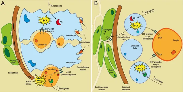
Figure I-5. Hormonal regulation of SCF/c-KIT system in the testis (A) and ovary (B). ... 22
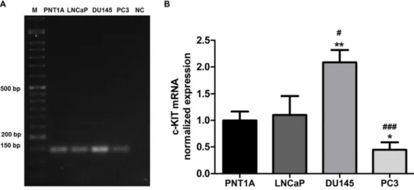
Figure IV-1. c-KIT mRNA expression in non-neoplastic (PNT1A) and neoplastic (LNCap, DU145 and PC3) human prostate cell lines. ... 39
Figure IV-2. Expression of full-length (145 kDa), and 50 and 30 kDa isoforms of c-KIT in non-neoplastic (PNT1A) and non-neoplastic (LNCap, DU145 and PC3) human prostate cell lines. ... 40
Figure IV-3. SCF mRNA expression in non-neoplastic (PNT1A) and neoplastic (LNCaP, DU145 and PC3) human prostate cell lines. ... 41
Figure IV-4. SCF protein expression in non-neoplastic (PNT1A) and neoplastic (LNCaP, DU145 and PC3) human prostate cell lines. ... 41
Figure IV-5. c-KIT mRNA (A) and protein (B) expression in rat prostate. ... 42
Figure IV-6. SCF mRNA (A) and protein (B) expression in rat prostate. ... 42
Figure IV-7. Effect of E2 (100 nM) on c-KIT mRNA expression in human prostate cell lines. . . 43
Figure IV-8. Effect of E2 (100 nM) on c-KIT protein expression (full-length 145 kDa, and 50 and 30 kDa isoforms) in human prostate cell lines. ... 44
Figure IV-9. Effect of E2 (100 nM) on c-KIT protein expression in prostate cell lines. ... 46
Figure IV-10. Effect of E2 (100 nM) on SCF mRNA expression in prostate cell lines. ... 47
Figure IV-11. Effect of E2 (100 nM) on SCF protein expression in prostate cell lines. ... 48
Figure IV-14. Effect of E2 on the mRNA (A) and protein (B) expression of SCF in rat prostate
tissue. ... 52
Figure IV-15. Effect of E2 treatment on animal weight (A) and prostate weight (B). ... 53
Figure IV-16. Proliferation in the prostate of control and E2-treated animals. ... 54
Figure IV-17. Apoptosis in the prostate of control and E2-treated animals. ... 55
Figure IV-18. Expression of apoptosis regulators in the prostate of control and E2-treated animals. ... 56
ADT Androgen Deprivation Therapies
AR Androgen Receptor
ATP Adenosine TriPhosphate BPH Benign Prostatic Hyperplasia CDK Cyclin-dependent kinase CK18 Cytokeratin-18
CS-FBS Charcoal-Stripped Fetal Bovine Serum DEPC Diethylpyrocarbonate
DHEA Dehydroepiandrosterone
DHEAS Dehydroepiandrosterone Sulfate
DHT Dihydrotestosterone DRE Digital Rectal Examination DTT Dithiothreitol
E1 Estrone
E2 17β-estradiol
E3 Estriol
ED Endocrine Disruptor
EDTA Ethylenediamine Tetraacetic Acid EGF Endothelial Growth Factor
ER Estrogen Receptor
ER-α Estrogen Receptor Alpha ER-β Estrogen Receptor Beta FBS Fetal Bovine Serum
FGF8 Fibroblast Growth Factor 8 GPR30/GPER G-Protein-Coupled Receptor-30 IGFs Insulin-like Growth Factors
IGFBP Insulin-like Growth Factor Binding Protein
JAK/STAT Janus Kinase/Signal Transducers and Activators of Transcription MAPK Mitogen-Activated Protein Kinase
mSCF Membrane Stem Cell Factor PAP Prostatic Acid Phosphatase PBS Phosphate Buffer Saline
PCa PCa
PIA Proliferative Inflammatory Atrophy PI3K/AKT Phosphatidylinositol 3-Kinase AKT PIN Prostatic Intraepithelial Neoplasia PMSF Phenylmethylsulfonyl Fluoride pNA p-Nitro-Aniline
PLC-γ Phospholipase-Cγ
PSA Prostatic Specific Antigen PVDF Polyvinylidene Difluoride
qPCR Real-time Quantitative Polymerase Chain Reaction RIPA Radioimmunoprecipitation
ROS Reactive Oxygen Species SCF Stem Cell Factor
SDS-PAGE Sodium Dodecyl Sulfate-PolyAcrylamide Gel Electrophoresis sSCF Soluble Stem Cell Factor
s-KIT Soluble c-KIT
TAP Transit Amplifying Population TGF Transforming Growth Factor Tr-KIT Truncated c-KIT
TRUS Transrectal Ultrasound
TUNEL Terminal Deoxynucleotidyl Transferase dUTP Nick-End Labeling
I. Introduction
Partially published in:
Figueira MI#, Cardoso HJ#, Correia S, Maia CJ, Socorro S (2014). Hormonal regulation of c-KIT receptor and its ligand: implications for human infertility? Progress in Histochemistry and Cytochemistry (in press). DOI: 10.1016/j.proghi.2014.09.001
IF:5.909
Cardoso HJ#, Figueira MI#, Correia S, Vaz CV, Socorro S (2014). The SCF/c-KIT system in the male: survival strategies in fertility and cancer. Molecular Reproduction and Development (in press). DOI: 10.1002/mrd.22430
IF: 2.812 # Both authors
1. Anatomy and Physiology of Prostate Gland: Brief Overview
The prostate is an accessory gland of the male reproductive tract, with a walnut shape and size (Fig. I-1). This gland is located dorsally to the symphysis pubis, anterior to the rectum, and at the base of the urinary bladder. It surrounds the first part of urethra, known as prostatic urethra, and the two ejaculatory ducts (Lee et al., 2011;VanPutte et al., 2014).Figure I-1. Localization of the prostate gland in the male reproductive system (adapted from
(VanPutte et al., 2014)).
The prostate gland consists of divided tubulo-acinar glands, surrounded by a fibromuscular stroma. It is composed by the posterior and lateral regions, enclosed by a partial capsule, and by the anterior and apical regions, enclosed by the anterior fibromuscular stroma (Sooriakumaran et al., 2012;Young et al., 2014).
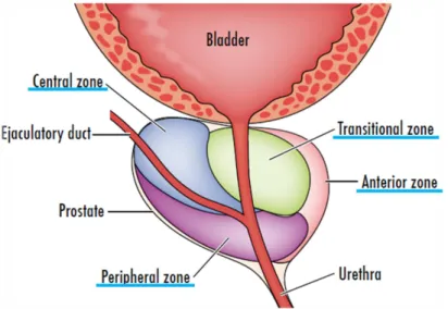
Several models were proposed to characterize the prostate anatomy. The currently most accepted divides the prostate in four distinct zones (Fig. I-2)(McNeal, 1981):
• A central zone, which surrounds the ejaculatory ducts, consisting of 20% of the prostate.
• A peripheral zone, consisting of, approximately, 70% of the gland. This zone makes up the bulk of the prostate.
• An anterior fibromuscular stroma, composed by fibromuscular tissue contiguous with the bladder, without glandular tissue.
• A transition zone, surrounding the proximal prostatic urethra, and consisting of about 5% of the glandular tissue.
Figure I-2. The zonal anatomy of the prostate gland (Eylert and Persad, 2012).
The epithelium of the prostate consists of three main types of cells: the secretory epithelial cells, the basal cells and the stem cells (Frick and Aulitzky, 1991). The presence of cells morphologically different from the smooth muscle was detected, more recently, and these cells were called interstitial cells of Cajal (Van der Aa et al., 2003;Shafik et al., 2005). The non-cellular stroma and connective tissue compose the extra-cellular matrix presenting elements that control prostatic development and function (Frick and Aulitzky, 1991).
The prostate is an exocrine gland, because it has the function of secrete components of seminal fluid, making this a nutritive and lubricating fluid medium in which spermatozoa are transported (Young et al., 2014). The prostatic fluid has alkaline properties that help to protect the spermatozoa from the acidic vas deferens fluid and the acidic environment of the female reproductive system (Guyton and Hall, 2006).
The prostatic secretions constitute, approximately, 30% of the semen (Guyton and Hall, 2006) and are related with its gelation, coagulation, and liquefaction, and some of the secreted proteins are involved in the coating and uncoating of spermatozoa and in interactions with cervical mucus (Aumuller and Seitz, 1990). Secreted products include ions like zinc and calcium, citrate, phosphate, citric acid, spermine, prostaglandins, cholesterol, seminin, clotting enzyme, profibrinolysin, the acid phosphatase, the prostatic acid phosphatase enzyme (PAP), used to assess prostate function, and the well-known prostatic specific antigen (PSA), the mainly indicator of prostatic disease (Frick and Aulitzky, 1991;Hayward and Cunha, 2000;Guyton and Hall, 2006). PSA is detected in bloodstream in case of pathology of the prostate, like chronic inflammation or cancer (Hayward and Cunha, 2000).
2. Prostate Cancer
Prostate cancer (PCa) is the third most frequent type of cancer after the cancers of the female breast and colorectal. Furthermore, PCa is the most common type of oncological disorder among men, in Europe, with an incidence of 214:1000 which has been increasing in last years. Moreover, 416.700 new cases has emerged and 92.200 men died, in Europe, in 2012, with PCa (Heidenreich et al., 2011;Ferlay et al., 2013). In the same year, only in Portugal, 6620 new cases of PCa emerged and it were estimated 1580 deaths (Ferlay et al., 2013;Miranda et al., 2013). The incidence and mortality of PCa differ with the geography, with many factors contributing to these variances (Ferlay et al., 2013;Tewari et al., 2014), but in all over the world, men that suffer from this terrible disease are losing its life quality.
PCa is a highly heterogeneous disease, both in terms of its pathology and clinical presentation, and presents several developmental stages. It starts to be localized in the prostate, with genetic aberrations, local invasion of extracellular matrix, but subsequently can progress to invasion of secondary organs. At the initial stages PCa is modulated by hormonal milieu and in later stages becomes resistant to hormonal modulation.
When cancer develops, the prostate structures become progressively less organized with smaller ducts and ultimately the gland may lose these structures entirely. With further growth, the tumors invade surrounding tissues and metastasize to the lymph nodes, bladder, and bone (Miller et al., 2003). In addition to structural changes, many molecular changes occur in PCa initiation and progression. The expression profile of many genes, namely, genes related with proliferation, apoptosis, and response to stress, is changed, due to genetic events that include epigenetic changes, chromosomal alterations, somatic mutations and alternative splicing (DeMarzo et al., 2003;Reynolds, 2008;Cheng et al., 2012). The cellular metabolism is also changed in PCa (Vaz et al., 2012;Zadra et al., 2013). Nevertheless, the metastatic potential of PCa is the most alertness problem of this type of cancer. Proliferation, neovascularization and extravasation at the primary site are events involved in metastasis (Clarke et al., 2009).
The majority of PCa cases are adenocarcinomas, which arise from the peripheral zone (approximately 70 %). On the other hand, the anterior fibromuscular stroma and the central zone are less related with cancer development, with, respectively, 25 % and 1-2 % of the cases (Hammerich et al., 2009;Lee et al., 2011;Sooriakumaran et al., 2012).
There are evidences that PCa arises slowly from proliferating stem cells or the transit amplifying population (TAP) from the basal compartment (Isaacs and Coffey, 1989). These stem cells differentiate into luminal cells in response to androgens. The TAP seems to expresses androgen receptor (AR). Moreover, in vitro stimulation of TAP with androgens promotes cells differentiation by inducing the expression of markers of luminal cells, such as
cytokeratin-18 (CK18), AR, PAP and PSA. Additionally, androgens down-regulate the α2β1
integrin expression, a molecule implicated in the maintenance of the immature basal cell phenotype (Heer et al., 2007). However, there is conflicting evidence regarding whether the tumors arise from basal cells or from the luminal epithelial cells (Wang et al., 2009;Lawson et al., 2010;Wang et al., 2013).
Indeed, PCa etiology remains largely unknown. Many factors can contribute to the development of this disease, but the best characterized are aging, ethnicity and familiar factors (Heidenreich et al., 2011;Tewari et al., 2014). The principal risk factor for PCa development is the aging (Tewari et al., 2014). Moreover, PCa is more frequently diagnosed in older men and they are more likely to die from the disease than younger men (Hoffman, 2012). In childhood, the human prostate is small, weighing around 2 g. Then, it undertakes exponential growth, at puberty, increasing to about 20 g of weight, due to the rise in serum testosterone levels, characteristic of this phase of life. After that, the weight of the prostate tends to become stable and remains constant until the 40-50 years old, age in which prostatic weight may begin to rise slowly (Berry et al., 1984;Guyton and Hall, 2006). This slowly increase in prostate weight frequently leads to the onset of Benign Prostatic Hyperplasia (BPH) (Berry et al., 1984), a condition characterized by the benign growth of the prostate gland from its normal size to a larger size that can be larger than 150 g (Powers and Marker, 2013). Although there are differences between BPH and PCa, these diseases have some commonalities, and there are evidences that men with BPH have an increased risk of PCa and PCa-related mortality (Orsted and Bojesen, 2013). The race/ethnicity also seems to be a PCa high risk factor, since African men present higher PCa incidence than other groups (Burks and Littleton, 1992;Mordukhovich et al., 2011). The heredity has an important role in PCa development, because the presence of affected first-degree relatives increases the PCa risk in men (Hemminki and Czene, 2002;Zeegers et al., 2003). Although these three factors are the main risk factors related with PCa, other may contribute to the disease only or in combination. Some of them include environmental factors, as the use of pesticides, dietary habits, lifestyle, inflammation and hormones (Giovannucci et al., 1997;Karan et al., 2008). In fact, exposure to pesticides showed to be related with PCa development (Lemarchand et al., 2014). Although some reports did not found correlation between obesity and PCa (Habel et al., 2000), others showed the opposite (Calle et al., 2003). Accordingly, it has been reported that a high-fat rich diet may contribute to PCa (Brasky et al., 2013). Some of the previous numbered factors like environmental, dietary and hormonal factors may contribute to PCa through inflammation (De Marzo et al., 2007;Karan et al., 2008). Some studies reported inflammation as a high risk factor for PCa (Roberts et al., 2004;Atala, 2014). Inflammation may contribute to the pathogenesis and development of cancers, since it can cause infiltrating cell, like leucocytes to release reactive oxygen species (ROS) that may destroy cellular structures and damage genome, which leads to activation of cell proliferation in order to supplant damaged cells, and to release of many cytokines that promote cell division,
angiogenesis and tissue repair. It can also contribute to destroy cell-cell and cell-matrix adhesion, important factors in cancer progression (Coussens and Werb, 2002;Alberti, 2008;Acharya et al., 2010). Moreover, inflammation damages the prostatic epithelium, cause lesions with atrophic morphology, but the cells remain able to proliferate. This condition is named proliferative inflammatory atrophy (PIA) (De Marzo et al., 1999). Morphological transitions can lead to histological lesions causing prostatic intraepithelial neoplasia (PIN), and it was shown that 40% of high-grade PIN lesions arise directly from PIA (Putzi and De Marzo, 2000). Thus, it is clear that PIA may be the linkage between inflammation and PCa.
PCa remains asymptomatic for a long time, which hinders the early screening of the disease. When symptoms become evident, it include urinary symptoms including hematospermia and hematuria, in localized PCA, and rectal obstruction, bone pain, and more systemic features of malignancy, in advanced disease (Tewari et al., 2014).
The PCa screening tests include, at first line, the physical examination with a digital rectal examination (DRE) and measurement of serum PSA. Based on these results, if there are suspicious findings, more sophisticated diagnostic techniques as transrectal ultrasound (TRUS) and guided systematic biopsy are used (Heidenreich et al., 2011;Tewari et al., 2014). The biopsy allows to determine the Gleason score, that measures the aggressiveness of the cancer, predicting prognosis and guiding treatment (Gleason and Mellinger, 1974). In fact, PSA is the mostly used biomarker to screen PCa (Ercole et al., 1987;Phillips, 2014). However, this test lacks some important features needed to a correct diagnosis, namely, sensitivity and specificity (Thompson et al., 2004;Eylert and Persad, 2012). This is a problem that may affect the successfully diagnosis and treatment with serious consequences. In last years, many studies aimed to found new biomarkers for PCa (Crawford et al., 2014).
Some therapeutic options are available to treat PCa. These include radical prostatectomy, brachytherapy, focal therapies (e.g. high intensity focused ultrasound, cryotherapy) and androgen deprivation treatment for local PCa (Eylert and Persad, 2012). In some cases of men with small tumors that have not spread beyond the gland it is recommend close monitoring but without applicate aggressive treatment, a strategy named “watchful waiting” (Eylert and Persad, 2012). In the case of metastasis, it may be used hormone therapy, chemotherapy or radiotherapy (Drudge-Coates and Turner, 2012). Nevertheless, these options are not always effective and investigation to better understand the molecular and cellular mechanisms involved in PCa, is still needed.
3. Hormonal Actions and Prostate Carcinogenesis
The hormonal factors, namely, steroid hormones have important roles on prostate development and physiology, but are also a predisposition factor to PCa development. According to this, several studies reported that administration of male sex hormones can induce PCa in animal models (Noble, 1977).
3.1. Androgens
Androgens are the most abundant sex steroids in men. They are responsible for the development of male organs and secondary sexual characteristics. The most well-known androgens are testosterone and its 5α-reduced metabolite, the potent androgen 5α -dihydrotestosterone (DHT). Testosterone and DHT are considered potent androgens, since even in low dosages they stimulate androgen-dependent structures, like prostate. Testosterone is the most important androgen in men, being mainly produced by Leydig cells on the testis, but also adrenal glands secrete low quantities of this steroid. Other common androgens include the dehydroepiandrosterone (DHEA), its metabolite the dehydroepiandrosterone sulfate (DHEAS) and androstenedione. These are weak androgens produced by the adrenal glands (Jones and Lopez, 2006).
Androgens exert their actions through binding to the intracellular AR, a member of the nuclear receptor superfamily that acts as a ligand dependent transcription factor (Mangelsdorf et al., 1995). This binding activates AR, allowing it to bind DNA and to recruit co-regulators for induction or repression of downstream gene transcription (Heinlein and Chang, 2002).
In the prostate, the biologically active androgen is the DHT that is produced by the local reduction of testosterone produced from the testis by the 5α-reductase enzyme. As other male organs, the prostate development and growth is regulated by androgens since the embryonic development to adulthood (Sensibar, 1995). Testosterone regulates the glandular morphogenesis in the developing prostate and the prostatic function and glandular maintenance in the adult prostate (Sensibar, 1995). Androgens are required for normal prostate development, acting directly on prostate epithelial cells to induce terminal differentiation, or acting at indirect way on prostate epithelium proliferation, through induction of secretory growth factors by the adjacent stroma (Isaacs and Coffey, 1989). In normal prostate, epithelial AR acts predominantly to produce the proteins secreted by the prostate and the stromal AR promotes growth (Lai et al., 2012).
Since androgens are so important in prostate development, hormonal changes may be related with prostate diseases, namely with PCa. Huggins and Hodges were the first to postulate that androgens promote prostate carcinogenesis (Huggins, 1967;Huggins and
Hodges, 1972). In fact, strong evidences reported a correlation between androgens and PCa, for example, African men are thought to have higher androgenic levels, leading to the higher incidence of PCa (Ross et al., 1998). Other evidences showed that estosterone and androstanediol glucuronide were statistically significant positively associated with PCa (Gann et al., 1996).
A comparative study of the effects of DHEA, testosterone and DHT in LNCaP cells revealed that all the androgens increased cell proliferation and PCa biomarkers, however, the DHEA showed delayed effects compared to the other two hormones (Arnold et al., 2005). Furthermore, animal studies also support this role: testosterone caused adenocarcinomas in the dorsolateral prostate of rats and a PCa incidence increase in different rat species (Bosland and Mahmoud, 2011).
The balance between proliferation and apoptosis is crucial to maintain the normal function of prostate. Androgens seem to act as regulators of the both processes through several mechanisms. One of the most common genetic alterations in PCa is the fusion between two genes, namely, the TMPRSS2 gene and the ETS transcription factor genes, ERG or ETV1. TMPRSS2 gene codifies a membrane-bound serine protease, which is regulated by androgens and overexpressed in PCa, and ETS transcription factor genes are involved in multiple processes, including cell proliferation and cancer cell invasion (Lin et al., 1999;Hsu et al., 2004;Tomlins et al., 2005). There are evidences that androgens may induce the translocation of TMPRSS2-ERG and TMPRSS2-ETV1 (Lin et al., 2009;Cai et al., 2010), suggesting a potential mechanism by which androgens promote prostate carcinogenesis through inducing gene translocation. Moreover, androgens regulate the expression of growth factors and their receptors, namely, insulin-like growth factors (IGFs), fibroblast growth factor (FGF8) and endothelial growth factor (EGF), that are involved in prostate cell proliferation, migration, and tumor angiogenesis, thereby facilitating prostate carcinogenesis and cancer progression (Rudra-Ganguly et al., 1998;Torring et al., 2003;Wu et al., 2007).
Androgens also seem to crosstalk with the downstream effectors of growth factor signaling, such as phosphatidylinositol 3-Kinase/AKT (PI3K/AKT), that plays a critical role in prostate carcinogenesis and its progression (Vivanco and Sawyers, 2002). At cell cycle, androgens may increase cyclin expression and promote the assembly of active cyclin/cyclin-dependent kinase (CDK) complexes (Xu et al., 2006;Balk and Knudsen, 2008). Androgens also may increase the cell proliferation through induction of autophagy and intracellular lipid accumulation (Shi et al., 2013) and also through increase of ROS in PCa cells to a level that potentiates cell growth (Lin et al., 2010;Lu et al., 2010).
Relatively to apoptosis, an important role of the androgens in its prevention has been also reported (Raclaw et al., 2008). Androgen treatment of the LNCaP hormone-dependent human PCa cell line induces increased expression of the anti-apoptotic Bcl-2 protein
(Berchem et al., 1995). The expression of the cyclin-dependent kinase inhibitor p21 gene was shown to be up-regulated by androgens and functioned as an apoptosis inhibitor to promote LNCaP cell growth (Lu et al., 1999). Moreover, androgens may suppress the TNFR family
(TNF-α/Fas)-induced apoptosis through inhibition of p53 expression and caspase-2 activation (Rokhlin et al., 2005). Other studies also showed that androgen could block apoptosis induced by Fas activation and TNF-α and reduce the Bax expression (Kimura et al., 2001).
Nevertheless, despite the pro-proliferative and anti-apoptotic effects of androgens, studies that report the opposite effect have been emerging (Wen et al., 2014).
The AR signaling pathway has been implicated in early PCa growth, metastatic disease, development of hormonal resistance and disease relapse. In the majority of PCa cases, the AR is expressed through all stages of development (Debes and Tindall, 2002).
Since AR is a critical effector of PCa development and progression, androgen deprivation therapies (ADT) by ablation of AR function through ligand depletion and/or the use of AR antagonists are the first line of therapeutic intervention. This triggers the cell death or cell cycle arrest of PCa cells (Knudsen et al., 1998;Agus et al., 1999;Koksal et al., 2010). Although these strategies are initially effective, recurrent tumors may arise as a result of inappropriately restored AR function (Feldman and Feldman, 2001).
Many AR alterations are related with PCa progression to more advanced stages, namely, the androgen-independent PCa. Some of these mechanisms involve the response of AR even at low levels of androgens, namely due to mutations, overexpression/amplification, activation by growth factors and cytokines, altered expression co-activators, calpain proteolysis or even by the increased local production of androgen by prostate cells (Linja and Visakorpi, 2004;Devlin and Mudryj, 2009;Saraon et al., 2011).
Somatic mutations of AR are mostly gain-of-function, which can lead to androgen hypersensitivity or decreased ligand specificity, contributing to PCa development (Han et al., 2005). In fact, AR gene mutations are rare in patients with primary PCa but have been reported with a higher frequency in bone marrow specimens from patients with advanced disease (Taplin and Balk, 2004). AR amplification/overexpression sensitizes cells to low concentrations of the ligand (Waltering et al., 2009), and is a process mainly present in tumors that recur after ADT (Bubendorf et al., 1999;Ford et al., 2003). Constitutively active AR splice variants, generated by gene splicing or genomic rearrangement, were detected in PCa cell lines and tumor samples (Guo et al., 2009;Hu et al., 2009), and may be responsible for recurrent PCa through alternative transcriptional output (Hu et al., 2012a). Posttranslational modifications of AR command activity, structure, and stability, including phosphorylation, ubiquitylation, acetylation and methylation. Overall, the majority of these modifications results in AR activation and is related with progression of PCa (McCall et al.,
2008). Other important question related with PCa progression is that, in some cases, the enzymes necessary for androgen synthesis are present and some of them elevated in PCa metastasis or recurrent tumors (Montgomery et al., 2008).
3.2. Estrogens
Estrogens are mainly known as “female sex hormones”. However, they are also present in male serum at low levels and their physiological action has been deserved attention. There are different estrogens, including the natural and synthetic compounds. The natural estrogens include estrone (E1), 17β-estradiol (E2) and estriol (E3). The most
well-known and potent is the E2 (Krolik and Milnerowicz, 2012).
In men, the estrogens production results from the testosterone conversion by aromatase. This phenomenon occurs mainly in the Sertoli cells of the testis, but also in other tissues, including the prostate, which express many of the steroidogenic enzymes, involved in estrogen biosynthesis and metabolism (Takase et al., 2006). In that way, E1 and E2 may be
produced locally within the prostate via aromatization of androstenedione and testosterone, respectively (Ellem and Risbridger, 2006). Thus, estrogens also seem to have an important role in normal development of prostate gland.
Despite the classical role of androgens as stimulating agents in PCa growth, currently, estrogens also have been implicated in the onset and progression of PCa. However, a duality for the possible role of estrogens in prostate cells has been gaining consistency over the last years. If some studies defend that estrogens are potential causative agents of PCa other strong evidences indicate that these steroids may be protective against PCa.
Estrogens have been associated with the uncontrolled growth and transformation observed in PCa (Carruba, 2007;Ho et al., 2011). In fact, the estrogens/androgens ratio increases with age in parallel with a decrease in testosterone levels. That coincides with an increasing prevalence of PCa in older men, suggesting, in fact, a role of estrogens in prostate carcinogenesis. This increased ratio seems to induce the development of inflammation upon aging and the onset of premalignant lesions (Vermeulen et al., 2002;Ellem and Risbridger, 2010).
An increased risk to develop PCa was found in patients with high E2 levels (Salonia et
al., 2011). Moreover, E2 exposure seems to neoplastically transform the rat prostatic
epithelial cells in vitro (Yu et al., 2011b). Other studies confirm that estrogens, alone or in combination with androgens are potent inducers of cell growth and differentiation in PCa (Ricke et al., 2007), and may induce squamous metaplasia of prostatic epithelium (Risbridger et al., 2001). Moreover, estrogens in combination to androgens showed to increase the PCa incidence in rodents in comparison to androgens alone (Bosland et al., 1995), and this
combination was thought to be necessary to PCa development (McPherson et al., 2001;Setlur et al., 2010), but now is known that even alone, estrogens may contribute to PCa development. Furthermore, estrogens also contribute to PCa progression and metastasis in nude mice (Ricke et al., 2006).
Also in vitro studies support these adverse effects of estrogens. Physiological concentrations of estrogens stimulate LNCaP cell proliferation and PSA expression (Castagnetta et al., 1995;Arnold et al., 2005). Accordingly, proliferation was also increased in normal prostate stromal cells (PrSC) and LNCaP cells treated with E2, and the ratio
estrogens/andogens seems to influence this proliferation (King et al., 2006). E2 treatment
also showed effects in cell invasion by up-regulating the production of matrix metalloproteinase 2 in PrSC and WPMY-1 cells, which was mediated by the TGFβ1 (Yu et al., 2011a) and induced stromal cell paracrine effects that promote PCa cell migration, through increasing stability of enolase 1, a critical enzyme for cellular energy metabolism, and promoting its secretion to the extracellular matrix (Yu et al., 2012).
Other mechanisms underlying the action of estrogens in carcinogenesis include the increase in the proliferation of epithelial cells; the up-regulation of growth factors-dependent signaling pathways that promote aberrant cell growth; the increase of prolactin-receptor signaling; the mitogen-activated protein kinase (MAPK) activation; the increased cell-survival potential through the overexpression of anti-apoptotic mediators; the elevation in oxidative stress-induced DNA damage; changes in gene-expression profiles related to cell proliferation, DNA damage, activation of proto-oncogenes and transforming factors; the breakdown of epithelial basement membrane, stromal extracellular matrix, the stimulation of inflammation and overexpression of anti-apoptotic mediators (Pandini et al., 2007;Ho et al., 2011). Other important question related to the estrogens is that they can be chemicals carcinogens, because they can be converted to reactive estrogens intermediates and cause damage to DNA and lipids causing mutations that may be involved in PCa development (Bosland, 2012). Moreover, catechol estrogens showed the ability of induces proliferation and malignant transformation in prostate epithelial cells (Mosli et al., 2013).
The endocrine disruptors (EDs) are a great threat to reproductive physiology and have been linked with the carcinogenesis of reproductive tissues. The majority of the well-studied EDs are estrogen agonists, which mimic the estrogen activity of endogenous hormones. EDs include certain pesticide residues on food, chemicals used in plastics production and phytoestrogens in dietary plant products (Prins, 2008;Hu et al., 2012b). The exposure to some EDs seems to contribute to prostate carcinogenesis (Hardell et al., 2006;Mahajan et al., 2006). The sensitivity of the prostate to ED seems to be higher during the fetal and neonatal development as well as during puberty (Prins et al., 2007;Lobaccaro and Trousson, 2014). However, neither all the EDs have a predisposing role for development of PCa, for example,
some phytoestrogens have showed a potential anti-carcinogenic effect with interest in PCa (Stephens, 1997).
On the other hand, there are evidences of the protective role of estrogens in prostate physiology. In rodents, low doses of estrogens have been shown to enhance prostatic growth, but high doses are generally growth inhibitory (vom Saal et al., 1997). Moreover, pharmacologic levels of estrogens inhibited the prostatic development, resulting in impaired growth and decreased responsiveness to androgens in adulthood (Naslund and Coffey, 1986;Prins, 1992). Other evidences have suggested that estrogens directly inhibit growth of PCa when administered in vitro in the absence of circulating hormones (Robertson et al., 1996). E2 showed to be able to suppress recurrent PCa growth and to delay mortality in
multiple castration resistant xenograft models in vivo (Corey et al., 2002). Accordingly, E2
caused inhibition of PCa growth in an animal model by mechanisms that are independent of androgen action (Corey et al., 2002). Other studies are in accordance with the anti-proliferative role of estrogens, showing that E2 inhibits growth of hormone-nonresponsive PC3
cells (Carruba et al., 1994;Kanagaraj et al., 2007). It is also the case of DU145 cells that showed decreased proliferation with the treatment with estrogens (Pravettoni et al., 2007).
Estrogenic actions are mediated by the classic intracellular receptor proteins (estrogen receptors alpha (ERα) and beta (ERβ), which act as transcription factors regulating the expression of target genes (Gibson and Saunders, 2012). Alternatively, estrogens may elicit signaling events by interaction with the G-protein-coupled receptor-30 (GPR30/GPER) (Prossnitz et al., 2008), but the expression and functionality of this receptor in prostate remains less characterized. ERα and ERβ are expressed in rodent and human prostate during development and into adulthood (Enmark et al., 1997;Prins et al., 1998). The classical ER, ERα, has been detected almost exclusively on the stroma and in subsets of basal cells, and ERβ seems to be expressed in the luminal and basal epithelial cells, with lower or none expression in stromal cells (Leav et al., 2001).
The ability of estrogens to suppress tumor growth and proliferation of prostatic cells may include receptor dependent mechanisms through inhibition of ERβ (Pravettoni et al., 2007), or receptor independent mechanisms such as induction of immune surveillance and metabolism of E2 to cytotoxic estrogens such as 2-methoxyestradiol (Robertson et al.,
1996;Qadan et al., 2001). Other reported mechanism was the inhibition of matrix metalloproteinases and increased levels of IGF binding proteins IGFBP-3 and IGFBP-4 associated with apoptosis, suggesting that estrogens may inhibit the proliferation of PCa cells by inducing apoptosis (Kanagaraj et al., 2007). The tumor suppressive role of E2 may also be
explained by the suppression of androgen levels by E2 (Montgomery et al., 2010), due to its
used as therapeutic options in PCa (Oh, 2002;Gomella, 2009). However, the application of estrogen based therapies is a theme with strong discussion associated (Oh, 2002).
More recently it was reported that the tumor effects may involve an anti-angiogenesis role of E2, explained by its ability to decrease the microvessel number in the
tumor tissues (Wen et al., 2013).
Currently, it is accepted that the opposite roles of estrogens are related with differential responses driven by ERα and ERβ (Risbridger et al., 2007;Ellem and Risbridger, 2009). Indeed, it has been shown that ERα and ERβ have opposite roles in PCa development. A recent study reported that ERα stimulates the genesis and progression of adenocarcinoma in the rat ventral prostate, while ERβ inhibits the onset of precancerous PIN lesions (Attia and Ederveen, 2012). Other evidences support the adverse effects of ERα and the protective role of ERβ, in respect to inflammation, proliferation and other mechanisms leading to prostate carcinogenesis (Pravettoni et al., 2007;Ellem and Risbridger, 2010;McPherson et al., 2010;Piccolella et al., 2014).
As AR, ERs also may undergo modifications, like genetic polymorphisms that may be responsible for different responses in PCa development (Tanaka et al., 2003). Both ERα and ERβ present splice variants. ERα displays, at least, five distinct mRNA isoforms, ERα-A-E (Ye et al., 2000). All these variants present deletions at the C-terminal ligand-binding domain, which is essential for receptor dimerization (Ye et al., 2000). These isoforms could be key regulatory elements or interact with other protein factors, regulating gene expression patterns and hormone sensitivity in normal and malignant prostate tissues (Ye et al., 2000). ERβ also presents five different mRNA isoforms (ERβ1-5). In this case the isoforms result from truncation or insertion in the ligand-binding domain (Lewandowski et al., 2002). These modifications are responsible for different roles of each isoform, for example, ERβ1 and ERβ2 showed opposite roles in regulating proliferation and bone metastasis genes, while the first has tumor-suppressing effects, the second has oncogenic capabilities (Dey et al., 2012). Moreover, several polymorphisms of ERα and ERβ were reported to be related with increased PCa risk (Tanaka et al., 2003;Thellenberg-Karlsson et al., 2006).
4. The Stem Cell Factor (SCF)/c-KIT System
The c-KIT and its ligand, the SCF, are expressed in a broad range of tissues, including brain, breast, testis, skin and prostate (Lammie et al., 1994)
The SCF/c-KIT system has been shown to play an important function in melanogenesis, hematopoiesis and gametogenesis (Nishikawa et al., 1991;Ratajczak et al., 1992;Sato et al., 2012) through the regulation of several biological processes, such as cell proliferation, differentiation, migration and apoptosis (Ronnstrand, 2004;Farini et al., 2007). The importance of SCF/c-KIT system in prostate also has been suggested (Leong et al., 2008).
Despite the expression and the important functions of SCF and c-KIT in normal tissues, they are also expressed in several types of cancer (Natali et al., 1992;Simak et al., 2000). Furthermore, they have been described as having important roles in cancer development and progression in many tissues, including lung, breast, pancreas and prostate (Krystal et al., 1996;Di Lorenzo et al., 2004;Ulivi et al., 2004;Wiesner et al., 2008;Zhang et al., 2011).
Moreover, given the close relationship of SCF/c-KIT system with cancer, the development of specific inhibitors interfering with c-KIT signal transduction pathways emerged as an exciting field in cancer treatment (Lennartsson and Ronnstrand, 2006;Ashman and Griffith, 2013).
4.1. The c-KIT Receptor: Molecular Biology and Signaling
The c-KIT receptor, also known as CD117, stem cell factor receptor or KIT receptor, was firstly described in 1986 as the transforming gene of the Hardy-Zuckerman 4 feline sarcoma virus, being identified as the proto-oncogene v-KIT (Yarden et al., 1987). The mouse
c-KIT gene is located in the dominant white spotted locus (W) (Chabot et al., 1988;Geissler et
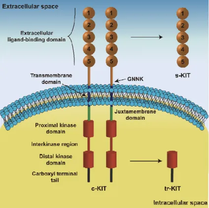
al., 1988) while the human gene is located on chromosome 4q11-q12 with a total length of 90 kb (d'Auriol et al., 1988). The main product of this gene is a single 5 kb transcript (Yarden et al., 1987) encoding a transmembrane glycoprotein with approximately 145-160 kDa that belongs to the type III receptor tyrosine kinase family (Ullrich and Schlessinger, 1990;Blume-Jensen and Hunter, 2001). This class of receptors shares a common structure encompassing three main functional regions (Fig. I-3): an intracellular kinase domain, a transmembrane region, and an extracellular ligand-binding domain (Lemmon and Ferguson, 2007). The cytoplasmic region of c-KIT, responsible for the signaling transduction, contains proximal and distal kinase domains, separated by an interkinase domain, and binding sites for ATP and magnesium ions (Mol et al., 2003;Roskoski, 2005). The transmembrane region is constituted for a short hydrophobic chain of amino acids, allowing the receptor to fix in the plasmatic membrane. The extracellular region is organized in five immunoglobulin-like domains and
have the function of recognize the ligand, but it also participates in receptor dimerization (Yuzawa et al., 2007;Paulhe et al., 2009).
Mechanisms of alternative splicing and others have been identified to originate c-KIT protein variants. One example is the c-KIT isoforms that differ in the presence or absence of the tetrapeptide Gly-Asn-Asn-Lys (GNNK) sequence in the juxtamembrane region of the extracellular domain of the receptor (Caruana et al., 1999;Voytyuk et al., 2003). Both isoforms of c-KIT bind the ligand with identical affinity (Caruana et al., 1999), but the effect of stimulation is faster and more pronounced in GNNK-negative c-KIT (Montero et al., 2008). Recently, it was demonstrated that the GNNK in the extracellular juxtamembrane domain of c-KIT (Fig. I-3) plays a relevant role regulating receptor activation and signaling (Phung et al., 2013).
A mechanism of alternative promoter usage and RNA transcription from a cryptic exon produces a truncated form of c-KIT protein (tr-KIT) with approximately 30-50 kDa which do not have the extracellular domain and the transmembrane region, and is located at cytoplasm (Rossi et al., 1992;Toyota et al., 1994;Albanesi et al., 1996;Takaoka et al., 1997;Muciaccia et al., 2010). tr-KIT do not have the first part of the kinase domain, lacking the kinase activity (Rossi et al., 1992), but retains the capability to induce signaling transduction (Sette et al., 1998). In fact, this tr-KIT can interact with other tyrosine kinase receptors, or with other receptor types (Sette et al., 1998), as a scaffold protein regulating multiple signaling pathways. This isoform of c-KIT has been detected in germ cells and human cancer cells (Rossi et al., 1992;Toyota et al., 1994).
c-KIT also can be proteolytically cleaved and released from the cell membrane originating a soluble isoform (Broudy et al., 1994;Turner et al., 1995), which could be detected in human serum (Wypych et al., 1995). It has only the extracellular domain and binds the ligand with the same affinity as the full-length c-KIT, suggesting a role in controlling ligand bioavailability (Wypych et al., 1995;Dahlen et al., 2001). More recently, serum levels of soluble c-KIT have been associated with the hematopoietic disorders, mobilization of hematopoietic stem cells to peripheral blood, asthma severity, and clinical outcome of patients with gastrointestinal stromal tumors (Kawakita et al., 1995;Nakamura et al., 2004;Deprimo et al., 2009;Makowska et al., 2009).
Figure I-3. Schematic representation of c-KIT structure. The five immunoglobin-like domains of the
extracellular domain are involved in ligand-binding and receptor dimerization. The transmembrane domain anchors c-KIT in the cytoplasmic membrane. The intracellular region, responsible for signaling transduction, contains proximal and distal kinase domains separated by an interkinase region, and a carboxyl terminal tail. Some alternatively spliced forms of c-KIT are characterized by the presence of the tetrapeptide GNNK in the extracellular juxtamembrane domain. The receptor can be cleaved and released from cell membrane originating a soluble c-KIT (s-KIT) only constituted by the extracellular domain. A truncated form of c-KIT (tr-KIT) originated by mechanisms of alternative promoter usage and RNA transcription from a cryptic exon lacks the extracellular and transmembrane domains retaining part of the kinase domain (Cardoso et al., 2014).
c-KIT signaling mechanisms depend of SCF binding to c-KIT, which results in receptor dimerization and activation of intrinsic tyrosine kinase activity (Blume-Jensen et al., 1991;Philo et al., 1996). The interaction of SCF with c-KIT leads to dimerization of receptor, activation of its tyrosine kinase activity and initiation of downstream signal transduction pathways (Blume-Jensen et al., 1991). The pathways activated by c-KIT signaling include the PI3-K, the Src, the Janus kinase/signal transducers and activators of transcription (JAK/STAT), the phospholipase-Cγ (PLC-γ) and the MAPK pathways.
Through the activation of the several signaling pathways, c-KIT is responsible for many effects at cellular level, including, control of cell proliferation differentiation, survival and apoptosis regulation (Ronnstrand, 2004). Therefore, it is highly understandable that deregulated actions of SCF/c-KIT system have been associated with carcinogenesis.
4.2. Molecular and Functional Aspects of SCF, the c-KIT Ligand
The SCF, also known as steel factor, c-KIT ligand or mast cell growth factor is a growth factor that binds to c-KIT, firstly identified in 1990 (Nocka et al., 1990;Williams et al.,
1990;Zsebo et al., 1990). It is codified on the Steel locus (Sl) of chromosomes 12 and 10, respectively, in humans and mouse (Zsebo et al., 1990;Geissler et al., 1991). The human, mouse and rat SCF genes consist of 9 exons (Martin et al., 1990) and encode a 45 kDa glycoprotein, which is located at the plasma membrane of different cell types (Gagari et al., 2006;Wiesner et al., 2008;Mansuroglu et al., 2009).
The SCF protein is constituted by three distinct regions (Fig. I-4): an intracellular domain (Langley et al., 1994;Zhang et al., 2000), an hydrophobic transmembrane domain, and an extracellular domain responsible for recognizing and binding to c-KIT (Langley et al., 1994). Besides the full-length membrane bound form of SCF (mSCF), with approximately, 45 kDa, soluble forms of SCF also have been identified. The soluble SCF (sSCF) is generated by the specific proteolytic cleavage of an alternative spliced form of SCF. A primary cleavage site is encoded by an alternative exon 6 and produces a 165 amino acid soluble protein (31 kDa) still containing the domain that recognizes c-KIT (Flanagan et al., 1991;Pandiella et al., 1992;Majumdar et al., 1994). Thus, both mSCF and sSCF proteins bind and activate the c-KIT receptor. However, it was verified that mSCF induces persistent activation and longer life span of c-KIT, while sSCF leads to a transient activation and faster degradation (Miyazawa et al., 1995). Miyazawa et al. (1995) proposed that sSCF can down-regulate c-KIT expression by triggering its degradation. The ratio of the two isoforms varies in different tissues, suggesting a tissue specific regulation of the SCF expression (Huang et al., 1992). Moreover, differences exist in the downstream pathways activated by c-KIT upon ligation of soluble or membrane isoforms of SCF (Kapur et al., 2002).
Figure I-4. Schematic representation of SCF structure. The SCF display an extracellular domain,
responsible for recognizing and binding to c-KIT, a transmembrane domain and an intracellular domain. The SCF exists as a membrane-bound homodimer (mSCF) or as a soluble protein (sSCF). The sSCF is originated by the proteolytic cleavage of an alternatively spliced variant of SCF that contains the alternative exon 6 (green) (Cardoso et al., 2014).