UNIVERSIDADE DE LISBOA
Faculdade de Ciências
Analysis of the Cooperative Action of PRC1 and Centralspindlin,
the Two Microtubule Bundling Factors Critical for Cytokinesis by
Advanced Live Cell Imaging.
Guilherme Pereira Correia
Mestrado em Biologia Molecular e Genética
i
UNIVERSIDADE DE LISBOA
Faculdade de Ciências
Analysis of the Cooperative Action of PRC1 and Centralspindlin,
the Two Microtubule Bundling Factors Critical for Cytokinesis by
Advanced Live Cell Imaging.
Guilherme Pereira Correia
Dissertação orientada pelo Prof. Doutor Masanori Mishima e pelo Prof. Doutor Rui Gomes
Mestrado em Biologia Molecular e Genética
2012
ii
Resumo
Citocinése em células animais requer um complexo jogo entre o fuso central pós-metafásico e uma série de proteínas regulatórias e moduladoras da dinâmica microtubular (Douglas and Mishima, 2010). Dois factores essenciais para a formação desta estrutura são a proteína PRC1, capaz de agregar microtúbulos, e o complexo centralspindlin (Jiang et al. 1998; Mishima et al. 2002) – um complexo heterotetramérico composto por duas subunidades diméricas: a proteína motora MKLP1 e a RhoGAP (Rho-family GTPase-activating protein) HsCYK-4. Neste trabalho, exploro como estes factores cooperam e qual a relevância da sua interacão para a divisão celular. Versões nativas ou mutadas de HsCYK-4, fundidas com GFP, foram empregadas para estudar a localização desta proteína e para determinar qual a influência da região C-terminal de HsCYK-4 (a região responsável pela interação com a proteína PRC1; Lee and Mishima, unpublished) na divisão celular. Determinou-se que a acumulação do complexo centralspindlin ocorre em duas fases distintas: 1) uma acumulação pré-ingressão da membrana celular, dependente da região C-terminal de HsCYK-4 e 2) uma acumulação pós-ingressão. Quando a primeira fase foi inibida, via ablação da região C-terminal, observou-se um aumento da proporção de células que falhavam a divisão. Isto ocorria devido a uma falha precoce durante o elongamento celular, ou devido a uma falha tardia devida a perda de integridade do corpo médio. Determinou-se que isto, pelo menos em parte, é causado pela formação de fusos centrais anormais com uma organização microtubular comprometida. Também nestas células foi observado que, apesar do complexo centralspindlin e da proteína PRC1 terem mecanismos de recrutamento independentes, a manutenção da sua localização requer a região de interação no C-terminus de HsCYK-4. Este trabalho dá relevância à interação entre centralspindlin e PRC1, tanto para a sua estável localização como para a manutenção da rigidez do fuso central e do corpo médio durante a divisão celular.
iii
Abstract
Cytokinesis in animal cells requires a complex interplay between the post-metaphase microtubule-based central spindle and several regulatory and microtubule modulating proteins (Douglas and Mishima, 2010). Two protein factors required for the assembly of the central spindle are the microtubule-bundling protein PRC1 and the centralspindlin complex (Jiang et al. 1998; Mishima et al. 2002) – an heterotetramer composed of the motor protein MKLP1 and the RhoGAP (Rho-family GTPase-activating protein) HsCYK-4 subunits. In this work I explore how PRC1 and centralspindlin cooperate and how their interaction leads to a successful cytokinesis. GFP-fused native or mutant versions of the HsCYK-4 protein allowed us to follow centralspindlin localisation and behaviour in vivo, and also determine what influence would the loss of the C-terminal region of HsCYK-4 - the region of PRC1 binding (Lee and Mishima, unpublished) - have on cell division. Centralspindlin accumulation at the middle of the central spindle was found to occur in two stages: a 1) pre-furrow ingression accumulation, heavily dependent on HsCYK-4’s C-terminal region, and a 2) post- furrow ingression accumulation. When the first stage was disrupted, by ablating the C-terminal region of HsCYK-4, an increase in the number of cell division failures was observed. These occurred due to an early failure during elongation of the pole-to-pole distance, or due to a late failure via loss of midbody integrity. This is, in part, caused by the formation of abnormal central spindles with a compromised microtubule organisation. Also, colocalisation between centralspindlin and PRC1 was impaired in these cells, showing that while these molecules have separate targeting mechanisms to the central spindle, the stability of that localisation is dependent on their interacting region. These data point to centralspindlin-PRC1 interaction being essential for the stability of their individual localisation and to maintain the rigidity of the central spindle and midbody during cell division.
iv
Resumo Extendido
Citocinése é o processo pelo qual uma única célula é capaz de se dividir em duas células filhas após segregação do material genético, perpetuando assim a sua linhagem. Apesar de ser um mecanismo celular fundamental, ubíquo a todos os organismos, é notável o quanto desconhecemos sobre este processo. Na célula, a anafase inicia-se com a segregação dos cromossomas, que são divididos equitativamente para pólos opostos da célula em divisão. Nesta fase é possível observar-se uma população de microtúbulos na região equatorial da célula – os microtúbulos interpolares. Estes têm origem de cada um dos pólos da célula e as suas regiões distais interagem de forma anti-paralela no centro da célula, levando à formação do fuso central, uma estrutura característica desta fase. Em células humanas, existe uma extensa rede de proteínas, como MAPs (microtubule-associated proteins; isto é, proteínas associadas aos microtúbulos), proteínas motoras e cinases, envolvidas na formação desta estrutura. Dois factores proteicos essenciais para este processo e conservados entre diferentes organismos são PRC1 e o complexo centralspindlin.
PRC1 é uma proteína pertencente a uma família de proteínas sem propriedade motora mas com capacidade de agregar microtúbulos (Schuyler 2003). Esta interage com a proteína motora KIF4 (Zhu and Jiang, 2005; Bieling et al. 2010), através da qual se movimenta para o centro do fuso central em formação, ligando preferencialmente microtúbulos com orientação oposta (oriundos de pólos opostos da célula) contribuindo para a formação do fuso central (Zhu and Jiang, 2005). O complexo centralspindlin é um complexo heterotetramérico composto por duas subunidades diméricas: a proteína motora MKLP1 e a proteína da família das RhoGAP (GTPase-activating protein) HsCYK-4 (Mishima et al. 2002). Após a transição metafase-anafase, este complexo migra através dos microtúbulos para a mesma posição central no fuso central onde interliga e agrega os microtúbulos do futuro fuso central, permitindo a sua formação e conferindo-lhe estabilidade (Mishima et al. 2002). Apesar de a interação entre PRC1 e o complexo centralspindlin já ter sido identificada, ocorrendo entre a região N-terminal de PRC1 e a C-terminal de HsCYK-4 (Lee and Mishima, unpublished), a sua função ainda está muito pouco estudada. Neste trabalho, exploro como estes dois factores cooperam e qual a relevância da sua interação para a divisão celular.
v
Este trabalho começa com a caracterização da divisão celular em células HeLa, utilizadas neste estudo como sistema modelo de células humanas/mamíferas, após depleção da proteína HsCYK-4 por RNAi. Esta depleção além remover a expressão desta proteína, evita que se forme o complexo centralspindlin. Tornou-se imediatamente clara a necessidade desta proteína para a divisão celular, pois a sua depleção levou a um aumento do número de células que falhavam a divisão celular (de 0% para ~20%). Estas células ultrapassavam a metafase, mas durante o elongamento do eixo maior da célula em divisão, que ocorre durante a anafase, ocorria uma regressão da célula e um retorno a um estado não-mitótico. Além disso, tornou-se evidente que mesmo as células que se dividiam, o faziam demorando mais tempo em todos os passos da divisão e atingindo um maior elongamento do eixo maior. Isto sugere que há um decréscimo da resistência da maquinaria que leva à divisão, contra as forças que se produzem durante a divisão. Maiores no sentido perpendicular ao da divisão durante os primeiros momentos da anafase – resultando nas regressões – e maiories no mesmo sentido que a divisão nos momentos subsequentes devido ao elongamento da célula em divisão.
A utilização de HsCYK-4 ou variantes sem a região C-terminal de interação com PRC1 (HsCYK-4 ΔTail), fundidas com GFP, permitiu a observação da distribuição e localização de centralspindlin na célula em divisão. A utilização de HsCYK-4 ΔTail permitiu a identificação de que a acumulação de centralspindlin ocorre em duas fases separadas temporalmente e no espaço. Uma primeira acumulação ocorre no centro do plano equatorial da célula imediatamente após a transição metafase-anafase. Esta é extremamente dependente da região C-terminal de HsCYK-4, possivelmente devido à falta de interação com PRC1 que serviria para estabilizar o seu recrutamento. De seguida, coincidentemente com a ingressão da membrana celular, uma acumulação de centralspindlin ocorre corticalmente, perto do nível da membrana celular. Esta acumulação parece estar dependente de uma ação cooperativa entre a região C-terminal de HsCYK-4 e a ingressão (ou factores associados com a ingressão, como a proteína anillin), como evidenciado pela utilização de inibidores da polimerização de actina – necessária para a ingressão. Duas vias de acumulação de centralspindlin haviam sido descritas em C. elegans, mas tinham escapado detecção em células de mamífero/humanas, possivelmente devido ao seu pequeno tamanho, até este trabalho.
Quando a primeira fase de acumulação foi inibida, via ablação da região C-terminal, observou-se um aumento da proporção de células que falhavam a divisão, de igual grau que quando havia depleção total de HsCYK-4 na célula. A região C-terminal de HsCYK-4 é portanto crucial para a função de HsCYK-4. Estas células falhavam a divisão celular devido a um impedimento do correcto elongamento da célula em divisão durante a anafase ou após
vi
formação da ponte intracelular (após completa ingressão da membrana) devido a uma fragmentação do corpo médio. Ambos os fenótipos evidenciam que a região C-terminal de HsCYK-4 é necessária para manter a estabilidade da maquinaria que suporta a divisão celular, muito provavelmente devido à mal-formação do fuso central e do corpo médio.
Transfecção das células expressando HsCYK-4-GFP com um plasmídeo com o gene codificando para mCherry-PRC1, permitiu a observação in vivo de ambas as proteínas durante a divisão. Como esperado. a elevada colocalisação entre PRC1 e HsCYK-4 observada quando a célula possui a totalidade da proteína HsCYK-4, foi drasticamente reduzida em células sem a região C-terminal desta. Observou-se a formação de foci com ambas as proteínas ou com apenas PRC1 ou HsCYK-4. Isto suporta que ambas as proteínas têm mecanismos de recrutamento para o fuso central independentes, mas que a sua estabilização nesta posição necessita da interacção entre elas. Esta falha de colocalisação irá certamente afectar a estrutura final do fuso central e corpo médio – explicando, pelo menos em parte o aumento no número de células que falha a divisão. Para comprovar isto, células foram fixadas e os microtúbulos foram marcados. Foi possível observar um grande número de células apresentando fusos centrais anormais nas células portadoras HsCYK-4 sem a sua região C-terminal. Este fenótipo foi ainda mais exacerbado após exposição a baixas temperaturas antes da fixação - um tratamento que é capaz de destabilizar microtúbulos. As células portadoras da totalidade de HsCYK-4 não mostraram ser tão sensíveis ao tratamento. Isto, evidencia a importância da região C-terminal de HsCYK-4 e da interacção com PRC1 para a formação do fuso central e manutenção da estabilidade dos microtúbulos que o compõem.
Este trabalho demonstra a relevância da interacção entre a proteína PRC1 e o complexo centralspindlin (via HsCYK-4) para uma progressão da divisão celular com sucesso. A região C-terminal de HsCYK-4 é necessária para tanto para a correcta localização e como acumulação de centralspindlin no centro do fuso central. Ao mesmo tempo contribui para uma colocalisação desta com a proteína PRC1. Esta localização correcta é necessária para a formação do fuso central através da sua capacidade de agregar microtúbulos, fornecendo-lhe também estabilidade e rigidez durante todo o processo de divisão celular. Sem que isto ocorra, torna-se frequente a falha de divisão celular, que culmina na geração de células com um número exagerado de cromossomas, o que pode facilmente levar a cancerigénese.
vii
Contents
Introduction 1
Results 4
HsCYK-4 is Required for Cell Division 4
Stable Accumulation of HsCYK-4 Requires its C-terminus 6 HsCYK-4 Accumulates in Two Spatial and Temporal Distinct Phases 9 HsCYK-4 C-terminus Is Required for HsCYK-4-PRC1 Colocalisation 13 Central Spindle Robustness Requires HsCYK-4-PRC1 Colocalisation 16
Discussion 19
Materials and Methods 22
References 23
Annex 26
1
Introduction
Cytokinesis, the final step in cell division, refers to the process by which a single cell divides into two daughter cells, after segregation of the genetic material, thereby maintaining its lineage. Being a mechanism absolutely essential to every organism it is crucial to reach a deeper understanding of this process. At anaphase onset the chromosomes are segregated, half to each pole of the cell. During this period we see a distinct population of microtubules in equatorial region of the cell. They originate from each of the two poles and their plus ends interact in an anti-parallel fashion in the centre of the cell, leading to the formation of the characteristic central spindle (for review see Eggert et al 2006.). In human cells, a very wide network of proteins, such as microtubule-associated proteins, motor proteins and kinases are involved in the formation of this structure (Douglas and Mishima, 2002). Two conserved and essential protein factors for central spindle formation are PRC1 and the centralspindlin complex (Kurasawa et al. 2004; Mishima et al. 2002). Each of them individually is able to bind and bundle microtubules in vitro, but both of them are required for a successful formation of the central spindle in vivo and for cell division (Mollinari et al. 2002; Mishima et al. 2002).
During anaphase and telophase, the central spindle remains in place as the chromosomes segregate, and determines the location where the contractile actomyosin ring (Barr and Gruneberg, 2007). This ring is directly responsible for inducing an ingression of the membrane, physically dividing the cell. As this happens, the central spindle is physically compacted, and at the same time the antiparallel microtubule bundles start to aggregate, eventually culminating in the formation of a single structure called the midbody (Mullins and McIntosh, 1982). It is this aggregate of microtubule bundles inserted into a net of mutually interacting proteins that apparently leads the final abscission step, although the exact mechanism by which it does so is still unknown (Barr and Gruneberg, 2007).
In mammals and other organisms, centralspindlin is a heterotetrameric complex, composed of a homodimer of HsCYK-4 (also known as MgcRacGAP; homologue of C. elegans’ CYK-4 and Drosophila’s Tumbleweed) and a homodimer of the MKLP1 (ZEN-4 in
2 C. elegans and Pavarotti in Drosophila) protein (Mishima et al. 2002). MKLP1 is a plus-end directed kinesin-6 type motor protein, on which the proper localization of centralspindlin to the middle region of the central spindle relies (Mishima et al. 2002). HsCYK-4 is a Rho-family GTPase activating protein (RhoGAP), which together with MKLP1 in the centralspindlin complex, is able to bind and bundle microtubules leading to the formation of central spindle. Besides this, HsCYK-4 is also known to interact with several other proteins involved in cytokinesis, such as PRC1 (Ban et al. 2004). MKLP1 is a plus-end directed kinesin-6 type motor protein, on which the proper localization of centralspindlin to the middle region of the central spindle relies (Douglas and Mishima, 2010). PRC1 is a microtubule-associated protein that dimerises and localizes to the middle of the central spindle on a manner that is dependent on the KIF4 motor protein (Zhu and Jiang, 2005; Bieling et al. 2010), although their direct interaction seems not to be essential for this (Hu et al. 2002). There, PRC1 bundles and cross-links the microtubules arranged in an anti-parallel fashion that compose the middle of the central spindle (Subramanian et al. 2010).
The master regulator of the entry into mitosis is the Cdk1 kinase. Before mitosis, cyclin B is expressed and binds to Cdk1, activating it (for review see Ma and Poon, 2011). Before the metaphase-to-anaphase transition, Cdk1 activity phosphorylates MKLP1, of the centralspindlin complex, inhibiting its binding to the microtubules (Mishima et al. 2004). After metaphase, cyclin B expression is down-regulated and the existing protein is signalled for degradation by the anaphase-promoting complex (APC; Ma and Poon, 2011). As the amount of cyclin B decreases in the cell, so does Cdk1 kinase activity. This lifts the inhibition of Cdk1 over centralspindlin. It is then able to bind to the microtubules. The centralspindlin molecules move towards the plus end of microtubules due to MKLP1 motor activity, on a manner that is dependent on the clustering of several centralspindlin molecules (Hutterer et al. 2009). By moving along the central spindle’s microtubules they reach the equatorial region of the cell, where they also associate with the microtubules on an opposite orientation (Mishima et al. 2002). This results in the cross-linking of the microtubules originating from opposite poles of the cell, leading to central spindle formation. Centralspindlin clustering into an higher-order structure in this region is thought to confer stability to the central spindle (Hutterer et al. 2009). Accumulated HsCYK-4 at the centre of the central spindle then promotes the formation of the actomyosin contractile ring through its GAP activity, although the mechanism through which it does so is still under debate. Small G-proteins cycle between an inactive GDP-bound state and an active GTP-bound state. GAP proteins accelerate the
3 hydrolysis of GTP in small G-proteins, while GEF (guanidine exchange factor) proteins promote the exchange of GDP for GTP (Naryuma and Yasuda, 2006). HsCYK-4 is able to inactivate Rac, Cdc42 or RhoA G-proteins (Canman et al. 2008; Jantsch-Plunger et al. 2000), but it is also able to interact with the GEF ECT2 and promote the localization of activated RhoA at a narrow region in the equatorial plane of the cell (Yüce et al. 2005; Nishimura and Yonemura, 2006; Somers and Saint, 2003). This positively regulates the assembly of the actomyosin contractile ring at the right position (Barr and Gruneberg, 2007).
Cdk1 also phosphorylates PRC1 (Mollinari et al. 2002), as in MKLP1’s case. When its activity decreases at the metaphase-to-anaphase transition, PRC1 is able to interact with the plus-directed motor protein KIF4 (Zhu and Jiang, 2005; Bieling et al. 2010). After which, it accumulates in the middle of the central spindle with a preference for the anti-parallel microtubule overlap region (Mollinari et al 2002). There, it cross-links the microtubules and contributes to central spindle formation (Kurasawa et al. 2004; Mollinari et al 2002). PRC1 was first found to to interact with HsCYK-4 via the latter’s C-terminal half (including the RhoGAP domain) and this was seen to diminish HsCYK-4 GAP activity towards the small G-protein Cdc42 (Ban et al. 2004). It was proposed that HsCYK-4-PRC1 interaction would be important to constraint HsCYK-4’s GAP activity early in division (Ban et al. 2004). A recent work in Mishima laboratory, done in C. elegans, reduced the region of interaction to HsCYK-4’s C-terminal ~60 amino acids tail region and demonstrated how it is absolutely critical for the stable maintenance of the mechanical integrity of the central spindle during the process of division (Lee and Mishima, unpublished). Ablation of this interaction lead to a decrease in the central spindle’s resistance against external mechanical forces applied to it and cytokinesis failure. However, the molecular details of how this interaction contributes to the formation and maintenance of the microtubule bundles in mammalian cells are still unclear.
4
Results
HsCYK-4 is Required for Cell Division
The exact mechanism by which centralspindlin contributes to cell division in mammalian cells remains an open question. To address this, we used human HeLa cells. These were cultured using standard culturing media and imaged under a microscope over the course of 1 or 3 hours. Using differential interfering contrast (DIC) microscopy the chromosomal state is clearly visible and this was used to determine the cell division stage. The imaging was started on cells that exhibited the characteristic metaphase plate (Figure 1A, 0 min; Figure 1B, 0 min). In untreated HeLa cells, cell division proceeds as normal (Figure 1A) in all of the observed cells (Figure 1E). In contrast, when these cells were depleted of HsCYK-4 by RNAi (Figure 1C), 17% of the imaged cells were seen to fail cytokinesis (Figure 1E) – consistent with the known function of HsCYK-4 (Mishima et al, 2002; Yüce et al, 2005) - and ~59% were arrested in metaphase. The latter phenotype seems to imply a role for HsCYK-4 in either a metaphase checkpoint, which has been described in Lagana et al. 2010 or a requirement of HsCYK-4 for the segregation to occur. Cells that failed cytokinesis did so through a contraction of the cell from a previously elongated state, when the distance between poles should be increasing (Figure 1B).
Upon further analysis of the HsCYK-4 depleted cells that were able to divide, some differences were obvious. To start with, the timing of every step of cell division since anaphase onset (0 min) was delayed (Figure 1D). Cleavage furrow ingression was considered to occur starting from the first frame where invagination of the cell membrane was observed to the frame where no more compaction of the recently formed intercellular bridge and midbody was observed. In control cells it took ~4 min 10 s for the cellular membrane to start ingression and ~4 min 50 s for it to conclude, in HsCYK-4 siRNA-treated cells it took ~5 min 15 s from anaphase onset to the start of ingression and 5 min for ingression to end.
At the same time, an increased elongation of the cell along the pole-to-pole axis was seen in HsCYK-4 siRNA-treated cells when compared to the control (Figure 1F). To obtain these measurements we have to consider that initial diameter before division is highly variable in HeLa cells and this will be reflected in the anaphase long axis length. Therefore,
5 Figure 1. DIC time-lapse images of the middle plane of a A) normally dividing control cell and B) early regressing HsCYK-4 depleted cell (bars: 10 µm). C) Western blot against HsCYK-4 and tubulin of cell lysates after treatment with control siRNA or HsCYK-4 siRNA, showing the amount of HsCYK-4 depletion after the treatment. D) Timing of division in control cells and in HsCYK-4 depleted cells; A.O.: anaphase onset. E) Bar plot indicating the fate of control or HsCYK-4 depleted cells. F) Plot of measured anaphase long axis length vs metaphase diameter of control or HsCYK-4 depleted cells. Modelled linear regression equations show the assumed linearity between x and y values. P-value obtained from a t-student statistical test.
6 we decided to consider a linear relationship between initial diameter measured in metaphase and long axis length at the end of anaphase. To this end, a linear regression was done with this assumption, so that all observed differences in the two lines are a result of the different treatments.
To confirm that all the previous results obtained from HsCYK-4 siRNA-treated cells were due to the absence of HsCYK-4, the same depletion was done in a transgenic cell line carrying the siRNA-resistant full-length HsCYK-4 gene (HsCYK-4 FL; Figure 2A). Taking into account only the cells that either successfully complete cytokinesis or fail the process, the phenotype resulting from the depletion of HsCYK-4 in HeLa cells was completely rescued when they were expressing HsCYK-4 FL (Figure 2E). Therefore we can exclude any non-specific effect of the siRNA. In contrast, the rescue was not observed in two independent transgenic cell lines carrying the siRNA-resistant HsCYK-4 gene lacking the C-terminal tail region (HsCYK-4 ΔTail/ΔTail*; Figure 2A; Figure 2E). More than that, the amount of cells failing cytokinesis in these cells is identical (ΔTail) or very close (ΔTail*) to the non-transgenic wild-type HsCYK-4 depleted cells. Therefore, this C-terminal tail region of 80 amino acids absent in both the ΔTail and ΔTail* transgenic cells is essential for HsCYK-4’s function during division. Putatively, this is due to the absence of interaction between PRC1 and HsCYK-4, as it is expected to occur via this site (Lee and Mishima, unpublished).
Stable Accumulation of HsCYK-4 Requires its C-terminus
The GFP-tagged constructs (Figure 2A) allowed for the live imaging of HsCYK-4 during cell division in the generated cell lines utilizing both DIC and in vivo fluorescence microscopy. This was done after depletion of the endogenous HsCYK-4 via RNAi (Figure 2D), which does not affect the transgenes as they carry a silent mutation that confers them resistance to this treatment. In the HsCYK-4 FL cell line, it is possible to observe the wild-type phenotype of HsCYK-4 distribution and dynamics (Figure 2B). At metaphase, HsCYK-4 is diffused throughout the cytoplasm, mildly and transiently associating with the mitotic spindle. After, at the same time as the metaphase-to-anaphase transition, an accumulation of HsCYK-4 emerges in rather broad bands in the middle of the segregating chromosomes. This signal concentrates, corresponding to the clustering of centralspindlin molecules in the equatorial plane of the cell (Hutterer et al. 2009), in the region of antiparallel microtubule overlap of the central spindle. It is from this central position that HsCYK-4 signals where the actomyosin
8 ring should assemble itself and coordinates the ingression of the cleavage furrow. As ingression occurs, the separate HsCYK-4 foci are compacted into a single very intense spot. This corresponds to the final compaction of all the microtubule bundles and central spindle associated proteins in order to form the future midbody. This structure maintains itself for a long period of time as the intercellular bridge extends and the cells return to the substrate after division.
Surprisingly, in the case of the cell lines carrying the HsCYK-4 ΔTail gene (ΔTail and ΔTail*) it is possible to observe 3 populations of cells: 1) cells that are apparently able to divide, and cells whose division fails at 2) an early stage before ingression is completed, or 3) at a later stage after the completion of ingression and formation of the midbody (Figure 2C; Figure 2F; Figure 2G). I will refer to these two latter phenotypes as early regression and late regression, respectively.
Early regressing cells exhibited a much weaker accumulation of HsCYK-4 ΔTail at the equatorial plane during the metaphase-to-anaphase transition (Figure 2F). This was also accompanied by the diffusion of some of the foci of HsCYK-4, giving rise to a transient accumulation, which was not able to maintain itself. In most cases, even this weak accumulation was enough to induce the ingression of the cleavage furrow, but not sufficient to maintain the ingression until completion. The regression of the recently ingressed furrow would then occur. At the same time, these cells return to a round up shape, through an apparent collapse of the central spindle, evidenced by the nearing of the recently segregated chromosomes.
Figure 2 (previous page). A) Domain structure of the used transgenic HsCYK-4 variants, showing its domains (CC: coiled coil, C1: putative membrane-binding; eGFP: enhanced green fluorescent protein) as well as the PRC1 binding region and RNAi resistant silent mutation. Time-lapse DIC and fluorescence max intensity z-axis projections of dividing B) HsCYK-4 FL and C) ΔTail, after endogenous HsCYK-4 depletion (bars: 10 µm). D) Western blot against GFP, HsCYK-4 and tubulin of cell lysates after treatment with control siRNA or HsCYK-4 siRNA, showing the amount of HsCYK-4 depletion after the treatment, and the maintenance of transgenic variants after HsCYK-4 RNAi treatment. E) Bar plot indicating the fate of control or HsCYK-4 depleted HsCYK-4 FL, ΔTail or ΔTail* cells that progressed past the metaphase-to-anaphase transition. F) Early regression phenotype of HsCYK-4 ΔTail cells (bar: 10 µm). G) Late regression phenotype of HsCYK-4 ΔTail cells (bar: 10 µm).
9 In late regressing cells, the accumulation of HsCYK-4 at earlier stages (metaphase-to-anaphase transition through to telophase) was very similar to the one exhibited by HsCYK-4 ΔTail-carrying successfully dividing cells. The difference being that after the formation of the midbody at the end of telophase, it is possible to observe some fragmentation of the recently formed midbody (Figure 2G). This could occur at any time from right after the completion of ingression to almost an hour later. It is evident that the midbody is not able to maintain its integrity, as it is connected to the recently distorted cellular membrane and therefore under great tension. The fragmentation would then culminate with a complete regression of the cleavage furrow (arrow heads in Figure 2G) leading to a re-fusion of both of the cells. Together, these regression phenotypes evidence the existence of a requirement for the C-terminal tail region of HsCYK-4-PRC1’s binding site – in HsCYK-4 recruitment and stabilisation at the equatorial plane of the dividing cell. If this is impaired, it will both decrease the intensity of the signalling cascade that leads to ingression or the mechanical integrity of the ingression. This interaction is again required, at a later stage, for the stability of the “midbody matrix” (Matuliene and Kuriyama, 2004), essential for cross-linking the microtubules and anchoring the membrane at the intercellular bridge.
HsCYK-4 Accumulates in Two Spatial and Temporal Distinct Phases
Looking at cells that were able to divide carrying the different transgenes –either the FL or the ΔTail version of HsCYK-4 – a clear difference was observed in the amount of HsCYK-4 accumulated immediately after the metaphase-to-anaphase transition. HsCYK-4 ΔTail was seen to accumulate less strongly at the equatorial region (Figure 2C). To allow for a proper comparison between the different transgenic strains studied, we proceeded to quantify the amount of fluorescent signal that would accumulate during the course of cell division. This was made via measuring the fluorescent intensity in a 20-pixel line overlaying the pole-to-pole axis, therefore crossing the equatorial region of the cell as it underwent division. By subtracting the background and choosing the max intensity grey values it is possible to get a good representation of the accumulation of HsCYK-4 (FL or ΔTail) through time at the centre of central spindle (example in Figure 3A). The measurements obtained were not due to differences in HsCYK-4 expression on the different cell lines as measured in Figure 3B.
The obtained measurements are represented in Figure 3C and 3D. The initial recruitment of HsCYK-4 (Figure 3D) was heavily impaired in HsCYK-4 ΔTail cell lines,
10 Figure 3. A) Scheme the measurement of fluorescence intensity done to obtain line plots C and D; yellow line corresponds to the 20-pixel averaged line; red rectangle corresponds to the max intensity spot that line plots represent. B) Average pixel intensities for HsCYK-4 FL, ΔTail and ΔTail*, measured from metaphase cells. C) Total measured pixel intensities at the location of HsCYK-4 (FL or ΔTail) accumulation. D) Enlarged version of the total plot showing the early moments of HsCYK-4 (FL or ΔTail) accumulation before ingression start. E) Timing of division in HsCYK-4 FL, ΔTail and ΔTail*; A.O.: anaphase onset.
amounting to less than half of that observed in the HsCYK-4 FL cell line. This points towards a role of the C-terminal tail region of HsCYK-4 in either its recruitment or maintenance of the accumulation at the centre of the central spindle. It is also reasonable to assume that either the
11 C-terminal tail region might have an influence in centralspindlin clustering or, if the interaction with PRC1 is disrupted in HsCYK-4 ΔTail, that a stabilising factor for HsCYK-4 at the central spindle is missing.
Paradoxically, the amount of HsCYK-4 measured at the centre of the equatorial plane (as in Figure 3A) in all cell lines used increases at around the same rate, coincidentally with the start of ingression (Figures 3C and 3E). A closer examination of the HsCYK-4 ΔTail cells lead to the observation that these would accumulate HsCYK-4 ΔTail in a characteristic pattern: a cortical ring near the cellular membrane (Figures 4A). This is very evident when measuring the pixel intensity perpendicularly to the long axis of the dividing cell at the equatorial plane during ingression (Figure 4B) and when reconstructing the fluorescence in 3D (Figure 4C). This hints at another possible mechanism of HsCYK-4 accumulation that would be independent of the C-terminal tail region of HsCYK-4, and coincidental with ingression. To study this possibility, we opted for the addition of an actomyosin contractile ring inhibitor, which would impede ingression. Latrunculin-A was chosen for this purpose since its ability to inhibit actin polymerisation (Morton et al. 2000; Yarmola, 2000) had already been previously used to study cell division and ingression. When inhibiting actin polymerisation with latrunculin-A in the HsCYK-4 FL cell line, the cortical accumulation of HsCYK-4 became obviously distinct from the central accumulation (Figure 4D). The central accumulation of HsCYK-4 was not affected by the drug treatment, and at the same moment in time as ingression was supposed to occur, an intense stabilisation of HsCYK-4 could be observed near the cell cortex. It is therefore a spatially and temporally distinct accumulation of HsCYK-4 during cell division.
Figure 4 (following page). A) DIC and fluorescence max intensity z-axis projections of the middle planes of a HsCYK-4 ΔTail cell, showing HsCYK-4 ΔTail cortical accumulation. The highlighted region is enlarged in B) as well as measured pixel intensity (bars: 10 µm). C) 3D reconstruction of the same cell as in A). D) DIC and max intensity z-axis projections of the middle planes of HsCYK-4 FL and ΔTail cells after latrunculin-A treatment (bars: 10 µm); time points correspond to anaphase onset, pre-ingression, and supposed ingression start; enlargement shows cell membrane (dashed line) and cortical accumulation site (arrow). E) Scheme of the measured regions used to obtain F) bar plots quantifying central and cortical accumulation in HsCYK-4 FL/ΔTail cells, with the calculated ratio between them.
12 Puzzlingly, in HsCYK-4 ΔTail cells that were exposed to the same drug treatment, the accumulation of HsCYK-4 ΔTail at the cortical level was heavily disrupted (Figure 4D). The amount of stabilised HsCYK-4 was quantified in both cell line using the max pixel intensities in the areas described in Figure 4E. It was found that in HsCYK-4 ΔTail cells the amount of protein that was stabilised near the membrane was more than five-fold lower than in HsCYK-4 FL cells (Figure HsCYK-4F). This seems to indicate that the C-terminal tail region is also implied in this second pathway of accumulation, which is possibly dependent on a cooperative action being this C-terminus and the ingression (or ingression-associated factors).
13
HsCYK-4 C-terminus Is Required For HsCYK-4-PRC1 Colocalisation
PRC1 is another key protein for the successful completion of cytokinesis (Zhu and Jiang, 2005) and the C-terminal tail region of HsCYK-4 had been implicated in the interaction between these two proteins (Lee and Mishima, unpublished). Therefore, we decided to use the previously used mutant cell lines to test the influence of the HsCYK-4 C-terminal region on the distribution of PRC1 during division. To this aim, we combined the observation of HsCYK-4-GFP cell lines after endogenous HsCYK-4 depletion with the transient expression of a fluorescent version of PRC1: mCherry-PRC1. This labelling of a subset of PRC1 molecules within a cell, allowed observing its location without much disruption of function.
Cells were transfected with a plasmid carrying the mCherry-PRC1 gene under the control of a constitutive human cytomegalovirus promoter (pCMV). Using 2 µg of this plasmid on the transfection mix lead to a severe overexpression of PRC1 (Figure 5A). PRC1 was seen to form large and thick perinuclear filaments on the cytoplasm of interphase cells, as previously described in PRC1-overexpressing cells (Mollinari et al. 2002). These abnormal structures ought to correspond to bundles of microtubules coated with the mCherry-PRC1 transgenic protein, in analogy with its bundling function in physiological amounts (Zhu et al. 2005). Nevertheless, even under this drastic phenotype cells were also seen in a mitotic state (Figure 5B), suggesting a possible regulatory mechanism acting on PRC1, that would allow microtubules to still assemble into a mitotic spindle and redirect PRC1 recruitment. These cells presented a very intensely mCherry-PRC1-coated mitotic spindle, with very robust filaments. These should arise from the increased bundling activity due to PRC1 overexpression. It is of note that these mitotic cells could not proceed past metaphase on the cell cycle, instead having the spindle eventually disassemble after a variable length of time. This should not correspond to a typical metaphase arrest derived from a mitotic checkpoint but due to a mechanical restriction upon the mitotic spindle separation. The reasoning being that it was possible to observe HsCYK-4 recruitment to these spindles (Figure 5B), which would only happen after decrease of Cdk1 activity, a signal for the cell to proceed past metaphase (Ma and Poon, 2011). HsCYK-4 recruitment to the central spindle was seen to be heavily impaired in the case of the HsCYK-4 ΔTail cells (Figure 5B). Although this is an abnormal state, this is exactly what we would expect if the C-terminal region of HsCYK-4 was crucial for its interaction with PRC1 and if this severely overexpressed PRC1 system would exaggerate the differences between the FL and the ΔTail HsCYK-4-PRC1 colocalisation. So, although care has to be taken when interpreting these non-physiological
15 results, they point at the dependence on the C-terminal region of HsCYK-4 for proper HsCYK-4-PRC1 colocalisation, possibly through their direct interaction.
In order to obtain a biologically relevant assessment of PRC1’s localisation during cell division, we opted to lower mCherry-PRC1’s expression via dilution of the gene-carrying plasmid. This was accomplished by mixing it with the empty CMV plasmid (without mCherry-PRC1 gene) in increasingly higher dilution factors, maintaining the total DNA amount at 2 µg. Then, this DNA was then used for the transfection mix as described in the Materials and Methods chapter. This would increase the probability of each DNA-transfection reagent complex having less mCherry-PRC1-carrying plasmids than in the previous situation, and increase the likelihood that cells would receive fewer copies of the plasmid – diminishing the expression. The dilution factor was optimized according to the number of cells that could be found expressing mCherry-PRC1 to reasonable levels without any abnormal PRC1 filaments/bundles. The dilution found to be the best compromise was 1/50, which corresponds to 0.04 µg of mCherry-PRC1-carrying plasmid diluted in 1.96 µg of empty vector.
At presumed normal expression levels and based on the distribution of mCherry-PRC1 (Figure 5C and 5D), PRC1, like HsCYK-4, starts to accumulate in distinct foci at the equatorial region of the cell after anaphase onset, in a cumulative manner. First appearing as a broad band, that is then compacted into distinct dots as the division progresses, reflecting an accumulation of PRC1 at a specific position, as they migrate towards the plus end of microtubules due to their interaction with KIF4 (Zhu and Jiang, 2005; Bieling et al. 2010). These foci are then compacted into a single larger and very bright spot as ingression occurs, which corresponds to the forming midbody. Overall they exhibit a very similar distribution to
Figure 5. DIC and fluorescence max intensity z-projections of mCherry-PRC1 transfected HsCYK-4 FL and ΔTail cells, exemplifying overexpression in A) interphase cells (dashed line: cell membrane) and B) mitotic cells (highlighted regions enlarged to the right). C) and D) Time-lapse of DIC and fluorescence images of mCherry-PRC1 transfected C) HsCYK-4 FL and D) ΔTail cells, showing the differences in colocalisation, as clear in the enlarged highlighted regions. E) Average HsCYK FL/ ΔTail and mCherry-PRC1 pixel intensity of the observed cells, showing that observed disparities are not due to expression level differences. F) Schematic of the regions used for calculation of the Pearson correlation coefficient, which are represented in G) bar plots for HsCYK-4 FL and ΔTail pre-ingression or ingressing cells; p-values determined through a Student’s t-test. All bars in figure: 10 µm.
16 the one HsCYK-4 has, during cell division. Closer examination revealed a drastic difference between the HsCYK-4 FL and the HsCYK-4 ΔTail cell line (enlarged in Figure 5C and 5D). While in the former, PRC1 and HsCYK-4 exhibit an almost perfect colocalisation throughout all stages of cell division, with PRC1 only exhibiting a slightly broader localisation at earlier stages. In the HsCYK-4 ΔTail cell line it was possible to observe a distinct difference in localisation between PRC1 and HsCYK-4 ΔTail, especially near the start of or during ingression of the cell membrane. Discrete foci of either only HsCYK-4 ΔTail, PRC1 or both form in the centre of the dividing cell. This should correspond to the formation of antiparallel microtubule bundles bundled by either only one or both molecules. This, once again, provides support to the idea that the C-terminal tail region of HsCYK-4 is required for an interaction between PRC1 and HsCYK-4 to occur that would be required for their correct colocalisation at the dividing cell’s middle plane.
To obtain more statistically significant results, the degree of colocalisation was quantified via measurement of the Pearson correlation coefficient (R-value) – a known method of colocalisation quantification (Manders et al. 1993). The R-value is a measure of the degree of colocalisation, resulting in a value from -1 to 1, with -1 corresponding to perfect anti-colocalisation, 0 being a random distribution of both molecules and 1 being perfect colocalisation of the two molecules. The measurements were done in a region corresponding to the zone of accumulation in either pre-ingression or ingressing cells, such as seen in Figure 5F, and were not due to differences in expression level of either HsCYK-4 or PRC1 in both cell lines (Figure 5E). Reflecting what was observed, the R-value for the HsCYK-4 FL cell line is much higher than the one for the HsCYK-4 ΔTail cell line (Figure 5G), either in pre-ingressing (0.85 in FL vs 0.59 in ΔTail) and pre-ingressing cells (0.90 in FL vs 0.79 in ΔTail). This is evidence for independent molecular mechanisms through which both HsCYK-4 and PRC1 localise to the centre of the central spindle, despite this their colocalisation and stable accumulation depend on their interacting region.
Central Spindle Robustness Requires HsCYK-4-PRC1 Colocalisation
The bundling of microtubules by only centralspindlin or PRC1 will definitely affect the structure and stability of the central spindle and midbody, and this should account for some of the previously seen failures in cytokinesis exhibited by HsCYK-4 ΔTail cells. The increased failure in cytokinesis completion, the higher anaphase length of the long axis and
17 the abnormal effect on PRC1’s distribution seen on the HsCYK-4 ΔTail cell line have been hinting at a possible anomalous structure or stability of the microtubule-based central spindle. To assess what effect the lack of the C-terminal region of HsCYK-4 might have on microtubules, we opted for fixing and staining cells with an anti-tubulin antibody. This was also associated with the staining of PRC1 (anti-PRC1 antibody), HsCYK-4-GFP (GFP-booster antibody) and DNA via Hoechst stain.
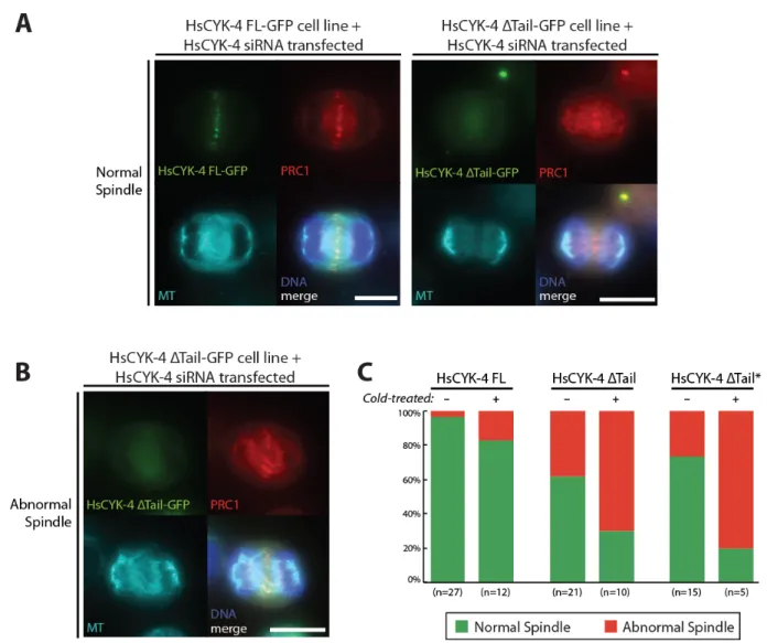
Figure 6. A) Fluorescence max intensity z-projections of fixed non-treated HsCYK-4 FL and ΔTail cells able to form normal central spindles (bar: 10 µm). B) Fluorescence max intensity z-projections of a fixed non-treated HsCYK-4 ΔTail cell showing an abnormal central spindle (bar: 10 µm). All cells stained with anti-GFP GFP-booster, anti-tubulin, anti-PRC1 antisera and Hoechst stain. C) Quantification of normal vs abnormal spindle morphologies found in HsCYK-4 FL, ΔTail and ΔTail* cell lines, both non-treated and cold-treated for 2 min prior to fixation.
18 Normal spindle morphologies were observed in all observed cell lines (Figure 6A), despite some problems in obtaining a correct microtubule fixing and staining in the HsCYK-4 FL cell lines, which exhibit a higher cytoplasmic background tubulin staining. The amount of abnormal spindles in HsCYK-4 ΔTail/ΔTail* cell lines, such as the example in Figure 6B, was much higher than in HsCYK-4 FL cells (non-treated cells in Figure 6C; 4% in FL cells against 38% and 27% in ΔTail and ΔTail* cell lines, respectively). The microtubules in abnormal spindles appear to be disorganized and not continuous, with gaps in distribution. This happens much more frequently in the HsCYK-4 ΔTail/ΔTail* cell lines, allowing us to securely conclude that the structure and stability of the central spindle is affected. If the microtubule stability is impaired on the cells lacking the C-terminal region of HsCYK-4 we would expect to see a decreased resistance to microtubule destabilizing treatments. So, we cold-treated the cells for 2 min before fixation – a treatment known to disrupt non-stabilised microtubules (Brinkley and Cartwright, 1975). This, as expected, increased the difference in the number of abnormal spindle morphologies between the HsCYK-4 FL and the HsCYK-4 ΔTail/ΔTail* cell lines (cold-treated cells in Figure 6C), with the former cells exhibiting a total amount of 17% abnormal spindles and the latter ones a total of 70% (ΔTail) and 80% (ΔTail*). This leads us to conclude that the C-terminal tail region of HsCYK-4 has a key role in the stabilisation of microtubules in the central spindle, although it still remains to elucidate if this is due to the binding to PRC1 or it stabilises the microtubules directly (eg. via cross-linking by promoting centralspindlin’s formation of higher-order structures).
19
Discussion
Historically, functional studies of the components involved in cytokinesis through a genetics approach have been confined to C. elegans and Drosophila. Only with the arrival of RNAi by protein-specific siRNA about a decade ago, was this kind of approach applied to mammalian cells. Given this fact, cytokinesis in mammalian cells is severely understudied. In this work we put the focus back on these cells, more specifically in human cells. In this work HsCYK-4 role as one of the main intervening proteins in cell division has been secured. The increase in cytokinesis failure when depleting HsCYK-4 in a wild-type background is witness to this fact. Also, our results give strength to the notion that HsCYK-4 is involved directly or indirectly in a mitotic checkpoint, given the high amount of metaphase arrest found when it was depleted.
HsCYK-4 and PRC1 have both been heavily implied in cell division, specifically in the assembly and maintenance of the central spindle. The interaction between them as been severely understudied so far, only having been described in Ban et al. 2004 and more recently by Lee and Mishima (unpublished). Unfortunately the biochemical study of this interaction (eg. immunoprecipitation) is not an easy task and could not be replicated in this work despite various attempts. The difficulty in doing so was due to two reasons. Firstly, the nature of the HsCYK-4 protein, which in the cell exists mostly in complex with MKLP1, forming the centralspindlin complex. This complex is very hard to solubilise in vitro (Hutterer et al. 2009), so in order to maintain solubility while extracting it you need to use a very high salt concentration. Alas, high salt concentrations are not compatible with the binding between PRC1 and HsCYK-4. It was technically challenging to get around this problem, and it could not be accomplished. Second, the rod-like nature of PRC1 and amount of exposed residues means that in vitro it very easily forms non-specific interactions (data not shown).
Bearing this in mind, we opted for in vivo labelling of HsCYK-4 and PRC1 as well as using the transgenic construct of HsCYK-4 lacking the PRC1 interacting region to gain some insight on the influence of the HsCYK-4-PRC1 interaction on cell division. Depletion of the endogenous HsCYK-4 protein in the cell line carrying the HsCYK-4 ΔTail or HsCYK-4 ΔTail* construct brought forth almost the same degree of cytokinesis failure as the one observed for HsCYK-4 depletion in wild-type cells. This shows that the C-terminal region of
20 HsCYK-4 is absolutely essential for its function in the cell. This is due to a requirement of this region for an initial (pre-ingression) accumulation of HsCYK-4 at the middle plane of the cell, the position of the antiparallel bundles of microtubules of the central spindle. Possibly because the interaction with PRC1 will contribute to its anchoring to the microtubules and stabilisation at the middle of the central spindle. If this does not occur, less centralspindlin is available to bundle and cross-link microtubules required for the formation of a stable central spindle. After this early phase of HsCYK-4 accumulation at the centre of the central spindle, HsCYK-4 starts to accumulate near the ingressing cleavage furrow. This was evidenced by the formation of a cortical ring of HsCYK-4 accumulation in the HsCYK-4 ΔTail and HsCYK-4 ΔTail* cell lines, and also the addition of latrunculin A. Indeed, this allowed us to identify that the later accumulation is not only temporally separate from the earlier one, but also spatially distinct from it. This hints at a possible distinct molecular mechanism of centralspindlin accumulation cortically during ingression, which is also dependent on the C-terminal tail region of HsCYK-4. The latrunculin-A treatment, by inhibiting the assembly of the actomyosin contractile ring, would also impair the recruitment of a series of ingression-associated factors, such as anillin (Piekny and Glotzer, 2008). So, it is reasonable to assume that either the presence of the C-terminal tail of HsCYK-4 or the ingression of the cleavage furrow is sufficient to promote the cortical accumulation of HsCYK-4 in the dividing cell.
Inhibiting the ingressing cleavage furrow with cytochalasin D – another actin polymerisation inhibitor (Godette and Frieden, 1986) – gave somewhat similar results, but altered HsCYK-4 pattern of distribution at the cortical side in HsCYK-4 FL cells (data not shown). This might be due to differences in the action of the inhibitors. These results were not included in the work because further optimisation of the procedure and a higher number of replicates are needed. Nevertheless, the current results provide the first supporting evidence for the existence of two phases of HsCYK-4 accumulation during human cell division. This has been known to occur in larger cells, such as the C. elegans one-cell embryo (Bringmann and Hyman, 2005; Verbrugghe and White, 2007), but due to the much smaller size of mammalian cells it was assumed that such distinct phases did not occur.
Colocalisation of HsCYK-4 and PRC1 was heavily impaired in the absence of HsCYK-4 C-terminal tail region. Also, the formation of distinct foci of either HsCYK-4 or PRC1 was observed. Both molecules are able to locate to the middle of the central spindle in these cells, via distinct pathways. Despite this they failed to localise at the exact same positions in HsCYK-4 ΔTail cells. Thus, this leads us to conclude that the C-terminal tail of HsCYK-4 is required for stabilisation of these molecules at the same position in the middle of
21 the central spindle, and that possibility this is due to their direct interaction. The central spindle’s microtubule distribution was also disrupted in the absence of HsCYK-4-PRC1 interacting region. This allows us to conclude that the C-terminal tail region of HsCYK-4 is required for the formation and maintenance of an organised central spindle.
HsCYK-4, according to its (and orthologues CYK-4 in C. elegans and Tumbleweed in Drosophila) known roles as an essential subunit of the microtubule-bundling centralspindlin complex (Douglas and Mishima, 2010), is implicated in maintaining the integrity of the mitotic microtubule-based central spindle. This is supported by our data, being that the increase in anaphase long axis length and the early regression phenotype, both can be explained by a loss of integrity of the central spindle. Two cytokinesis failure phenotypes were observed: 1) an early regression, and 2) a late regression after midbody formation. The former seems to be caused by the impaired recruitment/accumulation of HsCYK-4 to the central spindle. The failure in the first early pathway of accumulation, leads to a lesser amount of HsCYK-4 being present to stabilise and bundle the microtubules. Also, the compromised interaction with PRC1 would also decrease the degree of bundling of the microtubules. This leads to the formation of a very weakened central spindle that is not able to resist forces exerted on it. Also, less RhoA activation is probably occurring which will impair proper actomyosin ring assembly. The late regression phenotype observed in the HsCYK-4 ΔTail/ΔTail* cell line was not observed in HsCYK-4 depleted wild-type HeLa cells. Although, since this phenotype typically occurs very late after cell division is completed and most cells in this work were imaged for only 1 h, it is expected that not all late regressions were detected, leading to an underestimation of the occurrence of this phenotype. Nevertheless, these results show an involvement of HsCYK-4 and more specifically the C-terminal tail region of HsCYK-4 in midbody stability or correct maturation. The centralspindlin complex is known to be able to form higher-order structures (Hutterer et al. 2009), and evidence points to the midbody being composed of a matrix of centralspindlin molecules that confer rigidity to this structure (Matuliene and Kuriyama, 2004). So, a reduced amount of centralspindlin would reduce the midbody’s resistance to external tensions, such as the force the recently ingressed membrane exerts on it. Another possibility we like to consider is that this is a novel role for the HsCYK-4-PRC1 interaction, conferring yet another way of cross-linking the microtubules and midbody matrix components, which would increase its resistance to external forces. HsCYK-4 therefore, confers resistance to the central spindle and midbody against the various forces that act on it, during the drastic shape changes that occur during cell division.
22
Materials and Methods
Cell Culture, Synchronisation and Drug Treatment
HeLa were grown in DMEM (Dulbecco’s modified eagle medium) supplemented with 10% (v/v) fetal bovine serum and 10U penicillin/streptomycin mix (Life Technologies) at 37°C with 5% CO2 in a humidified incubator. Stable transgenic cell lines (HsCYK-4 FL-GFP,
HsCYK-4 ΔTail, and HsCYK-4 ΔTail*) had already been established in lab, for selection 0.25 µg/mL of puromycin was added. For imaging, cells were transferred to a 2 cm Fluorodish (World Precision Instruments) and allowed to adhere to the substrate. When synchronisation was required, 2.5 mM of thymidine was added to cells (S-phase block), 24 h later the cells were washed. 3 h after the wash, nocodazole was added to the cells (mitotic block) until most of them were in a mitotic state. If for biochemistry, cells were washed and allowed to release for 50 min under incubation and then snap-frozen until used. If for imaging, cells were washed and imaged immediately after. Latrunculin-A was previously diluted in cell medium and added to the cells immediately before observation to a final concentration of 2 µM.
siRNA and Plasmid Transfection
HsCYK-4 siRNA used (catalogue number: HSS120934) was ordered from Life Technologies (LT), as it had been previously successfully used in the lab. RNAi treatment the corresponding protocol from LT was used, with Oligofectamine (LT) as the transfection reagent. The protocol was optimised as follows: 12 µL from the stock 20 nmol siRNA was diluted in 200 µL of OptiMEM (LT) and vortexed, and 12 µL of Oligofectamine was diluted in 48 µL of OptiMEM. PRC1 transient expression and dilution was done using a human cell-expressible pCMV (cytomegalovirus promoter)-carrying empty plasmid or with the mCherry-PRC1 fusion construct. Both were available in the lab. For transfection, Lipofectamine 2000 (LT) was used as the transfection reagent and the accompanying protocol was used, with the following optimisations: 2 ng of DNA was diluted in 100 µL of OptiMEM (LT) and vortexed, and 4 µL of Lipofectamine 2000 was diluted in 100 µL of OptiMEM. Cells were incubated for at least 6 h after plasmid transfection or 48 h after the siRNA transfection.
Imaging
For live cell imaging, the cells were maintained in an humidified incubator chamber for microscopy at 37°C with 5% CO2. Time-lapse and fixed cells were imaged in a DeltaVision
microscope (Olympus) for 1 h (or 3 h in the case of some wild-type HeLa cells), at 1 min 30 s intervals with 14 planes on the z-axis with a 1 µm interval.
23
References
Ban, R., Irino, Y., Fukami, K. & Tanaka, H. Human mitotic spindle-associated protein PRC1 inhibits MgcRacGAP activity toward Cdc42 during the metaphase. The Journal of biological chemistry 279, 16394–16402 (2004).
Barr, F. A. & Gruneberg, U. Cytokinesis: placing and making the final cut. Cell 131, 847–860 (2007).
Bieling, P., Telley, I. A. & Surrey, T. A minimal midzone protein module controls formation and length of antiparallel microtubule overlaps. Cell 142, 420–432 (2010).
Braun, M. et al. Adaptive braking by Ase1 prevents overlapping microtubules from sliding completely apart. Nature cell biology 13, 1259–1264 (2011).
Bringmann, H. & Hyman, A. A. A cytokinesis furrow is positioned by two consecutive signals. Nature 436, 731–734 (2005).
Brinkley BR, Cartwright J Jr. Cold-labile and cold-stable microtubules in the mitotic spindle of mammalian cells. Annals of the New York Academy of Sciences 253, 428-39 (1975).
Desai, A., Bowerman, B. & Oegema, K. Inhibition of Rac by the GAP Activity of Centralspindlin Is Essential for Cytokinesis. Science 322, 1543–1546 (2008).
Douglas, M. E. & Mishima, M. Still entangled: assembly of the central spindle by multiple microtubule modulators. Seminars in cell & developmental biology 21, 899–908 (2010). Eggert, U. S., Mitchison, T. J. & Field, C. M. Animal cytokinesis: from parts list to mechanisms. Annual review of biochemistry 75, 543–566 (2006).
Goddette, D. W. & Frieden, C. Actin polymerization. The mechanism of action of cytochalasin D. The Journal of biological chemistry 261, 15974–15980 (1986).
Hirose, K., Kawashima, T., Iwamoto, I., Nosaka, T. & Kitamura, T. MgcRacGAP is involved in cytokinesis through associating with mitotic spindle and midbody. The Journal of biological chemistry 276, 5821–5828 (2001).
Hu, C.-K., Coughlin, M., Field, C. M. & Mitchison, T. J. KIF4 regulates midzone length during cytokinesis. Current biology : CB 21, 815–824 (2011).
Hutterer, A., Glotzer, M. & Mishima, M. Clustering of centralspindlin is essential for its accumulation to the central spindle and the midbody. Current biology : CB 19, 2043–2049 (2009).
Jantsch-Plunger, V. et al. CYK-4: A Rho family gtpase activating protein (GAP) required for central spindle formation and cytokinesis. The Journal of cell biology 149, 1391–1404 (2000). Jiang, W. et al. PRC1: a human mitotic spindle-associated CDK substrate protein required for cytokinesis. Molecular cell 2, 877–885 (1998).
Kitamura, T. et al. Role of MgcRacGAP/Cyk4 as a regulator of the small GTPase Rho family in cytokinesis and cell differentiation. Cell structure and function 26, 645–651 (2001).
24 Kurasawa, Y., Earnshaw, W. C., Mochizuki, Y., Dohmae, N. & Todokoro, K. Essential roles of KIF4 and its binding partner PRC1 in organized central spindle midzone formation. The EMBO journal 23, 3237–3248 (2004).
Lagana, A. et al. A small GTPase molecular switch regulates epigenetic centromere maintenance by stabilizing newly incorporated CENP-A. Nature Publishing Group 12, 1186– 1193 (2010).
Lee, J.-S., Kamijo, K., Ohara, N., Kitamura, T. & Miki, T. MgcRacGAP regulates cortical activity through RhoA during cytokinesis. Experimental cell research 293, 275–282 (2004). Ma, H. T. & Poon, R. Y. C. How protein kinases co-ordinate mitosis in animal cells. The Biochemical journal 435, 17–31 (2011).
Manders, E., Verbeek, F. & Aten, J. Measurement of co‐localization of objects in dual‐colour confocal images. Journal of Microscopy 169, 375–382 (1993).
Matuliene, J. & Kuriyama, R. Role of the midbody matrix in cytokinesis: RNAi and genetic rescue analysis of the mammalian motor protein CHO1. Molecular biology of the cell 15, 3083–3094 (2004).
Mishima, M., Pavicic, V., Gr backslash backslash uneberg, U., Nigg, E. A. & Glotzer, M. Cell cycle regulation of central spindle assembly. Nature 430, 908–913 (2004).
Mishima, M., Kaitna, S. & Glotzer, M. Central spindle assembly and cytokinesis require a kinesin-like protein/RhoGAP complex with microtubule bundling activity. Developmental cell 2, 41–54 (2002).
Mollinari, C. et al. PRC1 is a microtubule binding and bundling protein essential to maintain the mitotic spindle midzone. The Journal of cell biology 157, 1175–1186 (2002).
Morton, W. M., Ayscough, K. R. & McLaughlin, P. J. Latrunculin alters the actin-monomer subunit interface to prevent polymerization. Nature cell biology 2, 376–378 (2000).
Mullins, J. M. & McIntosh, J. R. Isolation and initial characterization of the mammalian midbody. The Journal of cell biology 94, 654–661 (1982).
Narumiya, S. & Yasuda, S. Rho GTPases in animal cell mitosis. Current Opinion in Cell Biology 18, 199–205 (2006).
Nishimura, Y. & Yonemura, S. Centralspindlin regulates ECT2 and RhoA accumulation at the equatorial cortex during cytokinesis. Journal of cell science 119, 104–114 (2006).
Piekny, A. J. & Glotzer, M. Anillin Is a Scaffold Protein That Links RhoA, Actin, and Myosin during Cytokinesis. Current Biology 18, 30–36 (2008).
Somers, W. G. & Saint, R. A RhoGEF and Rho family GTPase-activating protein complex links the contractile ring to cortical microtubules at the onset of cytokinesis. Developmental cell 4, 29–39 (2003).
Subramanian, R. et al. Insights into antiparallel microtubule crosslinking by PRC1, a conserved nonmotor microtubule binding protein. Cell 142, 433–443 (2010).
Verbrugghe, K. J. C. & White, J. G. Cortical centralspindlin and G have parallel roles in furrow initiation in early C. elegans embryos. Journal of cell science 120, 1772–1778 (2007). Yüce, O., Piekny, A. & Glotzer, M. An ECT2-centralspindlin complex regulates the localization and function of RhoA. The Journal of cell biology 170, 571–582 (2005).
25 Yarmola, E. G. Actin-latrunculin A structure and function: differential modulation of actin-binding protein function by latrunculin A. Journal of Biological Chemistry (2000).
Zhu, C., Lau, E., Schwarzenbacher, R., Bossy-Wetzel, E. & Jiang, W. Spatiotemporal control of spindle midzone formation by PRC1 in human cells. Proceedings of the National Academy of Sciences of the United States of America 103, 6196–6201 (2006).
Zhu, C. & Jiang, W. Cell cycle-dependent translocation of PRC1 on the spindle by Kif4 is essential for midzone formation and cytokinesis. Proceedings of the National Academy of Sciences of the United States of America 102, 343–348 (2005).
26
Annex
Supplemental Materials and Methods
Plasmid Expression and Purification
Plasmids used were already available in the lab and were amplified in E. coli cells of the DH5α strain. These cells were transformed by the addition of approximately 100ng of DNA via heat-shock (incubation on ice for 20 min followed by exposure to 42ºC for 45 s) and allowed to grow in LB (Luria-Bertrani) standard medium supplemented with an appropriate selection antibiotic (either kanamycin or ampicillin at 37°C. After selection and growth, a single colony was isolated and allowed to grow under the same conditions. Then, the plasmid was extracted and purified through the use of a QIAGEN Plasmid MidiKit as described in the manufacturer’s protocol. All amplified plasmids were verified by DNA electrophoresis of undigested and digested (with appropriate restriction enzymes; XhoI and XbaI were used) samples.
DNA electrophoresis
Standard 1% agarose gels supplemented with 0.1mg/ml ethidium bromide, to allow visualisation of the DNA, were used. These were prepared with 1x Tris-acetate-EDTA. The samples were mixed with loading buffer before loading into wells. Electrophoresis was done at 100V. DNA bands were observed under UV illumination.
SDS-PAGE and Western Blotting
If using cell lysates, these were pre-cleared by centrifugation prior to electrophoresis. Protein electrophoresis was done using a standard SDS-polyacrylamide gel (7.5% polyacrylamide mix). Then, the proteins were electrotransferred to a nitrocellulose membrane (HiBond ECL; Amersham). All antibody incubations, blocking and washing of membranes (occurred between each step) were done in a TBST solution (0.1%Tween). Membranes were blocked