Ana Rita da Silva Ferreira
Evaluation of immune-determinants associated with
resistance/susceptibility to
Paracoccidioides brasiliensis
:
a mouse model perspective
Avaliação dos determinantes imunes associados à
resistência/susceptibilidade a
Paracoccidioides brasiliensis
: uma
perspectiva do modelo de ratinho
Dissertação de Mestrado
Mestrado em Ciências da Saúde
Trabalho efectuado sob a orientação do
Doutor Egídio Manuel Pires Torrado
e do
Doutor Fernando José dos Santos Rodrigues
DECLARAÇÃO
Nome: Ana Rita da Silva Ferreira
Endereço electrónico: [email protected] Telefone: 915452409
Cartão do Cidadão: 14138777
Título da dissertação: Evaluation of immune-determinants associated with resistance/susceptibility to Paracoccidioides brasiliensis : a mouse model perspective
Orientadores:
Doutor Egídio Manuel Pires Torrado
Doutor Fernando José dos Santos Rodrigues
Ano de conclusão: 2015
Designação Ramo de Conhecimento do Mestrado: Ciências da Saúde
DE ACORDO COM A LEGISLAÇÃO EM VIGOR, NÃO É PERMITIDA A REPRODUÇÃO DE QUALQUER PARTE DESTA TESE.
Universidade do Minho, _____/_____/_________
The work presented in this thesis was done in the Microbiology and Infection Research Domain of the Life and Health Science Research Institute (ICVS), School of Health Science, University of Minho, Braga, Portugal (ICVS/3B’s- PT Government Associate Laboratory, Braga/Guimarães, Portugal).
ACKNOWLEDGMENTS
Durante estes dois anos, foram muitas as pessoas que, directa ou indirectamente, contribuíram para a realização deste trabalho. A todas elas, o meu mais sincero agradecimento.
Ao professor Fernando Rodrigues, por me ter recebido de braços abertos no seu grupo e por ter aceitado o desafio como meu orientador. Um muito obrigado por todo o apoio profissional e por ter acreditado em mim, mesmo nos momentos mais complicados. Agradeço todos os conselhos profissionais, todo o conhecimento científico transmitido ao longo deste percurso e principalmente por me ter mostrado novos caminhos.
Um agradecimento muito especial para o Egídio Torrado, pois, sem ele, este trabalho não seria possível. Obrigada por todos os ensinamentos transmitidos, pelo incentivo, pela confiança, paciência e principalmente por toda a dedicação dada a este projecto para que este fosse realizado com o maior sucesso! Agradeço todos os conselhos científicos e todas as conversas e desafios lançados ao longo deste ano e que me fizeram crescer profissionalmente. Vou certamente levar isso comigo para a vida. Obrigada!
Ao Professor Gil Castro, pela colaboração neste projecto e por toda a disponibilidade para discutirmos os resultados dando sempre valioso ponto de vista cientifico.
Ao Instituto de Ciências da Vida e da Saúde, nomeadamente à Professora Cecília Leão e ao Professor Jorge Pedrosa, pela oportunidade de iniciar a minha carreira cientifica neste instituto e pelo acolhimento e apoio recebidos.
À Cristina Cunha, ao João Menino a ao Agostinho Carvalho por todos os conselhos científicos e pessoais dados ao longo deste ano. Agradeço todo o apoio e disponibilidade que se revelaram preciosos ao longo deste percurso.
Agradeço também à Filipa por todo o apoio dado ao longo desta tese. Obrigada pela disponibilidade em me ajudares e em transmitires o teu conhecimento!
A todos aqueles que fazem parte do I3.01 e do I3.02, em especial à Jéssica e ao Mark por me terem integrado no laboratório no inicio desta aventura no ICVS. Obrigada pelos ensinamentos, pelo apoio e pela amizade! Um agradecimento especial também ao Henrique, à Julia, à Fernanda, ao Fabrício, ao Relber, à Belém, à Ana e à Ângela por todos os momentos bem passados e por todo o apoio, tanto pessoal, como profissional.
À Rosinha, à Maria Luís, à Suze, à Marques ao André, à Patrícia e à Isabel por todos estes anos (que já vão muitos!) de amizade incondicional! Obrigada pelas conversas animadas, pelos momentos inesquecíveis e por pelo apoio quando mais precisava!
À Coelho, a minha eterna “partner”! Uma das melhores coisas que levo destes últimos dois anos é a nossa amizade! Obrigada por todo o carinho, por todo o apoio e obrigada por aquela palavra amiga que me alegrou sempre que precisava. Esta é, sem dúvida, uma amizade de levo para a vida!
Aos meus primos, Ju e Pedro, por todos os incontáveis jantares e por todos os momentos e conversas animadas! Desejo-vos o melhor do mundo! Obrigada pelo carinho, pelo apoio e por serem, mais que primos, uns irmãos para mim! Agradeço também ao meu padrinho, por todos os passeios e sessões de cinema que tão bem nos fizeram! Obrigada pelo apoio e pelo encorajamento!
Ao Diogo, a quem devo um agradecimento mais do que especial. Obrigada por tudo o que fizeste ao longo deste últimos dois anos. Obrigada pelo carinho, pelo apoio, e pelas palavras que vieram sempre na altura certa para me tornarem mais forte e para me incentivarem a tentar sempre cumprir o meu objectivo! Muito obrigada pelo imenso apoio profissional e pelo importante conhecimento cientifico que me transmitiste!
À minha mãe…sem dúvida o agradecimento mais importante. Obrigada por seres o meu apoio e por me incentivares a ser cada vez melhor e a chegar cada vez mais longe! És um exemplo de força e determinação! Obrigada pelo carinho, pelas palavras e por todo o esforço que fizeste e que me tornou naquilo que sou hoje!
A
BSTRACTParacoccidioides brasiliensis is the etiological agent of paracoccidioidomycosis, one of the most prevalent systemic mycosis in Latin America. Infection by this fungus is thought to be initiated by the inhalation of conidia produced by the saprophytic phase of the fungus. Although many aspects of the host immune response against this pathogen are still unknown, several studies have suggested that macrophages play a key role in the defence against P. brasiliensis. Therefore, our aim is to characterize the macrophage response against strains of P. brasiliensis with different degrees of virulence in order to define the mechanisms required to control infection. To this end, we have established an in vitro model of bone marrow-derived macrophages (BMDM) infection. These cells were infected with P. brasiliensis 18, a highly virulent strain, or P. brasiliensis ATCC60855, a low virulent strain. Here, we show that P. brasiliensis morphology, assessed by scanning electron microscopy, has an important impact in the ability of BMDM to internalize P. brasiliensis. Indeed, while P. brasiliensis ATCC60855, a small strain with a reduced bud number, is easily internalized by BMDM, P. brasiliensis 18, a larger strain with multiple bud cells, is not efficiently internalized.
To determine the ability of macrophages to control infection, we assessed the ability of BMDM to control P. brasiliensis growth. We found that, after infection, stimulation of BMDM with interferon (IFN)-γ was not sufficient to activate the production of nitric oxide (NO), a key antimicrobial mediator, and to control P. brasiliensis growth. To define the mechanisms underlying this deficient activation in response to this pathogen, we measured the levels of several cytokines known to activate macrophages and we found that there was a reduced production of tumour necrosis factor (TNF), which was more evident after P. brasiliensis 18 infection. Indeed, it was only after stimulation of BMDM with both IFN-γ and lipopolysaccharide (LPS), a potent inducer of TNF, that these cells were able to produce large amounts of nitric oxide and to control infection. Therefore, our data suggest that P. brasiliensis modulates the signalling pathway that leads to the production of TNF to inhibit the activation of the macrophages fungicidal mechanisms. We also found that the reduced production of TNF by BMDMs is not mediated by interleukin (IL)-10, as both P. brasiliensis strains induced the same levels of IL-10. However, in vivo studies suggest that high levels of IL-10 during infection can be detrimental to the host by modulating the dynamics of the inflammatory response, stressing the importance of the regulation of this cytokine in the balance between protection and pathology during P. brasiliensis infection.
Altogether, our data support a model whereby the different induction of TNF by P. brasiliensis strains with different degrees of virulence may underlie the different forms or severity of the disease.
R
ESUMOParacoccidioides brasiliensis é o agente etiológico da paracoccidioidomicose, uma das micoses sistémicas mais prevalentes na América Latina. Pensa-se que a infecção por este fungo é iniciada aquando da inalação de conídios produzidos pela fase saprófita do fungo. Embora vários aspectos relacionados com a resposta imune do hospedeiro ainda sejam desconhecidos, estudos sugerem que os macrófagos têm um papel essencial na defesa contra P. brasiliensis. O nosso objectivo é caracterizar a resposta dos macrófagos contra estirpes de P. brasiliensis com diferentes graus de virulência de forma a definir os mecanismos necessários para controlar a infeção. Para isso, estabelecemos um modelo in vitro de infecção de macrófagos derivados da medula-óssea (BMDM). Estas células foram infectadas com P. brasiliensis 18, uma estirpe altamente virulenta, ou com P. brasiliensis ATCC60855, uma estirpe considerada menos virulenta. Através de uma análise por microscopia eletrónica de varrimento, observámos que a morfologia de P. brasiliensis tem um grande impacto na internalização deste fungo pelos BMDM. De fato, enquanto que P. brasiliensis ATCC60855, uma estirpe com células de dimensão reduzida e rodeadas por poucas células-filha, é mais facilmente internalizada, a estirpe P. brasiliensis 18, que é caracterizada por células de maiores dimensões e rodeadas numerosas células-filhas, dificilmente é internalizada. Para determinar a capacidade dos macrófagos para controlar a infecção, avaliamos a capacidade dos BMDM para controlar o crescimento de P. brasiliensis. Descobrimos que, após a infeção, a estimulação destes macrófagos com interferão (IFN)-γ não foi suficiente para activar a produção de óxido nítrico, um mediador chave na resposta microbicida dos macrófagos, nem para controlar o crescimento de P. brasiliensis. Para definir os mecanismos subjacentes a esta activação deficiente, medimos várias citoquinas que são produzidas em resposta â activação de macrófagos e observamos uma diminuição na produção de fator de necrose tumoral (TNF) que foi mais evidente após a infecção com P. brasiliensis 18. Assim, só depois da estimulação dos BMDM com IFN-γ e LPS, um potente indutor de TNF, é que estas células foram capazes de produzir grandes quantidades de óxido nítrico e de controlar a infecção. Desta forma, os nossos dados sugerem que P. brasiliensis modula a via de sinalização que leva à produção de TNF de forma a inibir a activação dos mecanismos fungicidas dos macrófagos. Também observamos que a produção de TNF não é mediada pela interleuquina (IL)-10, já que ambas as estirpes produzem as mesmas quantidades de IL-10 Contudo, estudos in vivo mostram que a IL-10 tem um impacto negativo na dinâmica da resposta inflamatória, o que demonstra a importância da regulação desta citoquina no balanço entre a inflamação e a patologia. Estes dados suportam um modelo no qual a indução de TNF
por estirpes de P. brasiliensis com diferente virulência pode ser a base das diferentes formas ou severidade da doença.
T
ABLE OF CONTENTSAcknowledgments ... iii
Abstract ... vii
Resumo ... ix
Figure index ... xiii
Abbreviations ... xv
Introduction ... 1
1.1 PARACOCCIDIOIDES BRASILIENSIS BIOLOGY ... 3
1.1.1 Phylogeny and cryptic speciation ... 3
1.1.2 Morphological characteristics ... 3
1.1.3 Ecology ... 4
1.2 PARACOCCIDIOIDOMYCOSIS (PCM) ... 6
1.2.1 Epidemiology: Demographics-Geographical Distribution ... 6
1.2.2 Pathobiology and clinical forms ... 7
1.2.3 Virulence factors ... 9
1.2.4 Diagnosis and treatment ... 10
1.3 IMMUNTITY TO P. BRASILIENSIS INFECTION ... 11
1.3.1 Innate immunity ... 11
1.3.2. Adaptive immunity ... 13
1.4 MODULATION OF THE IMMUNE RESPONSE: THE ROLE OF INTERLEUKIN (IL)-10 ... 16
1.4.1 IL-10 biology ... 16
1.4.2 IL-10 in Paracoccidioidomycosis ... 16
Aims ... 19
Materials and methods... 23
3.1 MICROORGANISMS AND CULTURE MEDIA ... 25
3.2 ANIMALS ... 25
3.3 CULTURE AND INFECTION OF BONE MARROW-DERIVED MACROPHAGES (BMDMs) ... 26
3.4 P. BRASILIENSIS PHAGOCYTOSIS ASSAYS ... 26
3.5 IMMUNOFLUORESCENCE ... 27
3.6 HEMATOXYLIN AND EOSIN STAINING ... 27
3.8 MEASUREMENT OF NITRIC OXIDE (NO) PRODUCTION ... 28
3.9 IN VIVO INFECTIONS ... 28
3.10 HISTOLOGICAL ANALYSIS ... 29
3.11 FLOW CYTOMETRY ANALYSIS ... 29
3.12 RNA EXTRACTION AND QUANTIFICATION ... 29
3.13 COMPLEMENTATY DNA (CDNA) SYNTHESIS ... 30
3.14 REAL-TIME POLYMERASE CHAIN REACTION (RT-PCR) ... 30
3.15 STATISTICAL ANALYSIS... 30
Results ... 33
4.1 PREPARATION OF P. BRASILIENSIS INOCULUM FOR IN VITRO AND IN VIVO INFECTIONS ... 35
4.2 TUMOR NECROSIS FACTOR (TNF) IS AN ESSENTIAL MEDIATOR FOR P. BRASILIENSIS CLEARANCE ... 37
4.3 BMDM WERE BETTER ABLE CONTROL P. BRASILIENSIS ATCC60855 INFECTION. ... 43
4.4 IL-10 DID NO IMPACT THE FUNGICIDAL ACTIVITY OF BMDM AGAINST P. BRASILIENSIS INFECTION ... 48
4.5 IL-10 OVEREXPRESSION IMPACTED CELL RECRUITMENT IN P. BRASILIENSIS INFECTED MICE ... 50
4.6 ESTABLISHING AN INTRANASAL MODEL OF INFECTION: MIMICKING THE NATURAL ROUTE OF P. BRASILIENSIS INFECTION ... 53
Discussion ... 55
Concluding remarks and future perspectives ... 63
FIGURE INDEX
Figure 1 Geographic distribution of the phylogenetic species of Paracoccidioides genus. ... 4
Figure 2 Paracoccidioides brasiliensis mycelia and yeast forms ... 5
Figure 3 Geographic distribution of Paracoccidioidomycosis. ... 7
Figure 4 Acute form of PCM. ... 7
Figure 5 Chronic form of PCM. ... 8
Figure 6. Proposed model to explain the immune responses observed in the different forms of PCM. . 15
Figure 7 P. brasiliensis 18 presented a large size and multiple budding-cells. ... 35
Figure 8 Longer periods of sedimentation resulted in a less complex inoculum ... 36
Figure 9 BMDM internalized a higher percentage of yeast cells at a MOI of 1:2 and a period of incubation of 6 hours ... 37
Figure 10 BMDM presented reduced ability to internalize larger P. brasiliensis cells ... 38
Figure 11 IFN-γ-activated infected BMDM did not control infection.. ... 39
Figure 12 IFN-γ-activated infected BMDM did not produce nitrites ... 39
Figure 13 IFN-γ-activated infected BMDM produced lower levels of TNF………..40
Figure 14 IFN-γ- and LPS- activated BMDM produced high levels of TNF and NO ... 41
Figure 15 IFN-γ- and LPS-activated infected BMDM exhibited high fungicidal activity ... 42
Figure 16 P.brasiliensis ATCC60855 presented smaller and less complex cells ... 43
Figure 17 The complexity and heterology of the P. brasiliensis ATCC60855 inoculum was reduced during sedimentation.. ... 44
Figure 18 P. brasiliensis ATCC60855 was easily internalized by BMDM ... 45
Figure 19 BMDM presented improved fungicidal activity against P. brasiliensis ATCC60855 ... 46
Figure 20 No differences were observed in TNF expression from 1hour to 24 hours post-infection by BMD infected with P. brasiliensis 18 or P. brasiliensis ATCC60855 ... 47
Figure 21 IL-10 production was not affected by distinct P. brasiliensis strains. ... 48
Figure 22 WT and IL-10-/- BMDM exhibited the same ability to control P. brasiliensis ATCC60855 infection. ... 49
Figure 23 pMT-10 mice overexpressing IL-10 developed a more severe inflammatory reaction at week 8 post-infection reaction in the liver.. ... 50
Figure 24 IL-10 overexpression in P. brasiliensis- infected mice resulted in the recruitment of inflammatory monocytes and neutrophils ... 51 Figure 25 IL-10 overexpression resulted in the accumulation of MHC-II+ inflammatory monocytes.. ... 52 Figure 26 Histological sections of the liver and lung stained with HE from WT mice showed no inflammatory reactions ... 53 Figure 27 Different P. brasiliensis strains induce distinct macrophage responses ... 61
A
BBREVIATIONS˚C- Degree Celsius µg- Microgram µL- Microliter
AG- Aminoguanidine hemisulfate APC- Antigen presenting cell
ATCC- American Type Culture Collection ATP- Adenosine-5'-triphosphate
BHI- Brain heart infusion
BMDM- Bone marrow-derived macrophages BSA- Bovine serum albumin
cDNA- Complementaty deoxyribonucleic acid CLR- C-type lectin receptor
CO2 – Carbon dioxide
DCs- Dendritic cells
DC-SIGN- Dendritic cell specific intercellular adhesion molecule-3-grabbing non-integrin (DC-SIGN)
DMEM- Dulbecco’s Modified Eagle Medium DNA- Deoxyribonucleic acid
ECM- Extracellular matrix
ELISA- Enzyme-Linked Immunosorbent Assay FBS- Fetal bovine serum
FITC- Fluorescein isothiocyanate FoxP3- Forkhead box P3
GTP- Guanosine-5'-triphosphate H&E- Hematoxylin and eosin H3PO4 – Phosphoric acid
HIV- Human immunodeficiency virus HRP- Horseradish peroxidase Hsp- Heat shock protein IFN-γ- Interferon-gamma IL- Interleukin
iNOS- Inducible nitric oxide synthase IRF- Interferon regulatory factor LCCM- L-929 conditioned medium LPS- Lipopolysaccharide
MHC- Major histocompatibility complex
MIP2- Macrophage inflammatory protein 2-alpha mL- Milliliter
mM- Millimolar
MOI – Multiplicity of infection mRNA- Messenger ribonucleic acid MT- Sheep metallothionein
MyD88- Myeloid differentiation primary-response protein 88
NF-κB- Nuclear factor kappa-light-chain-enhancer of activated B cells
NK- Natural killer ng- Nanogram NO- Nitric oxide
NOS2- Nitric oxide synthase II
PAMP- Pathogen-associated molecular patterns PAS- Periodic acid-Schiff stain
PBS- Phosphate buffered saline PCM- Paracoccidioidomycosis PCR- Polymerase chain-reaction PE-Phycoerythrin
PFA - Paraformaldehyde solution pg - Picogram
PMNs- Polymorphonuclear neutrophils PRR- Pattern recognition receptor RNA- Ribonucleic acid
RNI- Reactive nitrogen intermediate ROS- Reactive oxygen species RT- Room temperature
TGF-β- Transforming growth factor β Th - T-helper
TIR - Toll interleukin receptor TLR- Toll-like receptor
TMB- 3,3′,5,5′-tetramethylbenzidine TNF- Tumour necrosis factor Treg- Regulatory T-cell WT- Wild type
CHAPTER I
1.1 PARACOCCIDIOIDES BRASILIENSIS BIOLOGY
1.1.1 Phylogeny and cryptic speciation
Morphological and phylogenetic studies together with the recent development of molecular tools place Paracoccidioides brasiliensis in the phylum Ascomycota, order Onygenales and family Ajellomycetacea,
a fungal group common to most agents of systemic mycosis, including Bastomyces dermatitidis, Coccidioides immitis, Coccidioides posadassii and Histoplasma capsulatum (Bagagli, Theodoro et al. 2008).
In classic systematics, P. brasiliensis is considered to be an imperfect fungus due to lack of sexual structures and a teleomorphic phase, despite the high genetic variability among different isolates (Montoya, Moreno et al. 1997, Bagagli, Theodoro et al. 2008). Indeed, through the analysis of eight regions from five nuclear coding genes, Matute et al. found that P. brasiliensis is stratified in at least three distinct phylogenetic species: i) species 1 (S1) is paraphyletic and found in Brazil, Argentina, Paraguay, Peru and Venezuela; ii) phylogenetic species 2 (PS2) is monophyletic and found in Brazil and Venezuela; and iii) phylogenetic species 3 (PS3), also monophyletic, is found in Colombia (Figure 1). Isolates belonging to S1 and PS2 are sympatric and recombinant, whereas those belonging to PS3 are allopatric and clonal (Matute, McEwen et al. 2006). Teixeira et al. suggested the existence of 17 genotypically similar isolates, including P. brasiliensis 01, which were distinct from the three phylogenetic species described above. In this regard, these authors proposed a new “Pb01-like-cluster” as a new Paracoccidioides species named Paracoccidioides lutzii. Isolates of the P. lutzii species are endemic to the Brazilian Central-Western region and differ from P. brasiliensis species in its virulence, resistance to fungicides and proliferation (Teixeira, Theodoro et al. 2009).
1.1.2 Morphological characteristics
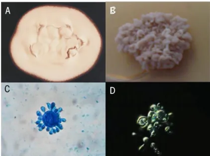
The morphological transformation of P. brasiliensis is based in the existence of two distinct morphological forms. At temperatures below 25ºC (in the environment), the most common form is the mycelium/conidial (Figure 2A), whereas at temperatures around 37ºC the mycelium/conidial form develops into the yeast form (Figure 2B), being the latter the pathogenic form. The mycelium/conidial form is composed by two layers: the outer layer, which is rich in β-1,3-glucan fibrils, and the inner layer, rich in rigid chitin fibrils (Brummer, Castaneda et al. 1993). In conditions of nutrient deprivation and low temperatures, different types of propagules, including arthroconidia or conidia, are formed
(McEwen, Restrepo et al. 1987, Edwards, Salazar et al. 1991). These propagules are thought to be
responsible for initiating infection after being inhaled by the host (Brummer, Castaneda et al. 1993).
Figure 1. Geographic distribution of the phylogenetic species of Paracoccidioides genus. Adapted from Teixeira et al., 2009
During mycelial-to-yeast transition, which is triggered by the temperature, there is a switch in glucan polymer linkage in the cell wall β-1,3-glucan to α-1,3-glucan. Indeed, the yeast form has a cell wall composed by α-1,3-glucan and small amounts of α-1,3- or α-1,6-glycosidic linkages (Kanetsuna, Carbonell et al. 1969). The most characteristic feature of the yeast form is the “pilot wheel” appearance (Figure 2C) that results from multiple budding cell divisions. However, single cells with only one bud, odd-like morphologies and transitional forms (cells arranged in short chains, balloon-shaped or broken cells) are also common. This form is multinucleated, with variable sizes ranging from 4 to 40 µm (Figure 2D) (Brummer, Castaneda et al. 1993).
1.1.3 Ecology
Although the environmental niches of P. brasiliensis are still largely unknown, the ecological characteristics of the endemic regions of the Colombian and Brazilian areas suggest that humid environments with rivers and rain forests are ideal for the maintenance of this pathogen in the environment (Bagagli, Franco et al. 2003). P. brasiliensis was firstly isolated by Adolfo Lutz from nine-banded armadillo (Dasypus novemcinctus), a wild mammal usually common in South America. This
suggesting active disease (Bagagli, Franco et al. 2003). However, the nature of the relationship between these two organisms is not yet completely understood (Naiff, Ferreira et al. 1986). Recently, P. brasiliensis was also detected in the naked-tailed armadillo (Cabassous centralis) and feces from fruit-eating bats (Artibeus lituratus) from Colombia and penguin excreta from Antarctica (Grose and Tamsitt 1965, Ferreira, Freitas et al. 1990, Giannini, Bueno et al. 1990, Corredor, Peralta et al. 2005).
Although advances have been made to define the ecological niche of P. brasiliensis, several environmental conditions, including temperature and the interaction with other soil organisms, remain largely unknown. It is therefore critical to define these conditions, as it will help determining how P. brasiliensis grows and disseminates in the host.
Figure 2. Paracoccidioides brasiliensis mycelia and yeast forms. Macroscopic characteristics of mycelial (A) and yeast form (B); Multiple budding cells observed in the yeast form (C); and Differential Interference Contrast (DIC) observation of yeast cells (D). Adapted from Menino, Saraiva et al. 2013
1.2 PARACOCCIDIOIDOMYCOSIS
Paracoccidioidomycosis (PCM) is a systemic endemic mycosis caused by P. brasiliensis. During the initial phase of infection, the pathogen affects the lungs subsequently disseminating to the mucosal membranes, skin, and many other organs including the liver and the spleen (Bonifaz, Vazquez-Gonzalez et al. 2011).
1.2.1 Epidemiology: Demographics-Geographical Distribution
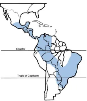
PCM is frequently reported in South- and Central-American countries. This disease is considered to be autochthonous from southern Mexico to northern Argentina with higher incidence in Brazil (accounting for more than 80% of the cases), Venezuela and Argentina (Figure 3). However, several cases have also been reported in Chile, Guyana, Belize and Caribbean Islands (Brummer, Castaneda et al. 1993). It has been reported an annual incidence rate of 1-3 per 100000 habitants and a mean mortality of 1.45 per million inhabitants, being the eighth cause of death from chronic infections and parasitic diseases (Calle, Rosero et al. 2001).
PCM affects mainly adults from ages between 30 and 60 years. In children and young adults, PCM is uncommon and indeed only 8% of patients are less than 20 years of age (Benard, Orii et al. 1994, Paniago, Aguiar et al. 2003). In a recent study of 5500 PCM patients, 5045 were male and only 455 were female (male-to-female ratio of 11.1 to 1), showing that gender is an important factor in this fungal disease (Shankar, Restrepo et al. 2011). In this regard, studies performed with experimental models of PCM infection showed that estrogens (17β-estradiol) inhibit P. brasiliensis mycelium-to-yeast transition, thus preventing infection (Shankar, Restrepo et al. 2011).
PCM infection is often (approximately 60% of the cases) observed in farmers or rural workers, and therefore it is thought to be associated with inhalation of dust (Calle, Rosero et al. 2001). Indeed, other occupations that are a risk factor for PCM are masonry, bricklaying and mining (Conti-Diaz, Calegari et al. 1979).
Although in some cases the propagules may become dormant, comorbidities including alcoholism, malnutrition, smoking or immunosuppressive diseases, including human immunodeficiency virus, may lead to active disease (Benard and Duarte 2000, Restrepo, Benard et al. 2008).
Figure 3. Geographic distribution of Paracoccidioidomycosis. Blue areas represent regions with high incidence, including Brazil, Venezuela and Colombia. Duane et al., 2013
1.2.2 Pathobiology and clinical forms
According to in vivo infections performed by McEwen et al., it is thought that infection by P. brasiliensis is initiated by the inhalation of fragments of mycelia or conidia, which are then phagocytosed by alveolar macrophages. The morphological transition to the pathogenic yeast form is started within only a few hours (12-18) followed by dissemination and induction of a progressive disease (McEwen, Bedoya et al. 1987). After this, and depending on host factors and virulence of the strain, the infectious process gives rise to asymptomatic infection or active disease.
Figure 4. Acute form of PCM. Lymphadenopathy and scars of previous suppurative lymph nodes on the cervical region (A). Ulcerative lesions on face (B). Adapted from Marques, 2012.
The asymptomatic infection (subclinical infection) is only detected by skin-tests and has no clinical manifestations. On the other hand, the active disease is characterized by pulmonary granulomatous lesions, where viable P. brasiliensis cells persist (named latent foci). During active disease, dissemination of the fungus can lead to the formation of extrapulmonary latent foci in the liver, spleen, lymph nodes, skin, and adrenal glands (Restrepo, Benard et al. 2008).
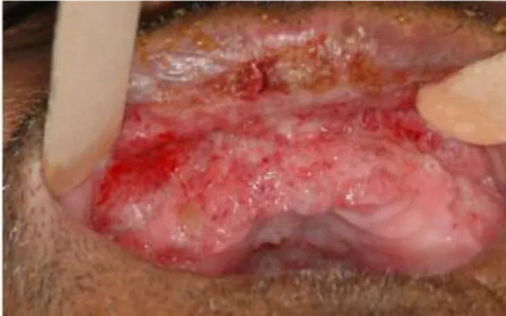
The two most common forms of the active disease are the juvenile form and the chronic form. The juvenile form is considered the most severe form of the disease representing only 3 to 5% of all cases. This form is characterized by a rapid course of infection (weeks to months) leading to reticuloendothelial system organ hypertrophy and bone marrow dysfunction, which results in a septicemic episode (Brummer et al., 1993). Fever, weight loss, lymph node enlargement and hepatosplenomegaly are the most common signs and symptoms (Figure 4). There are no clinical or radiological manifestations of this form of the disease in the lungs (de Mattos, Mendes et al. 1991).
Figure 5. Chronic form of PCM. Ulcerative and infiltrative lesions with multiple hemorrhagic dots and crusts on the upper gingiva and lip. From Marques, 2012.
The chronic form of the disease is more common occurring in more than 90% of the patients, most of them adult males. The disease progresses slowly, taking months or years to become fully established. Unlike the juvenile form, in the chronic form, pulmonary manifestations are evident in 90% of the patients. This form of disease can involve a single organ or system (unifocal) or several organs (multifocal) including lungs, oropharynx, skin, lymph nodes, adrenal glands and the upper respiratory tract. Other organs such as the eyes, central nervous system, bones and testicles are less frequently affected (Figure 5) (Colombo, Faical et al. 1994).
1.2.3 Virulence factors
Virulence factors are described as specific characteristics of a pathogenic microorganism that improve its ability to invade the host, leading to infection (Mendes-Giannini, Taylor et al. 2000).
Over the past few years, several virulence factors of P. brasiliensis have been described, including the fungus dimorphism, the adherence to host cells and the cell wall components. San-Blas showed that the cell wall polysaccharides play a crucial role in the protection of the fungus against host mechanisms (San-Blas, 1985). This author observed that, during mycelia-to-yeast transition, the chitin content of the cell wall increases 3-fold, being α-1,3-glucan the most abundant glucoside in the yeast form, while in the mycelial form, the most common glucoside is β-1,3-glucan. Since the reduction of β-glucan was associated with a reduction in virulence, these data suggest an important role of this polysaccharide in the virulence of P. brasiliensis (San-Blas, 1985).
The 43 kDa immunodominant glycoprotein Gp43 is expressed by most P. brasiliensis strains and has also been described as an important virulence factor (Torres, Hernandez et al. 2013). This protein is considered to be an adhesin that binds to laminin and fibronectin (Vicentini, et al. 1994). Several studies suggest that Gp43 is capable of inhibiting macrophage phagocytosis leading to P. brasiliensis dissemination (Popi, Godoy et al. 2008).
Another important virulence factor that has been extensively studied is Cdc42, a protein that belongs to a small Rho-like GTPase family responsible for important biological processes in several organisms (Osmani, Peglion et al. 2010). This protein has a key role as a polarity cue molecule during P. brasiliensis growth and morphogenesis. Almeida et al. suggested that an 88% reduction in PbCDC42 expression led to a more homogenous cell shape and size, and more importantly, to a significant reduction in P. brasiliensis virulence. This was observed upon increased survival after intravenous infection of mice as well as more efficient phagocytosis by bone-marrow derived macrophages (BMDM) (Almeida, Cunha et al. 2009). While these data point to an important role of P. brasiliensis cell size and shape in the virulence of the fungus, it has been shown that this pathogen has also developed mechanisms to protect itself against oxidative burst of the host macrophages. Indeed, the production of melanin, a multifunctional polymer ubiquitous to all P. brasiliensis strains, protects this pathogen from nitrogen and oxygen intermediate radicals (Taborda, da Silva et al. 2008).
1.2.4 Diagnosis and treatment
The most effective and inexpensive diagnosis method of PCM is the direct examination of clinical samples from bronchoalveolar lavage fluids or the outer edge of ulcers in tissue biopsies. In these samples, P. brasiliensis is stained with potassium hydroxide or calcofluor. Other forms of diagnosis are based on the histological examination of tissue after silver methenamine or periodic acid-Schiff staining (PAS) (Del Negro, Pereira et al. 2000). The isolation and growth of P. brasiliensis from PCM patients is done in Sabouraud medium, supplemented with antibiotic drugs and mold inhibitors, with an incubation time of 20 days (Brummer et al., 1993).
During the last decade, several methods have been developed to detect P. brasiliensis, including polymerase chain reaction (PCR) and real-time PCR. These methods are used to identify DNA in clinical and environmental isolates (Motoyama, Venancio et al. 2000), and serologic reagents to detect Gp43, Gp70 and Hsp70 from P. brasiliensis (de Camargo and de Franco 2000).
Until 1946, this disease was considered incurable. However, new therapeutic drugs were discovered and used in patients with PCM, including sulphonamide and amphotericin B. The use of sulphonamide started in 1946 and has several advantages, including low cost and low toxicity. This drug can be administrated for a long period of time (up to 5 years), with an efficacy of about 25%. Amphotericin B, introduced in 1958, is more frequently used due to its efficiency and duration of treatment (6 months to 2 years) (Brummer, Castaneda et al. 1993).
1.3 IMMUNTITY TO P. BRASILIENSIS INFECTION
The mammalian immune system is composed by the innate system and the adaptive system (Mogensen 2009). The innate immunity confers the first line of defense to infection and is initiated by the interaction between pathogens and pattern recognition receptors (PRRs). The adaptive immunity, in turn, is specific since it activates effector mechanisms in an antigen-specific manner (Medzhitov 2007).
1.3.1 Innate immunity
Innate immunity is the first line of defense, comprising both chemical elements and cells components of the immune system responsible for defending the host from invading pathogens (Calich, da Costa et al. 2008).
The first line of innate mechanisms are the physical barriers, including skin, mucous membranes and ciliated epithelium of the respiratory tract, that prevent pathogens from entering the organism (Beutler 2004, Calich, da Costa et al. 2008). However, if a pathogen breaches these barriers, the innate immunity provides a non-specific response that is characterized by the presence of cellular components such as phagocytes (neutrophils, macrophages and dendritic cells), innate lymphoid cells (natural killer (NK) cells and NKT cells), mast cells and eosinophils (Blanco and Garcia 2008, Mogensen 2009). These cellular components are capable of detecting invading organisms due to the presence of conserved, transmembrane or intracytoplasmatic receptors generally known as pattern recognition receptors (PRRs). PRRs recognize specific molecular structures shared by groups of microorganisms called pathogen-associated molecular patterns (PAMPs) (Janeway and Medzhitov 2002). Upon recognition, adaptor molecules, such as myeloid differentiation primary-response protein 88 (MyD88) and TIR domain-containing adaptor inducing interferon (IFN)-β (TRIF), transmit the signal downstream in order to activate pathways including nuclear factor kappa-light-chain-enhancer of activated B cells (NF-κB) and mitogen-activated protein kinases (MAPKs) (Akira, Uematsu et al. 2006, Kawai and Akira 2007). These cascades ultimately lead to the induction of antimicrobial responses (pro-inflammatory cytokines) and the activation of phagocytic cells (macrophages and polymorphonuclear cells) (Mogensen 2009).
Innate immunity to P. brasiliensis
In P. brasiliensis infection, there are two main groups of PRRs responsible for the recognition of this pathogen: Toll-like receptors (TLRs) and C-type lectin receptors. Among these, dectin-1, dectin-2,
mannose receptor, dendritic cell specific intercellular adhesion molecule-3-grabbing non-integrin (DC-SIGN), TLR2, TLR4 and TLR9 were shown to be involved in P. brasiliensis recognition (Menino, Saraiva et al. 2013, Richardson and Moyes 2015). Specifically, recognition is responsible for the activation of cells, including polymorphonuclear neutrophils (PMNs) and macrophages.
In recent years, it has been shown that P. brasiliensis is able to invade host cells due to the presence of laminin receptors and the glycoprotein Gp43 that behaves as an adhesin. These molecules interact with components of the extracellular matrix (ECM), such as fibronectin, laminin and fibrinogen, allowing P. brasiliensis to manipulate the host cells environment to favour their own growth and survival (Fortes, Miot et al. 2011, Torres, Hernandez et al. 2013). Indeed, it has been shown that, during P. brasiliensis infection, PMNs lose their ability to produce reactive oxygen species (ROS), one of the most important antimicrobial functions of these cells, leading to P. brasiliensis dissemination (Meloni-Bruneri, Campa et al. 1996). Although the mechanisms required to control P. brasiliensis are not completely understood, it is thought that macrophages play a critical role during P. brasiliensis infection. These cells produce tumour necrosis factor (TNF), a key cytokine for the activation of important macrophage antimicrobial mechanisms, including the production of nitric oxide (NO), and the formation and organization of the granuloma (Brummer, Hanson et al. 1989, Moscardi-Bacchi, Brummer et al. 1994, Murray and Nathan 1999, Gonzalez, de Gregori et al. 2000, Bernardino, Pina et al. 2013). In P. brasiliensis granuloma, macrophages appear to surround the yeast cells. These macrophages are then submitted to a process of maturation and structural modifications mediated by cytokines leading to their differentiation into epithelioid and giant cells. Neutrophils, NK cells and B cells are also present in the vicinity of P. brasiliensis yeast cells. Fava Netto and co-workers classified these lesions into two types: (1) a benign localized infection, where granuloma is compact and presents few P. brasiliensis cells; and (2) disseminated infection, with loose granulomatous inflammation and extensive areas of necrosis. The granulomatous structures generated after P. brasiliensis infection are composed of epithelial tubercles with central areas of necrosis, aggregates of polymorphonuclear leukocytes, a lymphomononuclear halo and fibrosis (de Camargo and de Franco 2000). Although the granulomatous response is thought to be required to control extrapulmonary dissemination of infection, this also leads to the formation of tissue scarring, often resulting in fibrosis (Moreno and Guzman de Espinosa 1976, Cock, Cano et al. 2000). During pulmonary P. brasiliensis infection, the development of fibrosis is initiated at week 8 and culminates at week 12-16 with the consolidation of the damaged tissue. In most cases this contributes to the death of the patient (Vilani-Moreno, Fecchio et al. 1998).
As described above, TNF is also critical to activate the antimicrobial activity of macrophages (Riipi and Carlson 1990, Jain, Evans et al. 2006). Indeed, several studies have suggested that monocytes stimulated with TNF produced more nitric oxide and exhibited more effective fungicidal activity (Carmo, Dias-Melicio et al. 2006, Moreira, Dias-Melicio et al. 2008). Nitric oxide (NO) is a potent macrophage antimicrobial mediator that is generated from amino acid L-arginine by the inducible isoform of the nitric oxide synthase (iNOS). In P. brasiliensis infection, the role of nitric oxide is still not completely understood. Indeed, while some studies point to an important role of NO in the control of P. brasiliensis growth, other studies suggest that NO may induce immunosuppression by inhibiting lymphoproliferation, MHC class II molecule expression and TNF production (Nascimento, Calich et al. 2002). It is therefore crucial to understand the relationship between TNF and NO in P. brasiliensis infection and to determine how these mediators impact macrophage ability to control infection.
1.3.2. Adaptive immunity
The adaptive immunity is initiated with the presentation of antigens by antigen-presenting cells (APCs), to naïve T-cells. APCs are sentinels of the immune system and include dendritic cells (DCs), macrophages and B cells (Hamilos 1989).
Upon pathogen recognition, APCs are activated and internalize pathogens as well as their antigens, migrating to the draining lymph nodes. During this process, APCs upregulate the expression of molecules presented in the surface of these cells, including the major histocompatibility complex (MHC) class II and costimulatory molecules (CD40, CD80 and CD84). Moreover, these cells also secrete cytokines such as interleukin (IL)-12 and IL-23 that will promote the differentiation of CD4+ T-cells into
different subpopulations (Parker 1993, Richardson and Moyes 2015). The T helper (Th) subsets Th1, Th2 and Th17 cells are considered to be the most important Th cells during fungal infections (Porter, Roberts et al. 2011, de Castro, Ferreira et al. 2013).
IL-12 produced by APCs, in combination with IFN-γ, leads to the induction of a Th1 phenotype (Mosmann and Coffman 1989, de Castro, Ferreira et al. 2013). Th1 cells are characterized by the expression of IFN-γ, TNF and IL-2 and are critical to control intracellular pathogens. On the other hand, Th2 cells are differentiated in the presence of IL-4 and produce mainly IL-4, IL-5 and IL-13, which are important in immunity to extracellular pathogens (Noelle and Snow 1992).
Th17 cells are the newest subpopulation of T-cells with an important role in fungal diseases (de Castro, Ferreira et al. 2013). These cells, which are differentiated in the presence of IL-1, transforming growth factor (TGF)-α and IL-6, are characterized by the production of IL-17A, IL-17F, and IL-22 (Mosmann and
Coffman 1989). Once activated, Th17 cells stimulate epithelial cells, macrophages and endothelial cells to produce cytokines and chemokines (CXCL8, IL-1β, IL-23, and IL-6), and also antimicrobial peptides (such as defensins) (Richardson and Moyes 2015). Although these CD4+ T-cell phenotypes are
important to control different infections, exacerbated Th1 responses induce immunopathology whereas excessive Th2 responses cause allergic inflammatory diseases (Zhu and Paul 2008). Nevertheless, the activity of these Th phenotypes is tightly regulated by regulatory T-cells (Treg). This specific subpopulation is characterized by the expression of the transcription factor Foxp3, and produce IL-10 and transforming growth factor (TGF)-β (Silva, Sotto et al. 2013).
Adaptive immunity to P. brasiliensis
To date, many aspects of the adaptive immune response against P. brasiliensis are still unknown. However, strong evidences suggest that, in humans, Th1 responses are associated with resistance to P. brasiliensis infection, whereas Th2 responses are associated with susceptibility (de Castro, Ferreira et al. 2013). Similarly, it has been shown that, in mice, resistance to P. brasiliensis infection is associated with high IL-2 and IFN-γ responses while susceptibility is associated with high secretion of IL-5, which inhibits IFN-γ production. Moreover, it has also been suggested that IL-10 is equally produced by both resistance and susceptible mice strains, suggesting the important role of this cytokine in regulating inflammation (Kashino, Fazioli et al. 2000).
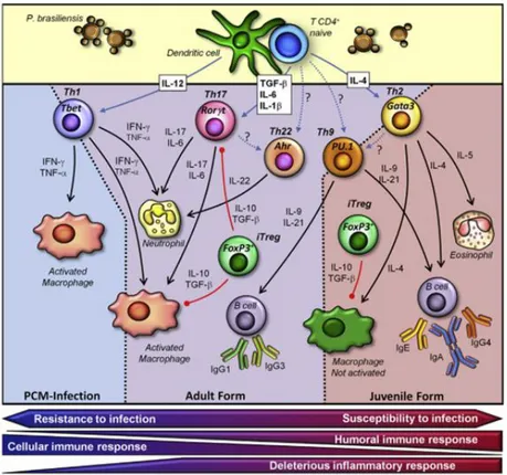
The different clinical forms of this disease are also associated with different Th-subsets. The juvenile form, the severest form of the disease, presents a predominant Th2 response with increased production of IL-4, IL-10, IL-5 and specific antibodies (such as IgG4, IgE and IgA). On the other hand, the sub-clinical form is characterized by a Th1 response, with the production of inflammatory cytokines such as IFN-γ, IL-12 and TNF. Patients presenting the chronic form of the disease develop a heterogeneous immunological response, as they produce cytokines associated with both Th1 and Th2 response, including TNF, IFN-γ, IL-12, IL-4 and IL-10 (Benard and Duarte 2000, Mamoni and Blotta 2006) (Figure 6).
In addition to Th1 and Th2 cells, Th17 cells are a new subpopulation of effector CD4+ T-cells that play a
key role during P. brasiliensis infection (de Castro, Ferreira et al. 2013). During C. albicans infection, several studies suggested that the activation of dectin-1 results in the induction of Th17 cells and the production of IL-23 and IL-17. These cytokines are responsible for mediating neutrophil recruitment to the peripheral inflammatory sites resulting in fungal clearance (Hernandez-Santos and Gaffen 2012). In
neutrophils to the site of infection and improved P. brasiliensis control (Loures, Araujo et al. 2014). However, it has been shown that an exacerbated Th17 response can also result in tissue damage and fibrosis, highlighting the existence of regulators that control the balance between protection and immunopathology (de Castro, Ferreira et al. 2013).
Figure 6. Proposed model to explain the immune responses observed in the different forms of PCM. Different forms of PCM present distinct immune responses elicited by P. brasiliensis infection. Adapted from de Castro, Ferreira et al. 2013.
1.4 MODULATION OF THE IMMUNE RESPONSE: THE ROLE OF INTERLEUKIN (IL)-10
When inflammation is exacerbated, the tissue is damaged resulting in serious consequences to the host. Therefore, it is essential to maintain a balance between inflammation and immunopathology. In this context, IL-10 has a crucial role in controlling the inflammatory reaction since it down-regulates the inflammatory response by developing protective immunity.
1.4.1 IL-10 biology
IL-10 is an anti-inflammatory cytokine that is recognized by several cell subsets, including macrophages, dendritic cells (DCs), neutrophils, B cells, eosinophils and NK cells. This cytokine is recognized through a specific surface receptor (IL-10R). IL-10R is composed by two subunits, IL-10R1 and IL-10R2. This interaction results in the activation of the Janus kinase/signal transducers and activators of transcription (JAK-STAT) signalling pathway and STAT transcriptional factors (Moore, de Waal Malefyt et al. 2001, Sabat, Grutz et al. 2010).
The anti-inflammatory nature of IL-10 is associated with the ability of this cytokine to inhibit the activity of the APCs, which include macrophages and DCs. In the early phase of infection, this cytokine impacts monocytes/macrophages effector functions by impairing the expression of MHC-II proteins, costimulatory molecules (CD80/CD86) and inflammatory cytokines, such as IFN-γ, 12, TNF and IL-1β. Therefore, by inhibiting the production of IL-12, this cytokine compromises the differentiation of specific naïve T-cells, including Th1-cells (Moore, de Waal Malefyt et al. 2001).
Despite its negative impact on inflammation, IL-10 has a positive effect mainly in the prevention of apoptosis of B cells and in the recruitment of NK cells and CD8+ T-cells. Moreover, IL-10 prevents
exacerbated responses since it re-establishes the balance between protection and immunopathology (Sabat, Grutz et al. 2010, Costa, Bazan et al. 2013).
1.4.2 IL-10 in Paracoccidioidomycosis
The role of IL-10 in fungal infections appears to be controversial. While it has been reported the beneficial role of this cytokine in Trypanosoma cruzi and Toxoplasma gondii infections, infections caused by C. albicans, and H. capsulatum are better controlled in the absence of IL-10 (Scharton-Kersten, Wynn et al. 1996, Hunter, Ellis-Neyes et al. 1997, Vazquez-Torres and Balish 1997, Guimaraes, Frases et al. 2009).
Specifically in P. brasiliensis infection, the role of IL-10 is still poorly understood. Nevertheless, in vitro and in vivo studies have suggested that IL-10 has a detrimental role in P. brasiliensis infection. Indeed, IL-10-deficient macrophages exhibited an improved fungicidal activity as a result of an increased production of NO, TNF, IFN-γ and MCP-1, resulting in a reduction in the number of viable yeast cells (Costa, Bazan et al. 2013).
In vivo studies also suggest a detrimental role of IL-10 in infection. In fact, IL-10-deficient mice showed lower fungal burden in the lungs and also a reduction in P. brasiliensis dissemination to other organs, including liver and spleen. Interestingly, improved control of P. brasiliensis was not associated with immunopathology (Costa, Bazan et al. 2013).
As previously referred, IL-10 seems to have a negative effect in the host immune response against P. brasiliensis. Therefore, we questioned whether a deregulation in the levels of IL-10 in different phases of infection could be responsible for the dissemination of the fungus and consequently severity of the disease.
CHAPTER II
P. brasiliensis is a thermodimorphic fungus and the etiological agent of PCM, a human systemic mycosis that affects more than 10 million individuals. Although some important advances have been accomplished to better understand the host immune response against this fungus, there is still a lack of knowledge regarding the interaction between the host and the fungus and the role of specific immunological elements such as NO and cytokines. Therefore, we are interested in understanding the mechanisms whereby macrophages control P. brasiliensis infection and also which mediators participate in this process. Moreover, we are also interested in understanding whether a deregulation in IL-10 levels could impact the balance between protection and pathology.
To address the impact of macrophage effector functions in P. brasiliensis infection, we established an in vitro model of BMDM infection. We also begin to establish an in vivo model of P. brasiliensis infection to study the mechanisms involved in the early and late phases of infection.
The main goals of this work are:
(i) To define the mechanisms whereby macrophages control infection by P. brasiliensis, and determine whether the virulence of different strains has an impact in modulating the macrophage response.
(ii) Establish an in vivo model of P. brasiliensis infection to characterize the innate and acquired immune response to this pathogen.
CHAPTER III
3.1 MICROORGANISMS AND CULTURE MEDIA
P. brasiliensis strains ATCC60855 and 18 were used in this study. P. brasiliensis ATCC60855 was registered at the American Type Culture Collection (Rockville, MD) whereas P. brasiliensis 18 was provided by the Corporación para Investigaciones Biológicas (Medellín, Colombia) Culture Collection and isolated from Brazilian patients with PCM. Yeast cells were maintained at 37˚C by subculturing in Brain Heart Infusion (BHI) (Conda) solid media supplemented with 1% glucose and gentamicin (50 µg/mL). For in vitro and in vivo assays, strains were cultured in BHI liquid medium supplemented with 1% glucose and gentamicin (50µg/mL) for 7-10 days at 37˚C with aeration on a mechanical shaker (200rpm). For inoculum preparation, sterile glass beads were added to the cell suspension and vortexed in 3 cycles of 10 seconds. After, glass beads were removed; the cells suspension was centrifuged for 5 minutes at 3500 rpm and washed twice with lipopolysaccharide (LPS)-free phosphate buffered saline (PBS). Cells were resuspended in 10 mL of LPS-free PBS and allowed to sediment for the required time determined for each strain. The upper part of the suspension containing the less complex cells was collected, counted using a Neubauer’s chamber and resuspended to the desired concentration and consequently diluted in cDMEM or LPS-free PBS for in vitro and in vivo infections, respectively. The viability of the cell suspension was determined using 0.4% Trypan blue solution.
3.2 ANIMALS
C57BL/6 (WT) mice were purchased from Charles River Laboratory and kept and bred in the Life and Health Sciences Research Institute (ICVS) animal housing facilities, at the School of Health Sciences, University of Minho. IL-10-/- animals (on a C57BL/6 background) were kindly provided by Anne O’Garra
and kept and bred in the same conditions as WT mice. Mice were housed with food and water ad libitum.
pMT-10 animals (on a C57BL/6 background) were generated by Paulo Vieira and António Gil Castro at the Gulbenkian Institute of Science (IGC). A p169ZT vector carrying the sheep metallothionein (MT) Ia promoter was used to clone the mouse IL-10 cDNA. The resulting vector was injected in C57BL/6 eggs and transgenic mice were confirmed by PCR using specific primers to MT and IL-10. IL-10 overexpression was induced by administrating 2% sucrose solution with 50 mM of zinc sulfate to animals ad libitum. Since the presence of zinc activates IL-10 expression considerably fast, IL-10 overexpression in mice was detected 3 days after administration and this was confirmed by measuring the serum levels of this cytokine. Experiments were performed with 8-12 weeks old males.
All mouse protocols were performed according to the European Union Directive 86/609/EEC, and previously approved by the national authority Direcção Geral de Alimentação e Veterinária.
3.3 CULTURE AND INFECTION OF BONE MARROW-DERIVED MACROPHAGES (BMDM)
BMDM were generated from 8-12 weeks-old WT and IL-10-/- mice. Initially, mice were euthanized with
CO2 asphyxiation and the femur and tibiae removed aseptically and flushed using a 25G needle with
complete Dulbecco's Modified Eagle Medium (cDMEM, DMEM supplemented with 10% of heat-inactivated fetal bovine serum (FBS), 1% HEPES 1M, 1% L-glutamine and 1% sodium pyruvate 100mM (Invitrogen)). Cells were counted using a Neubauer’s chamber and seeded during 7 days in Petri dishes (Sterilin) at a final concentration of 1x10⁶/mL in 8 mL of DMEM supplemented with 20% of L-929
conditioned medium (LCCM), obtained from L-929 cell cultures in cDMEM, at 37˚C in 5% of CO2
humidified air chamber. At day 7, macrophages were harvested from the Petri dishes using a cell scraper, counted and plated at a concentration of 1x10⁶/mL in 24-well plates and incubated for 2 hours
at 37˚C. After, supernatants were removed and BMDM infected with P. brasiliensis at a multiplicity of infection of 1:2 (yeast/macrophage ratio) and incubated at 37˚C. After 6 hours of infection, cells were washed with LPS-free PBS (6 times) to remove the non-internalized yeast cells and 1mL of cDMEM supplemented with IFN-γ (100U/mL), LPS (5ng/mL) or both was added to the respective groups and incubated for 1, 2 or 4 days. After the respective time periods, plates were centrifuged for 6 minutes at 1200 rpm and supernatants were filtered and stored at -80˚C for late cytokine and Nitric oxide (NO) analyses. 0.2 mL of sterile water were added to the wells for 30 minutes to allow macrophage lysis. Wells were then scraped to make sure that all BMDM were detached and the suspension was collected for yeast cell counting.
3.4 P. BRASILIENSIS PHAGOCYTOSIS ASSAYS
To determine the total percentage of phagocytosis (% of phagocytosis), BMDM were infected with P. brasiliensis at a MOI of 1:1 or 1:2 for 3 and 6 hours. After this period, wells were washed 6 times with LPS-free PBS to eliminate the non-internalized yeast cells and 0.2 mL of sterile water were added for 30 minutes to allow BMDM lysis. Yeast cells were then collected and counted using a Neubauer’s chamber and the cell viability determined using 0.4% trypan blue solution.
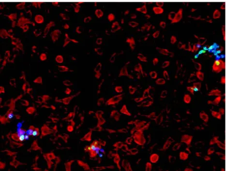
3.5 IMMUNOFLUORESCENCE
For immunofluorescence staining, P. brasiliensis cells were incubated with fluorescein isothiocyanate (FITC) for 1 hour at room temperature (RT). Cells were then washed twice with LPS-free PBS and adjusted at a concentration of 1.25x10⁶/mL. BMDM were seeded on poly-D lysine-coated coverslips and
infected with the labelled yeast cells for 6 hours. After this period, coverslips were washed 6 times and non-phagocytosed yeast cells stained with calcofluor for 10 minutes at RT. Cells were washed twice with LPS-free PBS and fixed for 30 minutes with paraformaldehyde (PFA) (4%). For latter macrophage staining, coverslips were incubated with blocking solution (PBS, 0.1% Triton X-100, 0.1% Tween-20, BSA 5% (p/v)) for one hour to allow blocking of unspecific binding sites to cells. CD11b-PE (clone M1/70, eBioscience) was diluted in blocking solution (1:100) and incubated for one hour at RT. Coverslips were washed 3 times for 5 minutes with washing solution (PBS, 0.1% Triton X-100, 0.1% Tween-20). Finally, the secondary antibody (Alexa fluor 594 goat anti-rat - Life Technologies) was added to bind to the primary antibody for one hour at RT and washed again 3 times before mounting coverslips with vectashield in microscope slides. Coverslips were observed with Olympus BX61 microscope and images were recorded with Olympus DP70 camera.
3.6 HEMATOXYLIN AND EOSIN STAINING
BMDM at a final concentration of 1x10⁶/mL were plated on poly-D lysine-coated coverslips in 24-well
plates and infected for 3 and 6 hours at a MOI of 1:1 or 1:2. After these time periods, coverslips were washed 6 times with LPS-free PBS to eliminate extracellular yeast cells and fixed with 4% PFA for 30 minutes. Then, coverslips were stained with haematoxylin and eosin (H&E) and mounted in microscope slides.
3.7 ELISA
Enzyme-linked immunosorbent assay (ELISA) sets for IL-10 7104), IL-6 7064) and TNF (88-7324) were obtained from eBioscience. This assay was performed to measure the levels of these cytokines in the supernatants collected from infected cell cultures. Firstly, 96-well plates were coated with cytokine-specific capturing antibody overnight. Then, non-specific ligations were blocked and the supernatants and the cytokine standard concentration solutions were added to the wells and incubated at RT for 2h. The specifically bound protein was detected with cytokine-specific biotin-labelled detection antibodies upon incubation for 1h at RT. Avidin-horseradish peroxidase (-HRP) was added to bind to the
detection antibodies for 30 minutes at RT. Finally, 3,3′,5,5′-tetramethylbenzidine (TMB) was added, resulting in a coloured end-product since this reagent interacts with HRP being metabolized by this last one. Cytokine concentration was determined by spectrophotometry using a plate reader (Bio-Rad- Microplate reader 680), at 450nm. The optical density was then converted to concentration values using the Microplate Manager 5.2.1.
3.8 MEASUREMENT OF NITRIC OXIDE (NO) PRODUCTION
The concentration of NO in the supernatants of BMDMsinfected with P. brasiliensis was determined by the colorimetric Griess reagent. Supernatants were mixed with an equal volume of Griess reagent (1% sulfanilamine, 0,1% nathylethylenediamine, 2,5% H3PO4) and incubated for 10 minutes in the dark. NO
concentration was determined by spectrophotometry using a plate reader (Bio-Rad- Microplate reader 680), at 550nm. The optical density was then converted to concentration values using the Microplate Manager 5.2.1.
3.9 IN VIVO INFECTIONS
Intraperitoneal infection: for intraperitoneal infection, inoculum was prepared as previously described and WT and pMT-10 mice were infected with 1x10⁶ P. brasiliensis 18 cells, resuspended in
0.2 mL of LPS-free PBS, by intraperitoneal route. At weeks 1, 4 and 8, animals were euthanized by CO2
asphyxiation and the lungs, liver and spleen removed for flow cytometry and histological analysis. Intranasal infection: for intranasal infection, animals were anesthetized and, with a loading tip, 1x10⁶ P. brasiliensis 18 yeast cells were carefully delivered to the respiratory tract in a total volume of
20µL. The suspension was administrated drop by drop in the nostril until the animals inhaled all the volume. Animals were monitored daily and at weeks 1, 2, 8 and 20 were sacrificed and their lungs, liver and spleen collect for histological analysis and flow cytometry analysis.
Intratracheal infection: WT mice were first anesthetized and placed in an angled platform hanging by its incisors on a wire and gently restrained with a rubber band. The mice tongue was pull out with forceps and a sterile bent gavage needle inserted until the bend of the needle was by the front incisors with the help of a laryngoscope. After the needle was in the correct position, 1x10⁶ P.
brasiliensis ATCC60855 yeast cells were injected into the trachea. Finally, animals were held upright for a few seconds to allow the inoculum inhalation into the lungs. Control of infection was performed at day
respiratory problems in the first 48 hours were sacrificed and theirs lungs removed for histological analysis.
3.10 HISTOLOGICAL ANALYSIS
Lungs, liver and spleen from sacrificed animals were harvested and fixed in 4% PFA for 7 days at 4˚C. Later, these organs were embedded in paraffin and tissue sections were stained with hematoxylin and eosin (H&E). Sections were observed under the microscope and the presence of granuloma or necrotic areas were assessed using Olympus BX61 microscope and images were recorded with Olympus DP70 camera with a magnification of 10x or 40x.
3.11 FLOW CYTOMETRY ANALYSIS
For extracellular staining, 2-3x10⁶ cells from the lung or the spleen were washed with FACS buffer (PBS
containing 2% of FBS, 0.01% of azide and 0.5% saponine) and stained for 30 minutes at 4˚C. The following antibodies were used: MHC II-FITC (M5/114.15.2. Biolegend), CD11b-PE (clone M1/70, eBioscience), Ly6C-PerCP/Cy5.5 (clone AL-21, Pharmingen), CD62L-PE/Cy7 (clone MEL-14, Biolegend), CD11c-BV421 (clone N417, Biolegend), Ly6G-APC (clone 1A8, Biolegend), F4/80-APC/Cy7 (clone BM8, Biolegend); CD8A-FITC (clone 5H10-1, Biolegend), CD44-PerCP/Cy5.5 (clone MI7, Biolegend), CD3-PE clone 17A2, Biolegend), CD4-BV421 (clone GK1.5, Biolgend), CD19-APC/Cy7 (clone HIB, Biolegend). After, cells were washed twice with FACS buffer and fixed in 100 ml of 4% paraformaldehyde (PFA) for 30 minutes at RT. Samples were acquired on a LSRII flow cytometry with Diva software. Data was analysed using FlowJo version 7 software. The number of cells was assessed taking into account the number of cells in the organ determined by Countess® Automated Cell Counter.
3.12 RNA EXTRACTION AND QUANTIFICATION
RNA from samples was extracted with NZYol Reagent (NZYTech) according to the manufacturer’s instructions. Initially, glycogen (20µg/µL, Roche) was added to each samples and incubated for 5 minutes at RT. After, 50 µL of chloroform were added to each samples and mixed by vortexing for 15 seconds and incubated on ice for 15 minutes. Samples were then centrifuged for 15 minutes at 13000 rpm at 4˚C and the RNA-containing aqueous phase collected. This phase was then mixed with an equal
volume of isopropyl alcohol (Sigma-Aldrich) in order to precipitate the RNA. Samples were incubated for 4 hours at -20˚C and centrifuged for 15 minutes at 13000 rpm. Supernatant was removed and 800µL of ethanol (Carlo Erba reagents) were added to the pellet to wash the RNA. Ethanol was removed by centrifugation for 5 minutes at 9000 rpm and the RNA was resuspended in RNase-free water (Gibco). RNA concentration was determined at 260nm and the purity determined according to the A260/A280 and
A260/A230 ratios (Nanodrop ND-1000 Spectophotometer).
3.13 COMPLEMENTATY DNA (cDNA) SYNTHESIS
cDNA was synthesized using NZY First-Strand cDNA Synthesis Kit (NZYTech). Briefly, RNA samples were adjusted to 100ng/µL. A reaction mix containing 10µL of NZYRT 2x Master Mix and 2µL of NZYRT Enzyme Mix was mixed with 10µL of RNA sample. cDNA synthesis was performed in a thermocycle (Eppendorf) with the next conditions: 10 minutes of incubation at 25˚C followed by an incubation period of 30 minutes at 50˚C and a period of 5 minutes at 85˚C to inactivate the reaction. cDNA was then used for gene quantification by real-time polymerase chain reaction (RT-PCR).
3.14 REAL-TIME POLYMERASE CHAIN REACTION (RT-PCR)
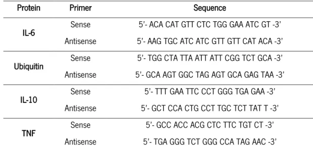
IL-10, TNF, IL-6 and ubiquitin expression was measured using SsoFast EvaGreen SuperMix (Bio-Rad). Briefly, 1µL of cDNA was mixed with 1 µL of 0.4µM sense and antisense specific primers (Table 1), 1µL of water and 5µL of SsoFast EvaGreen SuperMix. RT-PCR was performed in CF*96TM Real-time system (Bio-Rad) using the next conditions: 95˚C for 15 seconds, followed by 40 amplification cycles of 95˚C for 15 seconds, 58˚C for 20 seconds and 70˚C for 15 seconds.
3.15 STATISTICAL ANALYSIS
The results are given as means± standard deviation (SD) of at least 3 replicates per experimental group in in vitro infections, and 4 animals per experimental group in in vivo infections. Statistical analyses were performed using the GraphPad Prism 6 software. Student’s t-test, one-way analyses of variance (ANOVA) or two way analyses of variance (ANOVA) and the post-test Bonferroni were used to evaluate significant differences between different conditions. Values were considered significant when p<0.05.
Protein Primer Sequence
IL-6 Sense 5’- ACA CAT GTT CTC TGG GAA ATC GT -3’ Antisense 5’- AAG TGC ATC ATC GTT GTT CAT ACA -3’ Ubiquitin Sense 5’- TGG CTA TTA ATT ATT CGG TCT GCA -3’ Antisense 5’- GCA AGT GGC TAG AGT GCA GAG TAA -3’
IL-10 Sense 5’- TTT GAA TTC CCT GGG TGA GAA -3’
Antisense 5’- GCT CCA CTG CCT TGC TCT TAT T -3’
TNF Sense 5’- GCC ACC ACG CTC TTC TGT CT -3’
Antisense 5’- TGA GGG TCT GGG CCA TAG AAC -3’
CHAPTER IV
4.1 PREPARATION OF P. BRASILIENSIS INOCULUM FOR IN VITRO AND IN VIVO INFECTIONS
Different strains of P. brasiliensis can display different morphologies, including single cells, multiple budding cells and transitional forms (Brummer, Castaneda et al. 1993). These morphological differences have been suggested to be associated with virulence (Almeida, Cunha et al. 2009).
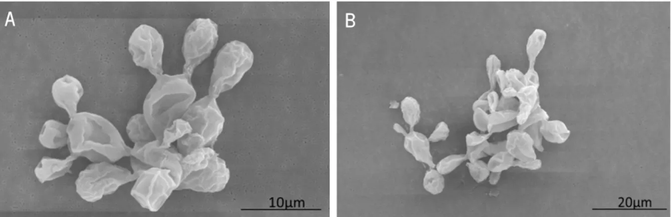
To determine the morphology of P. brasiliensis 18, a highly virulent strain, we begin by performing scanning electron microscopy. As represented in Figure 7, we found that, in addition to its large size that varies between 10 and 15 µm, this strain also presented several multiple budded cells (Figure 7A) that formed large aggregates after in vitro growth (Figure 7B).
Figure 7. P. brasiliensis 18 presented a large size and multiple budding-cells. Scanning electron microscopy showed that P. brasiliensis 18 had a spherical shape with larger and multiple budded cells (A). Cells tended to aggregate leading to the formation of clumps (B).
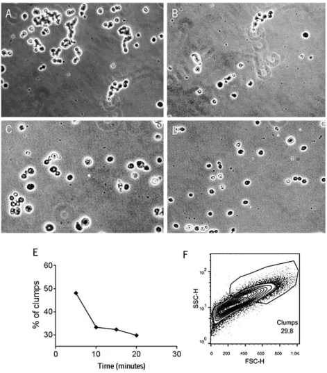
Taking into consideration the complexity of this P. brasiliensis strain, we first wanted to standardize the preparation of the inoculum for in vitro and in vivo infections. To this end, P. brasiliensis 18 was grown in Brain Heart Infusion (BHI) liquid medium supplemented with 1% glucose and gentamicin (50µg/mL) until the exponential growth phase (7-10 days). Next, the entire suspension was collected, glass beads were added and the suspension was vortexed in 3 cycles of 10 seconds to dissociate the clumps. Glass beads were then removed and the suspension was centrifuged for 5 minutes at 3500 rpm. The supernatant was discarded and the pellet was washed twice with lipopolysaccharide (LPS)-free phosphate buffered saline (PBS). At this point, sedimentation of larger cells and clumps was allowed for 5, 10, 15 and 20 minutes to determine the time required to obtain a homogeneous suspension (Figure 8, panels A-E).
As represented in Figure 8, we found that for P. brasiliensis 18, 20 minutes was the time required to obtain a homogenous population of single, smaller and less complex cells. We also assessed the P. brasiliensis cell complexity by flow cytometry analysis with similar data (Figure 8F).
Figure 8. Longer periods of sedimentation resulted in a less complex inoculum. Cells were washed twice with LPS-free PBS and sedimentation of clumps and larger cells was allowed for 5 (A), 10 (B), 15 (C) and 20 (D) minutes. The percentage of clumps was reduced throughout time (E). After 20 minutes, 29,8% of the inoculum was only composed by clumps (F). Results were from one representative experiment out of four independent experiments. Cells were photographed by phase contrast microscopy (DIC). Magnification 20x
Overall, these data showed that this approach allowed for the preparation of a homogeneous suspension of P. brasiliensis yeast cells that were used to infect BMDM and mice.