Antiproliferative effect of urolithin A, the ellagic
acid-derived colonic metabolite, on hepatocellular carcinoma
HepG2.2.15 cells by targeting Lin28a/let-7a axis
Zhenpeng Qiu
1*, Junxuan Zhou
1*, Cong Zhang
1, Ye Cheng
2, Junjie Hu
1and Guohua Zheng
3 1College of Pharmacy, Hubei University of Chinese Medicine, Wuhan, China
2
Hubei Engineering Research Center of Viral Vector, Wuhan Institute of Bioengineering, Wuhan, China
3
Key Laboratory of Chinese Medicine Resource and Compound Prescription, Ministry of Education, Hubei University of Chinese Medicine, Wuhan, China
Abstract
An abnormality in the Lin28/let-7a axis is relevant to the progression of hepatitis B virus (HBV)-positive hepatocellular carcinoma (HCC), which could be a novel therapeutic target for this malignant tumor. The present study aimed to investigate the anti-proliferative and anti-invasive effects of urolithin A in a stable full-length HBV gene integrated cell line HepG2.2.15 using CCK-8 and transwell assays. The RNA and protein expressions of targets were assessed by quantitative PCR and western blot, respectively. Results revealed that urolithin A induced cytotoxicity in HepG2.2.15 cells, which was accompanied by the cleavage of caspase-3 protein and down-regulation of Bcl-2/Bax ratio. Moreover, urolithin A suppressed the protein expressions of Sp-1, Lin28a, and Zcchc11, and elevated the expression of microRNA let-7a. Importantly, urolithin A also regulated the Lin28a/let-7a axis in transient HBx-transfected HCC HepG2 cells. Furthermore, urolithin A decelerated the HepG2.2.15 cell invasion, which was involved in suppressing the let-7a downstream factors HMGA2 and K-ras. Thesefindings indicated that urolithin A exerted the antiproliferative effect by regulating the Lin28a/let-7a axis and may be a potential supplement for HBV-infected HCC therapy. Key words: Urolithin A; Hepatocellular carcinoma; Lin28a; let-7a; Cell proliferation; HBx
Introduction
Liver cancer is one of the most malignant tumors globally, with high mortality rates, which commonly result from chronic inflammation and hepaticfibrosis (1). In high-incidence areas of hepatocellular carcinoma (HCC), such as West Africa approximately, 60% of HCC patients are infected with hepatitis B virus (HBV), suggesting that integrated HBV genetic material in hepatocytes may accelerate the process of cirrhosis and cell immortalization (2). Signaling pathways that activate cell proliferation and invasion are the effectors of HBx, a reverse transcription element in the HBV genome for fibrosis and oncogenesis, interacted by TGF-b or other transcriptional factors, such as nuclear factor-kB and Sp-1 (3–5). Consequently, although HBV replication in infected hepatocytes cannot be eradicated, acting on the HBx-triggered molecular networks and suppressing the cofactors of HBx might be a potent therapeutic strategy for HBV-infected HCC.
Dysregulated microRNA (miRNA) profiles are the collaborators of Hbx, which aggravate tumor progression
involved in carcinogenesis, cell invasion, and metastasis in human malignancies. Let-7a, one of the let-7 family members, was initially characterized in Caenorhabditis elegans with Lin28a, an RNA-binding protein that acted as a suppressor of let-7 expression and controller for development and differentiation (6). Let-7a is also down-regulated in HCC patients and its low expression in liver tissues may contribute to poor survival rates (7). More-over, Lin28-induced cancer cell EMT is dependent on the low let-7 level and overexpression of the EMT-associated let-7a downstream targets, such as K-ras and HMGA2 (8). Therefore, seeking an approach to alter the Lin28a/let-7a axis in hepatoma cancer cells may lead to the develop-ment of effective strategies for HCC therapy.
Urolithins, the dibenzopyran-6-one colonic metabolites derived from ellagic acid (EA) or ellagitannins (ETs), have been suggested to be beneficial for human health. Urolithins in target tissues and cells could act on sub-cellular compo-nents and activate signaling transduction. These cell responses
Correspondence: Guohua Zheng:<zgh1227@sina.com>
*These authors contributed equally to this work.
Received October 10, 2017 | Accepted February 2, 2018
contribute to the various biological potentials of EA- and ET-rich diets (9). Based on the anti-proliferative effects of urolithin A on HepG2 cells in our previous findings (10), the function of urolithin A in repressing HepG2.2.15 (HBV-integrated HepG2 cell line) cell proliferation and invasion are discussed in the present study. The results suggested that the regulating effects of urolithin A on the Lin28a/let-7a axis contributed to the inhibition of transcriptional factor Sp-1 and down-regulation of HMGA2 and K-ras in HepG2.2.15 cells.
Material and Methods
Chemicals
Urolithin A was synthesized as previously described (11). The purity (494%) of urolithin A was evaluated by HPLC, and its molecular weight was confirmed by mass spectro-metry analysis (Figure S1). A hydro-soluble tetrazolium salt WST-8 (2-(2-methoxy-4-nitrophenyl)-3-(4-nitrophenyl)-5-(2,4-disulfophenyl)-2H-tetrazolium, monosodium salt) was obtained from Dojindo Laboratories (Japan) for CCK-8 assay. All other chemicals and reagents were of analytical grade.
Cell culture
HepG2 and HepG2.2.15 cells (HepG2 cells integrated with a stable wild-type full-length HBV genome) were obtained from American Type Culture Collection and cultured in Dulbecco’s Modified Eagle Medium (Gibco, USA) supplemented with fetal bovine serum (10%), penicillin (1 mM) and streptomycin (1 mM) at 37°C in a humidified 5% CO2atmosphere.
Cell viability assay
The effect of urolithin A on HepG2.2.15 cell viability was evaluated using the CCK-8 assay with WST-8 accord-ing to manufacturer’s protocol (12). Cell viability values were normalized as follows: [Final absorbance (urolithin A) / Final absorbance (control)] 100%. IC50value for HepG2.2.15
cells was calculated according to a dose-response curve, which was plotted for each concentration.
Quantitative PCR assay for miRNA and mRNA
Total RNAs containing miRNAs in cells were pre-pared using mirVanatmiRNA Isolation Kit (Thermo Fisher Scientific, USA). Real-time quantitative PCR (qPCR) assay was performed on a MiniOpticont(Bio-Rad, USA) system using FastStart Universal SYBR Green Master (Roche, USA).
With initial denaturation at 95°C for 120 s, amplifications were performed for 40 cycles at 95°C for 5 s and 55°C for 25 s. Primers for qPCR are listed in Table 1.
Western blot
Western blot was performed as described previously (13). Briefly, separated proteins in SDS-PAGE were transferred onto polyvinylidenefluoride membranes and were sequen-tially immune-reacted with specific primary antibodies and secondary antibodies. Antibody-conjunct proteins were
quanti-fied using SuperSignalt West Pico Chemiluminescent Substrate (Thermo Scientific, USA). The primary antibodies cleaved caspase-3 (Cell Signaling Technology (CST, USA)), Bcl-2 (CST), Bax (CST), Lin28a (CST), Sp-1 (CST), HMGA2 (CST), K-ras (CST), Snail (CST), and Zcchc11 (Abcam, UK) were applied after the membranes were blocked in either 5% milk or 5% BSA. Anti-b-actin-peroxidase antibody was obtained from Sigma-Aldrich (USA) and used as an internal reference. The protein content was analyzed using Image Lab 5.1 (Bio-Rad, USA).
Vector and miRNA transfection
Cells were cultured in 6-well (qPCR and western blot) or 24-well (luciferase assay) plates and were then trans-fected with plasmids. The Lin28 open reading frame was inserted into the pcDNA3.1(+) vector (Invitrogen, USA) to express Lin28 (pcDNA3.1-Lin28) with Lipofectaminet
3000 (Invitrogen) in HepG2.2.15 cells. Let-7a inhibitor (20-O-methyl antisense oligonucleotide) and the non-specific control were commercially synthesized as previously described (14). For western blot assay, cells in 6-well plates with approximately 80% confluency were transfected with 50 nM of let-7a miRNA inhibitor negative control (MOCK) (Thermo Fisher, USA) or 100 nM of inhibitor using Lipofectaminet 3000 (Invitrogen, USA). Transfec-tion efficiency was validated by qPCR to directly assess the expression of let-7a.
Transwell invasion assays
The transwell assay was performed using Matrigel-coated (1:5, BD Biosciences) polycarbonatefilters (Corning Costar, USA) (15). Cells were subjected to urolithin A for 24 h before use, and the chemical was present throughout the assay. Invasive cells in the lower chamber were stained with crystal violet and visualized using an optical micro-scope. Cell invasion was quantitated via the absorbance Table 1.Primers used in real-time PCR.
Gene Forward primer 50to 30 Reverse primer 50to 30
Lin28a TTGTCTTCTACCCTGCCCTCT GAACAAGGGATGGAGGGTTTT
B-actin CCTGGCACCCAGCACAAT GGGCCGGACTCGTCATACT
Let-7a GGTGAGGTAGTAGGTTGTATAGTT Uni-miR qPCR primer (TaKaRa)
(600 nm) of eluted crystal violet with a microtiter plate reader as described previously (16).
Statistics
All in vitro assessments were performed at least in triplicate. The data are reported as means±SD.
Differ-ences between experimental groups were analyzed by Student’st-test or one-way analysis of variance (ANOVA), followed by Dunnett’st-test for multiple comparisons using SPSS (13.0) statistical program. P-values of less than 0.05 were considered statistically significant.
Results
Urolithin A inhibited cell proliferation and induced cytotoxicity in HepG2.2.15 cells
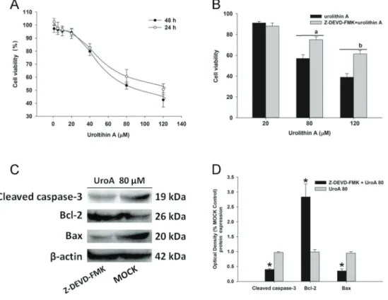
In the CCK-8 assay, cells were administrated with urolithin A and B by gradual concentrations (1–120mM) for 24 and 48 h. As shown in Figure 1A, urolithin A repressed cell proliferation in a dose-dependent manner. It was also found that urolithin A induced acute cytotoxicity from
80–120 mM, compared to the corresponding controls. However, urolithin B (1–120 mM) showed a moderately suppressing effect on HepG2.2.15 cells (Figure S2). Because of this, we chose urolithin A for further investi-gation. To identify whether urolithin A functions through caspase-3 signaling, cells were incubated in the absence or presence of Z-DEVD-FMK (20 mM) for 6 h before urolithin A treatment. Data showed that pretreatment with Z-DEVD-FMK abolished the inhibiting effect of cell pro-liferation induced by urolithin A (Figure 1B). Furthermore, we observed that the protein level of cleaved caspase-3 was up-regulated and the Bcl-2/Bax ratio was decreased by urolithin A in HepG2.2.15 cells (Figure 1C and D), suggesting that the suppressing effect of urolithin A on HepG2.2.15 viability could be involved in the activation of caspase-dependent apoptotic signaling.
Urolithin A altered the expressing pattern of Lin28a/ let-7a axis
Lin28a, which is a repressor of let-7a by recruiting Zcchc11, could be up-regulated by the HBx protein via
Figure 1.Urolithin A (UroA) suppressed HepG2.2.15 cell proliferation via caspase-3 dependent apoptosis.A, HepG2.2.15 cells were administrated with 0-120mM of urolithin A for 24 or 48 h. Cell viability was assessed by a CCK assay and the data were normalized to normal controls.B, The caspase-3 inhibitor Z-DEVD-FMK alleviated urolithin A-induced HepG2.2.15 cell death (80 or 120mM). The cells were incubated in the absence or presence of Z-DEVD-FMK (20mM) for 6 h prior to urolithin A treatment, and then incubated for 48 h. Data are reported as means±SD, n=3.a,bP
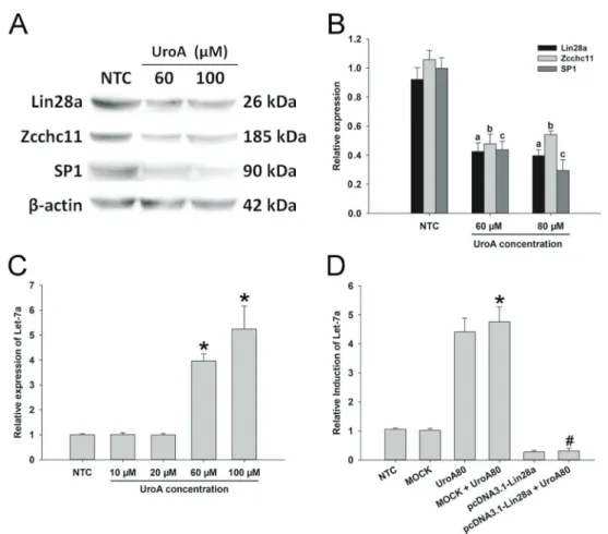
Sp-1 in HCC cells. Our data indicated that the protein expressions of Lin28a, Zcchc11, and Sp-1 were suppressed in the urolithin A group, compared to the control group (Figure 2A and B). We also observed that let-7a was up-regulated when the cells were subjected to urolithin A (Figure 2C). To further confirm the effects of urolithin A on the Lin28a/let-7a axis, overexpression of Lin28a (pcDNA3.1-Lin28a) by transient transfection was performed in HepG2. 2.15 cells (Figure S3). The results showed that the effects of urolithin A on the expression of let-7a were abolished by Lin28a transfection (Figure 2D), indicating that urolithin A increased the expression of let-7a by targeting Lin28a in HepG2.2.15 cells.
Urolithin A repressed Lin28a expression and elevated the expression of let-7a in transient HBx-transfected HepG2 cells
The effect of urolithin A on the Lin28a/let-7a axis was also confirmed in transient HBx-overexpressing HepG2
cells. In this section, HepG2 cells (4105cells/well) were seeded in 6-well plates for 24 h and transiently transfected with 2mg pcDNA-HBx (pc-HBX) plasmids (pcDNA3.1 (–) plasmid with the sequence encoding for the HBx protein; a gift from Prof. Fan Zhu at Wuhan University). After post-transfection for 24 h, cells were incubated with urolithin A (60 and 100 mM) for 48 h. As expected, qPCR assay showed that the elevated mRNA expression of Lin28a in the pcDNA-HBx group was repressed by urolithin A (Figure 3A). To examine whether urolithin A affected Lin28a at the protein level in the HBx-transfected HepG2 cells, western blot analysis was performed; the results demonstrated that Lin28a protein expression was suppressed (Figure 3B and C). Moreover, the HBx-induced let-7a decrease in the pcDNA-HBx group was alleviated by urolithin A treatment (Figure 3D). These data indicated that urolithin A delayed the HBx-induced change of the Lin28a/let-7a axis in HCC cells, further enhancing thefindings involved in the effects of urolithin A on Lin28a and let-7a in HepG2.2.15 cells.
Figure 2.Urolithin A (UroA) regulated the expression of Lin28a/let-7a axis.A, The protein expressions of Sp-1, Lin28a, and Zcchc11 were assessed by western blot assay.B, Sp-1, Lin28a, and Zcchc11 protein levels were semi-quantified by western blot assay. Data are reported as means±SD normalized to the correspondingb-actin values.a,b,cPo0.05vsthe protein expressions of Lin28a, Zcchc11, and Sp-1 in the
Urolithin A suppressed cell invasion by inhibiting K-ras/HMGA2 signaling in HepG2.2.15 cells
Since let-7a was increased by urolithin A, we assumed that downstream targets of let-7a could respond to urolithin A exposure. Accompanied by an elevation of let-7a (Figure 2C), the expression of HMGA2, a protein enhancing oncogenic transformation and epithelial-mesenchymal transition, was reduced by urolithin A compared to the MOCK group (Figure 4A and B). The effect of urolithin A on cell invasion was evaluated using a transwell assay. As shown in Figure 4C and D, cells in the control groups exhibited approxi-mately 3 times higher invasion efficiency than cells in the urolithin A (UroA100) group, indicating that urolithin A could suppress cell invasionin vitro. To further determine whether let-7a elevation contributed to the suppressing effect of urolithin A on cell invasion, the let-7a inhibitor was trans-fected for 24 h to abolish the expression of let-7a before urolithin A administration (Figure S4). In Figure 4E, the blockade of let-7a resulted in an increase in HMGA2 protein expression in the western blot assay, compared to
the urolithin A group. Moreover, K-ras, which was commonly considered to be a compatible factor of HMGA2 to accel-erate cell migration and invasion, was also down-regulated in the urolithin A group (Figure 4A). However, the protein expression of Snail showed no difference between the urolithin A and the MOCK groups. Hence, our data suggested that the regulation of the K-ras/HMGA2 signaling by urolithin A may contribute to the inhibiting effect on cell invasion.
Discussion
Accepted theory claims that the intestinal metabolism of ETs or EAs to dibenzopyran-6-one derivatives (urolithins) may play an indispensable role in the absorption of ETs. From this perspective, urolithins identified in tissues are important, and their molecular modulation in target cells can be more effective in interpreting the nutritional benefits for human health than that of ETs (17).
Urolithin A is an essential metabolite generated in humans after consumption of EA- and ET-rich food and Figure 3.Urolithin A downregulated Lin28a expression and elevated the expression of let-7a in HBX overexpressed hepatocellular carcinoma HepG2 cells. Urolithin A suppressed Lin28a mRNA expression (A) and protein expression (BandC) in HBX-overexpressed HepG2 cells. D, Urolithin A elevated the let-7a expression in HBX-overexpressed HepG2 cells (qPCR). *Po0.05 compared to the MOCK control.#P
healthy supplements (18). Cell signaling transduction (EGFR, b-catenin), essential regulators for cell cycle (cyclin D1, c-Myc, p21), and programmed cell death (p53, PARP, caspase-3)
urolithin A or C (20,21), the functions of urolithins in hepa-tocytes have not been adequately evaluated. Hence, to explore the capacity of urolithin A on HBx-relevant cell invasion, the Lin28a/let-7a axis and Sp-1, a transcriptional factor that could be elevated by HBx, were assumed in the present study to be potential targets of urolithin A in HepG2.2.15 cells.
Sp-1 is an HBx-combinative activator and a member of the Sp/KLF family of transcription factors, and is widely overexpressed in neoplasms, including liver carcinoma (22,23). Based on the anti-proliferative effects of urolithin A in HepG2.2.15 cells, we investigated whether urolithin A could affect the expression of Sp-1. The results revealed that the protein expression level of Sp-1 was repressed by urolithin A treatment. Lin28a, the transcriptional target of Sp-1, which could further elevate the levels of certain cancer-related miRNAs (24), was also suppressed by urolithin A in HepG2.2.15 cells. Therefore, we speculated that suppression of Sp-1 could be responsible for the decrease of Lin28a by urolithin A.
Let-7a functions as a tumor suppressor and is bio-logically deleted in several cancers, including in HCC patients with HBV infection (25). Moreover, Lin28a suppresses let-7a miRNA biogenesis in tumor cells by recruiting Zcchc11 (TUT4) in the cell cytoplasm to degrading pre-let-7 (26). In our study, urolithin A elevated the let-7a expression, which was delayed by Lin28a overexpression in HepG2.2.15 cells. The similar effects of urolithin A on the Lin28a/let-7a axis were further observed in HBx-overexpressed HepG2 cells. Some evidence has also highlighted that the Zcchc11 down-regulation is Lin28b-independent, and should be restricted to Lin28a-positive carcinomas, such as the T47D breast cancer cells (27). In the present study, the protein expression of Zcchc11 was reduced by urolithin A treatment, suggesting that Zcchc11 inhibition plays a bridging role for the effects of urolithin A on the Lin28a/let-7a axis.
High mobility group AT-hook 2 (HMGA2) is a transcrip-tion factor highly expressed in the embryonic stage. The exceptional re-expression of HMGA2 by chromosomal rearrangements at chr12q13-15 in neoplasia of mice or
humans, such as HCC and lung cancer, is involved in the let-7 deletion or loss of let-7-binding sites (28–30). In this study, the western blot analysis supported the notion that urolithin A can down-regulate HMGA2 expression and the capacity is let-7a-dependent. We also found that the invasive potential of HepG2.2.15 cells could be repressed by urolithin A treatment. Let-7a also affected oncogenic K-ras expression by binding the 3’-UTR of K-ras. Exoge-nous let-7a miRNA in HepG2 cells could specifically reduce abundant K-ras expression (31). The protein expression of K-ras was decreased by urolithin A in HepG2.2.15 cells at the repressible concentration in the transwell assay. Therefore, it is possible that the functions of urolithin A in HepG2.2.15 cells may partly match the above-identified proposal involved in let-7a/HMGA2/K-ras signaling.
In the present study, we described the effects of urolithin A in repressing the proliferation and invasion of the HBV-overexpressed HepG2.2.15 HCC cell line. In fact, the virus replication in HBV-infected patients could not be eliminated, but only alleviated with antiviral agents, such as adefovir. The data in this study could be considered an update of our previous data (10) on HepG2 cells because urolithin A has suppressed cell invasion in HepG2.2.15 cells. In other words, ourfindings are not only involved in the targets of carcinogenesis but are also based on the initial observation of the HBx-interactional cancerous trans-criptional factor, Sp-1. Therefore, our data demonstrated that urolithin A suppressed the HepG2.2.15 cell proliferation and invasion via regulating the Lin28a/let-7a axis and EMT-involved targets, such as HMGA2 and K-ras.
Supplementary Material
Click here to view [pdf]
Acknowledgments
This study was supported by a grant from Research Project of Hubei Provincial Department of Education (Q20162001) to Dr. Zhenpeng Qiu.
References
1. Chen X, Calvisi DF. Hydrodynamic transfection for genera-tion of novel mouse models for liver cancer research.Am J Pathol2014; 184: 912–923, doi: 10.1016/j.ajpath.2013.12.002 2. Kirk GD, Lesi OA, Mendy M, Akano AO, Sam O, Goedert JJ, et al. The Gambia liver cancer study: Infection with hepatitis B and C and the risk of hepatocellular carcinoma in West Africa. Hepatology2004; 39: 211–219, doi: 10.1002/hep.20027 3. Arbuthnot P, Kew M. Hepatitis B virus and hepatocellular
carcinoma.Int J Exp Pathol2001; 82: 77–100, doi: 10.1111/ j.1365-2613.2001.iep178.x
4. Tang H, Da L, Mao Y, Li Y, Li D, Xu Z, et al. Hepatitis B virus X protein sensitizes cells to starvation-induced autophagy
via up-regulation of beclin 1 expression.Hepatology2009; 49: 60–71, doi: 10.1002/hep.22581
5. Ng SA, Lee C. Hepatitis B virus X gene and hepatocarcino-genesis. J Gastroenterol2011; 46: 974–990, doi: 10.1007/ s00535-011-0415-9
6. Reinhart BJ, Slack FJ, Basson M, Pasquinelli AE, Bettinger JC, Rougvie AE, et al. The 21-nucleotide let-7 RNA regulates developmental timing in Caenorhabditis elegans. Nature 2000; 403: 901–906, doi: 10.1038/35002607
8. Wang T, Wang G, Hao D, Liu X, Wang D, Ning N, et al. Aberrant regulation of the LIN28A/LIN28B and let-7 loop in human malignant tumors and its effects on the hallmarks of cancer. Mol Cancer 2015; 14: 125, doi: 10.1186/s12943-015-0402-5
9. Tomas-Barberan FA, Gonzalez-Sarrias A, Garcia-Villalba R, Nunez-Sanchez MA, Selma MV, Garcia-Conesa MT, et al. Urolithins, the rescue of "old" metabolites to understand a "new" concept: Metabotypes as a nexus among phenolic metabolism, microbiota dysbiosis, and host health status. Mol Nutr Food Res2017; 61, doi: 10.1002/mnfr.201500901 10. Wang Y, Qiu Z, Zhou B, Liu C, Ruan J, Yan Q, et al. In vitro antiproliferative and antioxidant effects of urolithin A, the colonic metabolite of ellagic acid, on hepatocellular carci-nomas HepG2 cells.Toxicol In Vitro2015; 29: 1107–1115, doi: 10.1016/j.tiv.2015.04.008
11. Bialonska D, Kasimsetty SG, Khan SI, Ferreira D. Urolithins, intestinal microbial metabolites of Pomegranate ellagitan-nins, exhibit potent antioxidant activity in a cell-based assay. J Agric Food Chem2009; 57: 10181–10186, doi: 10.1021/ jf9025794
12. Ji Q, Hao X, Meng Y, Zhang M, Desano J, Fan D, et al. Restoration of tumor suppressor miR-34 inhibits human p53-mutant gastric cancer tumorspheres. BMC Cancer 2008; 8: 266, doi: 10.1186/1471-2407-8-266
13. Zhou B, Wang J, Zheng G, Qiu Z. Methylated urolithin A, the modified ellagitannin-derived metabolite, suppresses cell viability of DU145 human prostate cancer cells via targeting miR-21.Food Chem Toxicol2016; 97: 375–384, doi: 10.1016/ j.fct.2016.10.005
14. Khodayari N, Mohammed KA, Goldberg EP, Nasreen N. EphrinA1 inhibits malignant mesothelioma tumor growth via let-7 microRNA-mediated repression of the RAS oncogene. Cancer Gene Ther 2011; 18: 806–816, doi: 10.1038/cgt. 2011.50
15. Zhang L, Tu Y, He W, Peng Y, Qiu Z. A novel mechanism of hepatocellular carcinoma cell apoptosis induced by lupeol via Brain-Derived Neurotrophic Factor Inhibition and Glyco-gen Synthase Kinase 3 beta reactivation.Eur J Pharmacol 2015; 762: 55–62, doi: 10.1016/j.ejphar.2015.05.030 16. Petitclerc E, Boutaud A, Prestayko A, Xu J, Sado Y, Ninomiya
Y, et al. New functions for non-collagenous domains of human collagen type IV. Novel integrin ligands inhibiting angiogenesis and tumor growth in vivo.J Biol Chem2000; 275: 8051–8061, doi: 10.1074/jbc.275.11.8051
17. Espin JC, Gonzalez-Sarrias A, Tomas-Barberan FA. The gut microbiota: a key factor in the therapeutic effects of (poly) phenols.Biochem Pharmacol2017, doi: 10.1016/j.bcp.2017. 04.033
18. Cerda B, Tomas-Barberan FA, Espin JC. Metabolism of antioxidant and chemopreventive ellagitannins from straw-berries, raspstraw-berries, walnuts, and oak-aged wine in humans:
identification of biomarkers and individual variability.J Agric Food Chem2005; 53: 227–235, doi: 10.1021/jf049144d 19. Espin JC, Larrosa M, Garcia-Conesa MT, Tomas-Barberan
F. Biological significance of urolithins, the gut microbial ellagic Acid-derived metabolites: the evidence so far.Evid Based Complement Alternat Med 2013; 2013: 270418, doi: 10.1155/2013/270418
20. Sharma M, Li L, Celver J, Killian C, Kovoor A, Seeram NP. Effects of fruit ellagitannin extracts, ellagic acid, and their colonic metabolite, urolithin A, on Wnt signaling.J Agric Food Chem2010; 58: 3965–3969, doi: 10.1021/jf902857v 21. Kang I, Kim Y, Tomas-Barberan FA, Espin JC, Chung S.
Urolithin A, C, and D, but not iso-urolithin A and urolithin B, attenuate triglyceride accumulation in human cultures of adipocytes and hepatocytes.Mol Nutr Food Res2016; 60: 1129–1138, doi: 10.1002/mnfr.201500796
22. Shen Q, Uray IP, Li Y, Krisko TI, Strecker TE, Kim HT, et al. The AP-1 transcription factor regulates breast cancer cell growth via cyclins and E2F factors. Oncogene2008; 27: 366–377, doi: 10.1038/sj.onc.1210643
23. Safe S, Abdelrahim M. Sp transcription factor family and its role in cancer. Eur J Cancer 2005; 41: 2438–2448, doi: 10.1016/j.ejca.2005.08.006
24. You X, Liu F, Zhang T, Lv N, Liu Q, Shan C, et al. Hepatitis B virus X protein upregulates Lin28A/Lin28B through Sp-1/c-Myc to enhance the proliferation of hepatoma cells.Oncogene 2014; 33: 449–460, doi: 10.1038/onc.2012.618
25. Li J, Shi W, Gao Y, Yang B, Jing X, Shan S, et al. Analysis of microRNA expression profiles in human hepatitis B virus-related hepatocellular carcinoma.Clin Lab2013; 59: 1009– 1015, doi: 10.7754/Clin.Lab.2012.120901
26. Hagan JP, Piskounova E, Gregory RI. Lin28 recruits the TUTase Zcchc11 to inhibit let-7 maturation in mouse embryonic stem cells. Nat Struct Mol Biol 2009; 16: 1021–1025, doi: 10.1038/nsmb.1676
27. Piskounova E, Polytarchou C, Thornton JE, LaPierre RJ, Pothoulakis C, Hagan JP, et al. Lin28A and Lin28B inhibit let-7 microRNA biogenesis by distinct mechanisms. Cell2011; 147: 1066–1079, doi: 10.1016/j.cell.2011.10.039 28. Mayr C, Hemann MT, Bartel DP. Disrupting the pairing between let-7 and Hmga2 enhances oncogenic transforma-tion.Science2007; 315: 1576–1579, doi: 10.1126/science. 1137999
29. Lee YS, Dutta A. The tumor suppressor microRNA let-7 represses the HMGA2 oncogene. Genes Dev2007; 21: 1025–1030, doi: 10.1101/gad.1540407
30. Heo I, Joo C, Cho J, Ha M, Han J, Kim VN. Lin28 mediates the terminal uridylation of let-7 precursor MicroRNA. Mol Cell2008; 32: 276–284, doi: 10.1016/j.molcel.2008.09.014 31. Tsang WP, Kwok TT. Let-7a microRNA suppresses