The biotechnological potential of the seaweed
Gracilariopsis longissima (Rhodophyta, Gracilariales):
assessment of growth performance and nutraceutical
value of a natural resource
Marta Alexandra dos Santos Veríssimo Silvestre de Freitas
The biotechnological potential of the seaweed
Gracilariopsis longissima (Rhodophyta, Gracilariales):
assessment of growth performance and nutraceutical
value of a natural resource
Marta Alexandra dos Santos Veríssimo Silvestre de Freitas
Dissertation Report for obtaining the Master's Degree in Marine Resources
Biotechnology
Dissertation carried out under the guidance of Doctor Teresa Margarida Lopes da Silva Mouga and co-supervision of Doctor Clélia Correia Neves Afonso
iii
Title: The biotechnological potential of the seaweed Gracilariopsis longissima (Rhodophyta, Gracilariales): assessment of growth performance and nutraceutical value of a natural resource
Copyright© Marta Alexandra dos Santos Veríssimo Silvestre de Freitas
Escola Superior de Turismo e Tecnologia do Mar Instituto Politécnico de Leiria
2017
Escola Superior de Turismo e Tecnologia do Mar and the Instituto Politécnico de Leiria have the right, perpetual and without geographical limits, to archive and publish this dissertation/project work/internship report through printed copies reproduced in paper or digital form, or by any other means known or to be invented, and spread through scientific repositories and admit your copying and distribution educational or research purposes, non-commercial purposes, provided that credit is given to the author and Publisher.
v
Acknowledgements
My fellow reader, know that this document you are now holding, would not see the light of day if it were not for the care of many people, who guided and supported me through the whole process, in so many unique, but treasured ways. Thus each and every one of the following deserve my heartfelt, deepest Thank You, namely:
Doctor Teresa Mouga and Doctor Clélia Afonso, for proposing to me such an exciting challenge, which flawlessly matched what I was looking for, and for all the valuable teachings, supervision, guidance, patience, and friendship offered along the way. João Carneiro e Sílvia Gomes, for all the appreciated support in keeping my macroalgae growing healthy and happy, for all the friendship, and amazing working environment. Doctor Susana Bernardino, João Reboleira and João Francisco for all the patience and assistance regarding the determination of antioxidant activities and nutritional profiles. Catarina Miranda and Beatriz Trindade for all the help regarding organic and ash content analysis, and for being so amazingly fun to work with.
Adriana Januário e Inês Franco, for being always supportive and caring, since the very first day we started working together.
Vera Severiano, Hugo Morais, and Pedro Ramalho for all the precious assistance regarding the technical and logistical aspects involving laboratory work.
The amazing teachers and entire staff of Escola Superior de Turismo e Tecnologia do Mar de Peniche, and the Cetemares incredible family, for all the treasured lessons given with contagious enthusiasm and for making me feel right at home.
All my colleagues from both Biotechnology and Aquaculture Master and Undergraduate Degree classes, for being made of epic awesome sauce.
Ana Neves, for the lifelong support. Inês Mourão for shining so brightly.
My family, who threaded every single step of this journey with me, but especially my mother Umbelina, for that unwavering belief in me.
vii
Abstract
In recent times, humankind has increasingly explored the oceans in the search of novel, natural, and sustainable sources of bioactive compounds, such as those from macroalgae, which nowadays occupy a noticeable place within the multifaceted fields of Science and Healthcare. Seaweeds feature prominently in the history and tradition of many cultures, and are now harvested and farmed for many purposes, according to species and country, but especially for food and hydrocolloid production. The seaweed
Gracilariopsis longissima (Rhodophyta, Gracilariales) belongs to a family of red
seaweeds widely valued as agarophytes, while the species itself was recently acknowledged as an effective bioremediator in IMTA systems. The G. longissima seaweed from Lagoa de Óbidos was hereby the focus of the present study, being the whole work divided in two main chapters, concerning research with distinct objectives. Chapter One is mainly focused on the assessment of G. longissima growth rates according to salinity and under controlled conditions, where it was uncovered that this seaweed presents the best growth rate and performance at 35‰ salinity (with 1.611%.day-1 of length increase, and the emergence of 16 new ramifications), while
being unable to survive at salinities of 15‰ and below. Chapter Two establishes a nutritional profile for G. longissima, namely by defining seasonal variations of protein content and determining the fatty acids profile, as well as providing insights on seasonal variations of antioxidant activity. Seasonal variations of antimicrobial activity were also tested, against the pathogenic bacteria Escherichia coli, Bacillus subtilis, and Vibrio
alginolyticus. Results show promise regarding the protein content (ranging from 11.19 to
27.04% of dry weight), and a fatty acid profile rich in arachidonic and palmitic acid (36.78 and 43.84% of total FAs, respectively), while presenting weak antioxidant activity (with total phenolic content ranging from 0.54 to 1.90 mg GAE.g-1, and DPPH scavenging
effect ranging from 3.93 to 8.92%). G. longissima also holds a remarkable antibacterial activity against the bacteria tested, with no seasonal significant differences detected. Overall, G. longissima stands as a potential natural source of biologically active compounds, being thus theoretically relevant for a wide range of biotechnological applications. Although being distinct works, the results obtained from Chapter One and Chapter Two must be considered synergistically, in the sense that they offer a contribution regarding the growth conditions for G. longissima, and also insights on interesting compounds the seaweed may provide, that can eventually justify future attempts to a large-scale and thriving cultivation system dedicated to this seaweed. Keywords: salinity range, growth rate, nutritional value, antioxidant, antimicrobial.
ix
Resumo
Recentemente, a humanidade tem sondado os oceanos na procura de fontes naturais de compostos bioativos, tais como os provenientes de macroalgas, que hoje em dia ocupam lugares de destaque em campos da Ciência e Medicina. As macroalgas figuram na história e tradição de várias culturas, sendo hoje em dia exploradas e cultivadas para diversos propósitos, dependendo da espécie e país, mas geralmente como alimento ou produção de ficocolóides. A alga Gracilariopsis longissima (Rhodophyta, Gracilariales) pertence a uma família de macroalgas vermelhas amplamente autenticadas pelo seu valor como agarófitas, sendo a espécie em si ultimamente reconhecida em sistemas IMTA como biorremediadora. A alga G. longissima da Lagoa de Óbidos foi assim o objeto do presente estudo, estando este dividido em dois capítulos principais, relativos a pesquisa com objetivos distintos. O Capítulo Um foca-se sobretudo na avaliação das taxas de crescimento de G. longissima em salinidades distintas e sob condições controladas, e onde se inferiu que a macroalga apresenta o melhor crescimento e performance a uma salinidade de 35‰ (com um aumento de comprimento de 1.611%.dia-1, e 16 novas ramificações), não sobrevivendo contudo em salinidades de
15‰ e inferiores. O Capítulo Dois estabelece um perfil nutricional para G. longissima, nomeadamente deteção de sazonalidade no conteúdo em proteína e determinação do perfil lipídico, fornecendo também um parecer sobre variação sazonal na sua atividade antioxidante. Foi igualmente testada sazonalidade na atividade antimicrobiana, contra as bactérias Escherichia coli, Bacillus subtilis e Vibrio alginolyticus. Os resultados indicam um considerável conteúdo em proteína (desde 11.19 a 27.04% de peso seco), e um perfil de ácidos gordos rico em ácido araquidónico e ácido palmítico (36.78 e 43.84 % do total de ácidos gordos, respetivamente) mostrando, no entanto, uma fraca atividade antioxidante (conteúdo total em polifenóis desde 0.54 a 1.90 mg EAG.g-1, e
atividade sequestradora do DPPH desde 3.93 a 8.92%). A atividade antibacteriana de
G. longissima é notável, não apresentando variação sazonal contra qualquer uma das
bactérias testadas. De modo geral, G. longissima apresenta-se como uma potencial fonte de compostos biológicos ativos, com apreciável relevância em um vasto leque de aplicações biotecnológicas. Apesar de distintos, os resultados obtidos nos Capítulos Um e Dois devem ser considerados mutuamente, pois oferecem contribuições respeitantes a condições de crescimento para G. longissima, e um parecer sobre compostos fornecidos pela macroalga, que poderão eventualmente justificar o futuro cultivo em larga-escala dedicado a esta espécie.
Palavras-chave: salinidade, taxa de crescimento, valor nutricional, antioxidante,
xi
Index of Subjects
Introduction ... 1
Seaweed Industry and Biotechnological Applications ... 2
The Red Seaweed Gracilariopsis longissima... 5
Species Profile ... 5
The current standoff in Gracilariaceae taxonomy ... 5
Study Site: Lagoa de Óbidos, Caldas da Rainha, Portugal ... 7
Objectives ... 9
Chapter One ... 11
Abstract ... 11
Introduction ... 11
Materials and Methods ... 13
Sampling and Acclimatization ... 13
Selection and Isolation of Healthy Tips ... 13
Growth Experiments ... 14
Growth Measurements ... 15
Contamination Control ... 16
Statistical Analysis ... 17
Results and Discussion ... 17
Growth Rate Assessment ... 17
Control of Contaminants ... 23 Additional Considerations ... 28 Conclusion ... 30 Chapter Two ... 33 Abstract ... 33 Introduction ... 34
Materials and Methods ... 37
Sampling and Storage ... 37
Moisture, Organic, and Ash Content ... 37
Total Protein Content ... 38
Total Fatty Acid Content ... 38
Fatty Acid Profile ... 39
Preparation of Ethanolic Extracts ... 40
xii
Antimicrobial Activity ... 41
Statistical analysis ... 42
Results and Discussion ... 42
Moisture, Organic, and Ash Content ... 42
Total Protein Content ... 44
Fatty Acid Content and Profile ... 46
Antioxidant Activity ... 51
Antimicrobial Activity ... 56
Conclusive Remarks ... 60
Final Remarks and Future Research ... 63
Bibliography ... 67
Appendix ... 81
Appendix A ... 81
xiii
Index of Figures
Figure 1.1: Satellite image of Lagoa de Óbidos ... 7
Figure 1.2: Photographic image of the sample site in Lagoa de Óbidos ... 8
Figure 2.1: Healthy frond of Gracilariopsis longissima from Lagoa de Óbidos ... 14
Figure 2.2: Culture setup of Gracilariopsis longissima assay ... 15
Figure 2.3: Gracilariopsis longissima daily growth rates ... 18
Figure. 2.4: Gracilariopsis longissima tips photographed at day 0 and 44 ... 18
Figure 2.5: Gracilariopsis longissima ramifications ... 19
Figure 2.6: Gracilariopsis longissima tips photographed at 5 and 10‰ salinity... 20
Figure 2.7: Representative images of Gracilariopsis longissima thalli following two days of incubation in potato dextrose agar, and a representative image obtained by microscopy (400 x) showing a section of a heavily infected thallus ... 24
Figure 2.8: Gracilariopsis longissima thalli infected by epiphytes ... 27
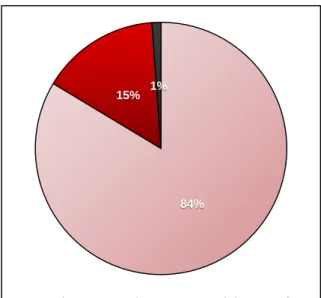
Figure 3.1: Moisture, organic, and ash content proportions obtained from Gracilariopsis longissima samples collected in February 2017 ... 43
Figure 3.2: Total protein content obtained from Gracilariopsis longissima samples collected throughout the year ... 44
Fig. 3.3: Chromatogram of the fatty acid methyl esters of Gracilariopsis longissima samples collected in February 2017 ... 49
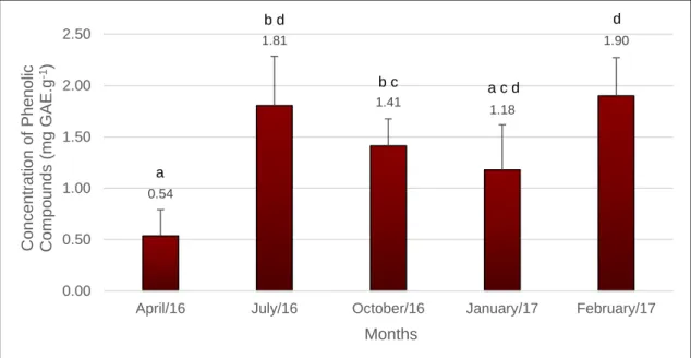
Figure 3.4: Total phenolic compounds obtained from Gracilariopsis longissima samples collected throughout the year ... 53
Figure 3.5: DPPH Scavenging effect obtained from ethanolic extracts of Gracilariopsis longissima samples collected throughout the year ... 54
Figure 3.6: Antibacterial activity of Gracilariopsis longissima extracts ... 58
Figure 3.7: Inhibition halos obtained with extracts of Gracilariopsis longissima samples, against the growth of Escherichia coli and Bacillus subtilis ... 58
xiv
Index of Tables
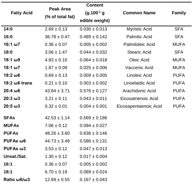
Table III.I: Fatty acid profile of total lipids extracted from Gracilariopsis longissima
samples collected in February 2017. ... 48
Table A.I: Composition of the stock solutions required for Von Stosch’s Enriched
Seawater Medium, for use with red algae ... 81
Table A.II: Length of Gracilariopsis longissima tips determined at the beginning and
ending of each salinity trials ... 82
Table A.III: Daily Growth Rate, according to salinity, determined for Gracilariopsis
longissima ... 83
Table A.IV: t-student statistical analysis performed upon the Daily Growth Rate data
determined for Gracilariopsis longissima... 83
Table B.I: Values obtained for the moisture, organic, and ash content determined for
Gracilariopsis longissima samples collected in February 2017 ... 85
Table B.II: Protein content determined by the Kjeldahl method, in Gracilariopsis
longissima collected in 2016 and 2017. ... 85
Table B.III: t-student statistical analysis, performed upon the protein content determined
in Gracilariopsis longissima ... 85
Table B.IV: Total phenolic compounds obtained from extracts of Gracilariopsis
longissima samples collected throughout the year ... 85
Table B.V: t-student statistical analysis performed upon the total phenolic compound
values obtained from Gracilariopsis longissima samples ... 85
Table B.VI: DPPH Scavenging effect obtained from extracts of Gracilariopsis longissima
samples collected throughout the year ... 85
Table B.VII: t-student statistical analysis, performed upon the DPPH scavenging effect
values obtained from Gracilariopsis longissima samples ... 85
Table B.VIII: Antibacterial activity of extracts of Gracilariopsis longissima samples
collected in April, July, October, and January, against the infectious bacteria Escherichia
coli, Bacillus subtilis, and Vibrio alginolyticus. ... 85
Table B.IX: t-student statistical analysis performed upon the antibacterial activity of the
1
Introduction
The oceans are home to amazingly diverse and interesting life forms, providing home to nearly 90% of the organisms inhabiting Earth, yet surprisingly, and although covering more than two-thirds of the Earth’s surface, its depths are still widely unknown. This is, in main part, due to the extreme environments that characterize the deep blue, rendering human life impossible and research a genuine challenge. It is generally known, however, that oceans provide many unique environments and resources, with plentiful and diverse marine organisms with great potential to produce bioactive compounds with applications in several fields of science and healthcare. Within this group, macroalgae stand prominently as one of the most interesting resources of such compounds (Ibañez and Cifuentes 2013; Andrade et al. 2013; Kim and Chojnacka 2015), as they are perfectly adapted to a wide range of ecological aquatic niches. Algae produce diverse and unique compounds and secondary metabolites in order to survive and compete successfully in their environment, having built perfectly fine-tuned defence strategies and metabolic pathways as a response to their perpetual adaptation and evolution over the last 2.45 billion years (Cardozo et al. 2007; Wang et al. 2015). Reflecting such potential, research has already chemically determined over 15.000 novel compounds from both macro and microalgae (Cardozo et al. 2007).
Macroalgae are, in the broadest definition, macroscopic and multicellular aquatic plants that lack the highly specialized structures of higher plants, such as true leaves, stems and roots. Also known as seaweeds, their modest structure consists of a thallus, sometimes provided with a leaf-like lamina or blade, and a holdfast to provide attachment to a surface (Gallardo 2015). Marine macroalgae can thrive in a great variety of ecological niches, namely intertidal, shallow, and deep sea areas up to 180 m depth, and also in estuaries, either freely floating or attached to a solid subtract (Vijayan et al. 2016). Macroalgae perform photosynthesis, and possess biological and ecological functions similar to those of higher plants. All seaweeds have the photosynthetic pigments chlorophyll a, carotenoids, and xanthophylls and are classified into three main groups, according to the visible colour conveyed by the nature and combination of their dominant pigments: the green algae Chlorophyta contain the chlorophylls a and b, and carotenoids; the brown algae Phaeophyceae (phylum Heterokontophyta) has the chlorophylls a and c, carotenoids, and xanthophylls with fucoxanthin as the dominant pigment, and the one responsible for their characteristic brown colour; and the red algae Rhodophyta contain the chlorophylls a and d, carotenoids, and a number of phycobilins such as phycoerythrin, which gives these seaweeds their typical reddish hues (Rowan 2011; Gallardo 2015; Pereira 2016). The selective presence in these pigments is related
2
to the sea habitat of the seaweed, due to their different light intensity requirements to perform photosynthesis. Green algae generally occupy niches with large amounts of sunlight energy available, such as coastal areas, whereas brown algae are mainly found in intermediate depths; red algae, however, are better adapted to greater depths or sheltered niches were sunlight radiance is limited (Barsanti and Gualtieri 2014).
Seaweed Industry and Biotechnological Applications
Historically, seaweeds are harvested from nature, yet since the early 20th century there
has been a fast and significant growth of the seaweed cultivation, not only as a response to continuous and increasing demands in food and pharmaceutical industries (Yang et al. 2015), but also to counter the risk of resource exhaustion. The depletion of natural stocks is an issue especially pertinent to coastal regions still lacking proper management of the sustainable exploitation of seaweed natural stocks (Rebours et al. 2014). Forster and Radulovich (2015) list several species which currently are the selected targets of the existing seaweed farming industry, known to perform well in culture systems, namely the brown seaweeds Laminaria japonica and Saccharina lattisima, and various Porphyra (Rhodophyta) species for temperate waters; for tropical waters, farming industries prefer the red seaweeds Kappaphycus alvarezii and a number of Euchema species, being the latter grown mainly for hydrocolloid production. Other seaweed species are grown commonly for food, though in far smaller amounts, such as species belonging to the genus Gracilaria (Rhodophyta), Ulva and Caulerpa (Chlorophyta), and Sargassum (Phaeophyceae). The abovementioned authors call to attention, however, that it is too premature yet to settle on preferences from among this list.
Seaweeds have been part of the human diet for thousands of years, based on archaeological evidence in Chile dating back to 14 000 years before present, and early written records from China (300 A.D.) and Ireland (600 A.D.) (Wells et al. 2017). Seaweeds now currently hold worldwide recognition as an important living natural provider of various chemicals, being one of the richest and most promising source of bioactive primary and secondary metabolites. Phycocolloid compounds such as agar, carrageenan, and alginate are examples, being widely used in food, cosmetics, and biomedical industries as gelling, thickening, and stabilizing agents (Samaraweera et al. 2011; Kasanah et al. 2015; Pereira 2016). Polysulfated polysaccharides are other examples, namely galactosyl glycerol and fucoidan, to name a few, whose strength lies in their use as antioxidant, antiallergic, anti-HIV, anticancer, and anticoagulant agents (Jiao et al. 2011).
3
Many Asian countries highly regard seaweeds as a valuable resource, and use them as diet staples on a daily basis (Sahoo and Yarish 2005; Pereira 2011; Pereira 2016). Japan was the first country to coin the term “functional food” in the 1980s, to name food products fortified with components promoting the human health, and endorsing this way a further understanding of the relationship between nutrition and health which stands as the basis of this concept (Hamed et al. 2015). This traditional view seeped into the western culture and is now extensively supported by algal researchers worldwide, who brand seaweeds as potential candidates as an healthy food source, with low calorie content, rich in polysaccharides, minerals, vitamins, essential amino acids and dietary fibers (Mabeau and Fleurence 1993; Lordan et al. 2011; Syad et al. 2013; Carvalho and Pereira 2015; Debbarma et al. 2016; Wells et al. 2017). Macroalgae are acknowledged to fight obesity, tackle free radicals, reduce the incidence of cardiovascular diseases, and promote a healthy digestion (Plaza et al. 2008; Cardoso et al. 2015; Roohinejad et al. 2016). Moreover, recent studies also stress the potential value of seaweeds as therapeutic agents, with reportedly demonstrated biological properties such as antibacterial (Kasanah et al. 2015), antifungal (Peres et al. 2012), antiviral (Chen et al. 2013), antioxidative (Yang et al. 2012; Yeh et al. 2015; Raja et al. 2016; Pinteus et al. 2017), anti-inflammatory (D’Orazio et al. 2012; Lee et al. 2013), anti-tumour (Horta et al. 2014; Pádua et al. 2015; Rodrigues et al. 2015a), and antihypertensive activities (Suetsuna et al. 2004; Fitzgerald et al. 2012; Paiva et al. 2016). A detailed review by Vijayan et al. (2016) focus on seaweeds as a novel and eco-friendly source in bionanotechnology, relevant to the fields of medicine, environmental monitoring, and electronics.
Seaweeds even take part in human day-to-day life on a regular basis, in certain ways: soap, toothpaste, shampoo and air freshener, are just a small sample of products that often contain compounds of macroalgae origin. Recent awareness of the state of the planet, under anthropogenic pressure and depletion of its natural resources, as well as suspicion over products with chemical ingredients, has led most of the general public into heeding a more environmental-friendly lifestyle and preferring basic, natural and environmentally sustainable products. This relatively new tendency has promoted research into the potential of marine algae as cosmeceuticals (Wang et al. 2015), with the encouraging results driving cosmetic companies to increasingly advertising seaweed extract based products among others (Carvalho and Pereira 2015), soundly promoting their benefits to a progressively attentive audience.
Seaweeds play a critical role as ecosystem engineers (Jones et al. 1994), by protecting shorelines and shaping reefs, providing shelters and serving as feeding/nursery ground of marine organisms (Carvalho and Pereira 2015), ultimately creating habitats and
4
promoting species diversity (Haywood et al. 1995). Seaweeds are also known to reduce eutrophication, control harmful microalgal blooms, absorb nutrient excess, remediate contaminants and sequestrate CO2 (Aresta et al. 2005; Singh et al. 2011; Chen et al.
2015; Cabral-Oliveira et al. 2016; Krause-Jensen and Duarte 2016), ultimately contributing to the improvement of coastal environments. Both wild and farmed seaweed communities can potentially mitigate global warming by dampening wave energy, buffering ocean acidification, and providing oxygen to the waters (Roleda and Hurd 2012; Castro and Huber 2016; Duarte et al. 2017). Yang et al. (2015) discuss in their review not only the aforementioned benefits, but also state that the large-scale cultivation of seaweed holds the key to eco-friendly water quality improvement in the coastal waters of the world. Algal pigments have been applied as trophodynamic indicators in biogeochemistry and ecology studies, with research focused mainly on chlorophylls and carotenoids (Kleppel 1988). Current research also investigates the applicability of algae as a potential biofuel source, shedding light upon standpoints on macroalgae-based biorefinery technology (Chen et al. 2015; Suutari et al. 2015; Kumar et al. 2016). More recently, seaweeds are an increasingly valued component in Integrated Multi-Trophic Aquaculture (IMTA) systems (Abreu et al. 2011b; Carvalho and Pereira 2015; Samocha et al. 2015), in which two or more trophic levels grow in one farm, and the by-products provided by one species can feed another; fed aquaculture is combined with inorganic and organic extractive aquaculture to create a synergistically balanced system, as close as possible to natural schemes (Neori et al. 2004). Therefore, there is a growing tendency of the research related to the improvement of seaweed (Raikar et al. 2001; McHugh 2003; Reddy et al. 2008; Baweja et al. 2009), essential to the development of the commercial seaweed industry. However, to this date, merely about 5% of the roughly ten thousand identified algae species worldwide is being explored, especially as human food or animal feed. The highest contributor to this low percentage are the Asian countries, where seaweeds are used as sea vegetables, being an inherent part of their culture and routine (Schmid et al. 2014). Conversely, in Southern Europe edible seaweeds have still a meagre contribution to feeding habits and nearly no place in regional recipes; the assumption that seaweeds are unfit for consumption is still deeply rooted in the European mindset, and Portugal is no exception to this. As research is being released and updated, however, this trend is shifting into a slowly but steady recognition of the potential seaweeds present due to their nutritional, pharmaceutical, and cosmetic value (Pereira 2011).
5
The Red Seaweed Gracilariopsis longissima
Species Profile
The marine Rhodophyta Gracilariopsis longissima (S. G. Gmelin) (Steentoft et al. 1995) is widely known by several common names: cabelo de velha in Portuguese; thin dragon beard plant and Ceylon moss in English; hai mien san, fen tsai, hunsai, hai tsai, and hoi tsoi in Chinese; ogo and ogo-nori in Japanese; nuoc-mam, rau-cau, and xoa xoa in Vietnamese (Pereira 2016; Guiry and Guiry 2017). Some of these common names may possibly derive from its exceptionally bushy appearance, given by the repeatedly division of the plump branches (up to 2 mm in width) which protrude from the cylindrical shaped terete thallus. Healthy specimens of G. longissima usually hold a deep dark red colour, being nearly black in the base of the thalli; similarly to many other seaweed species, the rich colour fades to a complete greyish white when the individual perishes. Colour variants such as green and yellow may also appear, and it is likely the result of ordinary genetic controls, as observed for the Gracilaria genus (Santelices and Doty 1989). The male and the female gametophytes can be identified by the presence of cystocarps in the latter, which are fruiting bodies which appear as distinct semi-circular dark lumps scattered all over the thalli (Sahoo and Yarish 2005; Pereira 2016). G. longissima occupies a variety of habitats both in tropical and temperate latitudes, on intertidal or shallow-subtidal surfaces with low water motion, and where the sediment is sandy or muddy. Having a worldwide distribution, G. longissima is found in SW and SE Atlantic, Indian Ocean, SW Asia in Iran, Israel and Sri Lanka, SE Asia in Vietnam, NE and E Pacific, Australia, and Pacific Isles in Hawaii, Polynesia and Samoa (Pereira 2016).
The current standoff in Gracilariaceae taxonomy
Extensive research, reports and reviews attest the genus Gracilaria exceptional value regarding several fields of health and industry (Santelices and Doty 1989), rendering the research and findings in Gracilariopsis genus very poor in comparison. Mendeley database currently includes 2.321 entries under the name Gracilaria and only 245 entries under the name Gracilariopsis; a quick NCBI research yields results filed under the name
Gracilaria in 22 databases, whereas the name Gracilariopsis is presented in 15
databases, and in a comparatively lower number of entries in each database (for example, in the nucleotide database there are 3.480 sequences submitted for Gracilaria, and only 721 for Gracilariopsis) (NCBI Resource Coordinators 2017). The hypothesis of identification irregularities is put forward, where there is the possibility that all
Gracilariopsis spp. might have been identified as Gracilaria spp. in several studies and
even FAO reports, given not only the outstanding similarity between both genera, but also the fact that the genus Gracilariopsis was only recently reinstated by Fredericq and
6
Hommersand in 1990 (Guiry and Guiry 2017). Knowledge of the reproductive structures permits the trustworthy taxa identification of gracilarioids, however, some populations are sterile and grow merely by vegetative fragmentation (Santelices and Doty 1989). Morphologically barely indistinguishable from other Gracilariopsis species, and even from species belonging to the entire family, the identification of the species requires a well-trained eye. Consequently, the entire taxonomy of Gracilariaceae, especially between the Gracilariopsis and Gracilaria genus, is currently terribly chaotic (Bird 1995) and occasionally put under revision (Santelices and Doty 1989). Confusion often arises regarding any given species distribution, life-history, and even annual raw production values, or biotechnology value assessments, when said species do not hold a guaranteed identity. Therefore, all the reports which concern Gracilaria spp. and
Gracilariopsis spp., whose identification was not established with confidence but
performed having morphology observations alone as basis instead, place the entire findings and reports for both genera at jeopardy and may not reflect the reality. The currently untidy state of the taxonomy of Gracilaria and Gracilariopsis has even led to the release of a statement by M. D. Guiry, from AlgaeBase (Guiry and Guiry, 2017) found in a few Gracilaria and Gracilariopsis individual species page (including G. longissima), declaring that specimens reported to date as Fucus verrucosus, Fucus confervoides, and all terete Gracilaria from the NE Atlantic and elsewhere, actually require individual examination in order to define whether they belong to Gracilaria or Gracilariopsis genus, before assigning an individual species identity. Therefore, and although often confused with Gracilaria gracilis and once known as Gracilaria verrucosa, G. longissima is the current taxonomically accepted name for the species (Guiry and Guiry 2017), and the acknowledged identity of the individuals studied in the current work.
7
Study Site: Lagoa de Óbidos, Caldas da Rainha, Portugal
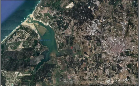
The Lagoa de Óbidos (Caldas da Rainha, Portugal) is a semi-enclosed lagoon situated in a shallow depression, which forms a natural barrier placed between the Atlantic Ocean and the riverine ecosystem of Foz do Arelho. The lagoon is extended by two branches, the Braço do Bom Sucesso by West and the Braço da Barrosa by East (Fig.: 1.1), and is bordered by the municipalities of Caldas da Rainha by North, and Óbidos by South.
Being the widest tidal inlet in the Portuguese shore (6.9 km2 area, 2 m average depth),
Lagoa de Óbidos provides shelter to a rich diversity of wildlife, including the red seaweed
G. longissima, which grows and thrives as unattached, entangled mats on mud, and
often underneath a dense meadow of sea lettuce (Ulva lactuca). The sampling site where
G. longissima was collected is shown in Fig.: 1.2.
Figure 1.1: Satellite image representing Lagoa de Óbidos and the main anthropological
8
Due to its natural tendency of closing its connection to the sea, Lagoa de Óbidos is at risk of disappear completely, and thus it currently depends on sensible human intervention to reverse this situation. The lagoon continually shifts its morphology, with channels and sandbanks under constant dynamism, mainly due to tide and wave action (Malhadas et al. 2009) although it is limited to a few hundred meters upstream of the mouth (Fortunato and Oliveira 2007). The upper lagoon is hardly affected by tides, and with freshwater also playing a minor role, there is an overall and progressive reduction of the average depth and the surface area, further aggravated by accretion and large residence times, of up to several years (Fortunato and Oliveira 2007). A number of authors suggest that such conditions do not favour pronounced variations on salinity on Lagoa de Óbidos (Carvalho et al. 2005; Santos et al. 2006).
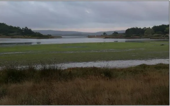
Figure 1.2: Sample site, corresponding to the red marker depicted in fig. 1. The seaweed
9
Objectives
The main objective of the present work was to study the red seaweed Gracilariopsis
longissima occurring in Lagoa de Óbidos, Portugal, in order to determine the growth
rates of the seaweed under laboratory conditions and to evaluate its nutritional profile and bioactive capacity throughout the year.
Specific objectives:
1. To study the growth rate of G. longissima under distinct salinity conditions, ranging from 0 to 35‰.
2. To establish the best decontamination protocol of G. longissima under laboratory conditions.
3. To assess G. longissima nutritional profile, namely seasonal variation of protein content, and fatty acids profile.
4. To determine seasonal variations of antioxidant capacity, by total phenolic content and the DPPH radical scavenging activity.
5. To evaluate seasonal variations of antimicrobial value, namely antibacterial resistance against the Gram negative bacteria Escherichia coli, the Gram positive bacteria Bacillus subtilis, and the Gram negative marine bacteria Vibrio
alginolyticus.
The present manuscript is structured in two main chapters, Chapter One and Chapter Two, each corresponding to a paper ready to be submitted to a scientific peer-reviewed journal. The main conclusions achieved throughout the course of this work are outlined at the end of each chapter, whereas conclusive remarks pertaining both chapters are delineated at the end of the document. In order to achieve a clean and organised layout for ease of reading, the Bibliography section encompasses references that were cited throughout the entire manuscript, and was thus placed solely at the end, instead of following each chapter. Regarding the Appendix section, it follows the Bibliography, and it is divided in two parts, Appendix A and Appendix B to match, respectively, Chapter One and Chapter Two, from where they are relevant to.
11
Chapter One
The role of salinity on the survival and growth performance of
Gracilariopsis longissima (Rhodophyta, Gracilariales): an assessment
performed on a laboratory scale
Abstract
The adoption of the ideal conditions is of utmost importance in any seaweed farming system, in order to achieve high biomass production and lucrative yields for the species target, which will ultimately translate into a successful and profitable business. In this sense, the survival and growth performance of the red seaweed Gracilariopsis
longissima was evaluated, under a wide range of salinities and in small-scaled, controlled
laboratory conditions. Clean G. longissima thalli was grown under controlled conditions (24 ± 1ºC, photoperiod set at 12:12 (Light:Dark) and provided by daylight cool white fluorescent lamps (10-15 µmol photons m-2 s-1), in Von Stosch Media enriched seawater
at distinct salinities, ranging from 0 to 35‰, for 44 days. Daily growth rates were assessed having the initial and final length of the thalli, measured at day 0 and day 44, respectively, and number of new ramifications were also counted. The seaweed G.
longissima is highly tolerant to a wide range of salinities, namely from 20 to 35‰,
although being unfit to survive at salinities below 20‰. The seaweed presented the maximum fitness at 35‰, observed by a growth rate of 1.611%.day-1, and also revealed
16 new ramifications throughout the length of the assay. Insights on the influence and possible solutions on contaminations such as those caused by fungi and epiphytes, which are unquestionably hazardous to any seaweed culture system, are also provided. The present study offers therefore the first steps towards a throughout assessment of the ideal conditions, which must be applied into a culture system dedicated to the growth of G. longissima, in order to ensure that the maximum performance for the species is achieved.
Keywords: Red macroalgae, growth rate, ramifications, cultivation, contamination control.
Introduction
Algal culturing techniques have been described in books and papers since the early 19th
century, from which Preisig and Andersen (2005) and Barsanti and Gualtieri (2014) briefly list the most noticeable advances, especially in microalgae culturing. Several methods and concepts such as media formulations, as well as reports on keeping healthy
12
and pure axenic cultures, have been developed and published as earlier as the late eighteen century. In regards to macroalgae, however, until the 1950s nearly all economically important seaweeds were still harvested from wild populations only, with the exception of the macroalgae Porphyra, the commonly known nori, which holds a long cultivation history dating back as early as the 17th century (Chen and Xu 2005). Concerns
eventually arose regarding the risk of resource depletion due to overharvesting from 1950 onwards and the full disclosure of the complete life history of Porphyra has led to refinements and development of culture techniques and industry for this macroalgae, while also paving the way for the establishment and expansion of culture industries dedicated to many other economically important seaweeds (Tseng 1981; Sahoo and Yarish 2005). The overall aquatic plant farming, overwhelmingly of seaweeds, has been growing rapidly since then and it is now current practice in about 50 countries, having reached 27.3 million tonnes of production in 2014, which constituted one-quarter of the worldwide total aquaculture production by volume for this year (FAO 2016). Nowadays, large-scale seaweed cultivation techniques are standardized, routine, and economical; a successful culture takes into account several factors such as morphology and regeneration capacity of the thallus, and the synergy between irradiance, temperature, nutrients, salinity, and water movement (Sahoo and Yarish 2005). Depending on the chosen farming method, challenges may arise in the form of epiphytes (Msuya et al. 2014), fouling, and expensive infrastructures and nutrient requirements. Full knowledge of the species is crucial as well, as different taxa require distinct farming methods. China is currently the largest mariculture producer in the world, undergoing rapid development during the last three decades, and reaching annual productions as high as 45 469.0 thousand tonnes in 2014, contributing thus to 61.62% of the world total production (FAO 2016). Gracilarioid species, to which both Gracilaria and Gracilariopsis are a part of, significantly account to these numbers, as they are one of the most cultivated seaweeds worldwide: regarding the production of farmed aquatic plants in the world, Gracilaria spp. reached values around the 3752 thousand tonnes in 2014, only surpassed by Kappaphycus alvarezii and Eucheuma spp. (10 992 thousand tonnes), and
Laminaria japonica (7 655 thousand tonnes) (FAO 2016). The reasoning underlying such
high values is the great value of Gracilariaceae species mainly as an agarophyte, being a tremendously valued source of hydrocolloids (Bixler and Porse 2011; Abreu et al. 2015), but also as a food and feed component (Chopin et al. 2001; Hernández et al. 2006; Mantri et al. 2009; Pereira 2011; Yarish et al. 2012; Kim and Yarish 2014; Samocha et al. 2015; Yang et al. 2015). Particularly Gracilariopsis longissima (S. G. Gmelin) (Steentoft et al. 1995) has been widely studied in Integrated Multi-Trophic
13
Aquaculture (IMTA) systems (Yang et al. 2015), with recent studies endorsing the several potential contributions this seaweed may provide to such systems, namely high performance as a biofilter agent (Hernández et al. 2005; Hernández et al. 2006; He et al. 2014), high tolerance to excessive levels of micronutrients such as copper (Brown and Newman 2003; Brown et al. 2012), ability to withstand ultraviolet radiation (Álvarez-Gómez et al. 2017), and thermal requirements for survival and growth (Steentoft and Farnham 1997).
To this date, however, and to the best of the knowledge and information gathered, studies reporting the growth performance of G. longissima, and even of the entire genus are scarce, when compared to the research already performed for the Gracilaria genus. In an attempt to fill this gap, the objectives of the present studies were to study the red seaweed G. longissima occurring in Lagoa de Óbidos, Portugal, in order to evaluate growth rates under distinct salinity conditions, ranging from 0 to 35‰, and therefore to determine both its salinity range of survival and the salinity value for optimal growth. The results obtained will contribute to a full assessment of the ideal set of conditions required to undertake a successful and profitable culture of this seaweed.
Materials and Methods
Sampling and Acclimatization
Specimens of G. longissima (Rhodophyta, Florideophyceae, Gracilariales) were harvested from Lagoa de Óbidos, in Caldas da Rainha, Portugal (39°24'18.93"N, 9°11'13.05"W) in September 2016, during low tide and transported to the laboratory in plastic containers. The classification of this seaweed was based on AlgaeBase (Guiry and Guiry 2017) and confirmed by botanists. In the laboratory, each plant was first washed with running seawater and cleaned thoroughly to remove debris, necrotic parts, epiphytes, and other organisms from the thalli surface. The seaweeds were then kept in constantly aerated seawater (25-30‰) during the following week, in a climatic room (24 ± 1ºC) for adjustment purposes. The photoperiod was set at 12:12 (Light:Dark), with the irradiance being provided by daylight cool white fluorescent lamps (10-15 µmol photons m-2 s-1).
Selection and Isolation of Healthy Tips
A successful culture initiation requires the establishment of an axenic culture, by either spore or tip isolation. The selection, isolation, and cleaning of healthy seaweed tips was performed according to Yarish et al. (2012), being this the most critical step in culturing any seaweed. Therefore, from the previously acclimated G. longissima stock, fronds
14
exhibiting a deep dark-red colour and fleshy thalli, traits that are indicative of healthy individuals, were chosen (Fig.: 2.1). The fronds were rinsed in a series of vessels containing clean seawater, followed by a final and quick rinse of no longer than 60 seconds in clean freshwater, enough to induce osmotic shock to any adhering organisms without straining the seaweed.
Starter thalli were obtained by carefully cutting the tips (1-2 cm) from each cleaned and rinsed parent frond, as it is the area corresponding to the apical tissue where new and active growth happens. Each tip was individually and meticulously wiped down with sterilized cotton-tipped swabs, and subsequently dragged through an agar gel previously prepared in Petri dishes (1.0% bacteriological agar, VWR, Radnor, PA USA, in 1:1 distilled water/seawater ratio) to pull off any remaining microscopic contaminants. The agar drag was performed three times for each tip, and always through unused portions of the agar gel. All tools, seawater and distilled water used in cleaning process were previously sterilized by autoclave (121 °C, 15 minutes).
Growth Experiments
Selection of healthy fronds and isolation of tips followed the aforementioned Yarish et al. (2012) methodology. Healthy and clean seaweed tips were placed into 250 ml flat bottom flasks (8 tips per flask) and provided with sterilized seawater enriched with Von Stock solution (VSE), prepared according to Redmond et al. (2014). The components comprising the VSE media are the macronutrients nitrate and phosphate, the micronutrients iron and manganese, EDTA, and the vitamins B12, thiamine, and biotin
(Appendix A, table A.I). Salinity tests were carried out at 0, 2, 5, 10, 15, 20, 25, 30 and
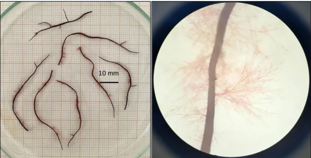
Figure 2.1: Healthy frond of Gracilariopsis
longissima collected from Lagoa de Óbidos, Portugal. The deep dark red coloration is particularly noticeable, especially in the thicker thalli.
15
35‰, being these values achieved by adding distilled water to filtered water (particles, sand, and UV filters were used), and checked with a refractometer (E-line refractometer, Bellingham and Stanley, China). The water was then sterilized by autoclave (121ºC, 15 minutes) and further enriched with Von Stosch solution. Germanium dioxide was also added to the medium to prevent the growth of diatoms, and nystatin (Mycostatin® 100
000 UI.ml-1, Bristol-Myers Squibb) to prevent and control fungi contamination (Yarish et
al. 2012). Medium was changed weekly throughout the duration of the experiment, whereas nystatin was supplemented in the first week only. Cultures were kept in a climatic room (24 ± 1ºC) under constant filtered aeration, with photoperiod set at 12:12 (Light:Dark) and provided by daylight cool white fluorescent lamps (10-15 µmol photons m-2 s-1) for 44 days, according to Yarish et al. (2012) and Hayashi et al. (2007b) (Fig.:
2.2). All the specimens were transferred to progressively lower or higher salinities (being 25‰ the starting point) on a weekly basis, in order to allow their adaptation and prevent stress caused solely by abrupt salinity shifts. Triplicates and controls were performed for all the assays, and clean stocks were kept as backup during the experiment. Tips that exhibit loss of pigmentation, visually observed by partial or total tip discoloration, were considered to be under stress or dead, and were thus removed from the assay.
Growth Measurements
Growth was recorded as changes in tip length and number of ramifications. Therefore, measures were performed along the main branch of each tip, and total ramifications per tip were counted; initial measurements were taken at the beginning of each assay, and final measurements taken after 44 days, for the salinities where seaweeds were
Figure 2.2: Culture setup of Gracilariopsis longissima assay at different salinities, assembled
in a climatic room (24 ± 1ºC) with photoperiod set at 12:12 (Light:Dark) and provided by daylight cool white fluorescent lamps (10-15 µmol photons m-2 s-1).
16
successfully thriving. Measures were determined upon full-scaled photos of G.
longissima tips placed upon millimetric paper, in Adobe Photoshop® CC software (Adobe
Systems, San Jose, CA USA). Daily growth rate calculations were performed based on the equation below, according to Mtolera et al. (1995), Gerang and Ohno (1997), Aguirre-Von-Wobeser et al. (2001), Bulboa et al. (2007), Hayashi et al. (2007a), Hayashi et al. (2007b), Hung et al. (2009) and Hayashi et al. (2011), whose formula is the one recommended to be used as the standard for seaweed growth rate determination (Yong et al. 2013).
Daily Growth Rate (% day-1)
= [ (
𝐿𝑡𝐿0
)
1 𝑡− 1 ] × 100
Where L0 and Lt stand as, respectively, the length measured at day 0 and day 44 of the trials, and t corresponds to the duration of the assay, in days.
Contamination Control
Seaweed cultures have often long timeframes from the excising of tips to the outplanting of individuals, and they can be easily ruined by contaminations of other microalgae, epiphytes, cyanobacteria, or fungi, which force the start of a new culture from the very beginning (Redmond et al. 2014). In this sense, contamination studies are quite helpful, as they frequently give tools and methods to prevent infection surges and subsequent damage of a seaweed culture, thus preventing financial and time losses. Therefore, in the present study additional assays were performed, to establish effective procedures against fungi contaminations.
Selection of healthy fronds and isolation of tips followed the method by Yarish et al. (2012) as previously described. After cleaning, each seaweed tip was individually immersed during 20 seconds in a fungicide or disinfectant solution, or 10 seconds for each solution when performing successive immersions in different agent solutions. Each seaweed tip was then independently placed in a test tube with 5 ml of Von Stosch media enriched seawater at 30‰ salinity and kept for four weeks. Agents tested were the antifungal medication Nystatin (Mycostatin® 100 000 UI.ml-1, Bristol-Myers Squibb, New
York City, NY USA) further dilluted at 5, 10, and 20 ml.L-1, the systemic fungicide Tocsin
WG (70% p/p thiophanate-methyl, Sipcam, Lisbon, Portugal) at 1 g.L-1, the preventive
fungicide Pomarsol (80% p/p of thiram, Bayer, Carnaxide, Portugal) at 0.2 g.L-1, and the
disinfectant hydrogen peroxide (H2O2 10 volumes, Continente, Matosinhos, Portugal)
17 g.L-1) then Nystatin (5 ml.L-1), H
2O2 then Nystatin (5 ml.L-1), and H2O2 then Tocsin WG
(1 g.L-1). An additional experiment was also performed where Nystatin was added to the
enriched seawater instead (at 1 ml.L-1), therefore allowing the treatment to act
continuously throughout the assay period. For plant fungicides, all the initial working concentrations were achieved based on manufacturer recommendations. Media was changed weekly, and controls and quadruplicates for each assay were kept. After two days, and on a weekly basis throughout the length of the four weeks assay, a selection of healthy seaweed tips was individually placed upon a potato dextrose agar (VWR, Radnor, PA USA) plate, which was then sealed and incubated at 30ºC. The present of fungi was visually confirmed following the two days of incubation on the agar. Effectiveness of any given fungicide concentration was evaluated by the seaweed fitness while on trial, and by the presence of fungi following the incubation period.
Statistical Analysis
Data is expressed as means ± standard deviation. The Student’s t-distribution test was performed to detect significant differences on the calculated daily growth rates according to salinity, considering the level of significant difference of p < 0.05. The software Microsoft Office Excel 2013 was used to perform all the statistical analysis.
Results and Discussion
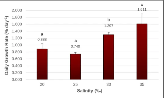
Growth Rate Assessment
Specimens of G. longissima were grown under controlled laboratory conditions at salinities ranging from 0 to 35‰, with constant surveillance of their performance during 44 days, and daily growth rates were determined. Fig.: 2.3 represents the daily growth rates calculated for seaweed growing at 20‰ and above only, corresponding to individuals which survived through the 44 days trial (see also Appendix A, Table A.II, Table A.III, and the corresponding p values of the t-student statistic test in Table A.IV). The figure 2.4 stands as an example of tips which remained healthy after the 44 days, where the successful performance is visually observed by the permanence of the dark rich colour throughout the assay, and by the fragmentation through vegetative propagation occurring in some tips. The highest daily growth rate value corresponds to seaweed growing at 35‰ (1.611%.day-1), being the significantly highest value measured
(t-student, p < 0.05), followed by seaweed growing at 30‰ (1.297%.day-1). Seaweed
kept at 20 and 25‰ yielded significantly lower daily growth rates than those obtained for seaweeds growing at 30 and 35‰ salinity, yet presenting no significant difference between each other (respectively, 0.888 and 0.740%.day-1) (t-student, p = 0.182).
18
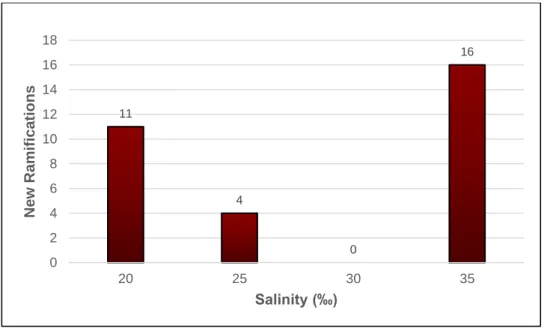
The number of new ramifications was also determined, according to salinity, with the results presented in Fig.: 2.5, again pertaining only to salinities where the individuals survived through the 44 days trial. The seaweed kept at 35‰ presented the highest number of new ramifications (16), followed by those kept at salinities 20 (11), 25 (4), and 30‰, the latter presenting no ramifications.
0.888 0.740 1.297 1.611 0.000 0.200 0.400 0.600 0.800 1.000 1.200 1.400 1.600 1.800 2.000 20 25 30 35 Daily G ro w th Rat e (% d ay -1) Salinity (‰)
Figure 2.3: Gracilariopsis longissima daily growth rates (% day-1) determined after 44 days,
at the salinities of 20, 25, 30 and 35‰. Results are expressed as mean ± standard deviation. n = 3. Letters a-c indicate statistical differences according to the t-student statistical test, with p < 0.05.
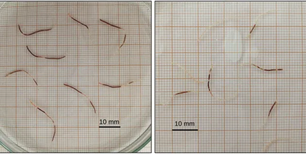
Figure. 2.4: Gracilariopsis longissima tips thriving at 25‰ photographed at day 0 and day 44
of the trials. The successful fitness is visually observed by the unchanging deep red colour and vegetative propagation of some tips.
a a b c 10 mm 10 mm
19
Seaweeds kept at 15‰ and below perished within the 44 days trial, and thus their daily growth rates were not determined and data is not shown. Salinities of 15‰ and below proved to be progressively lethal to the seaweed, as the algae segments died within the first three weeks of the experiment. Salinities of 10 and 5‰ were lethal to the segments after the first two weeks of culture, whereas at salinities of 2‰ the segments died after two days in culture. Distilled water also proved to be lethal within the first 24 hours in culture, as proven by an assay with specimens transferred directly from 5 to 0‰. Figure 2.6 shows examples of the colour of seaweed tips kept at stressful conditions, exhibiting partial or total tip discoloration which corresponds to necrotic parts. The bleaching occurred starting either at the tips or in the middle section of the thalli, eventually spreading to the whole individual. Dying individuals also lost their characteristic fleshy texture, becoming flaccid and breaking down to the touch.
11 4 0 16 0 2 4 6 8 10 12 14 16 18 20 25 30 35 New Ramif ica tion s Salinity (‰)
Figure 2.5: Gracilariopsis longissima number of new ramifications that appear throughout the
essay, according to salinity. Numbers on top of each bar represent the total number of new ramifications counted for each salinity. n = 24.
20
The present study shows that G. longissima growth rates were expressively reliant on salinity changes. G. longissima appears to be better adapted to the highest salinity studied (35‰), shown by the highest daily growth rate and the number of new ramifications counted, yet it is shown that at 20 and 25‰ these organisms are capable of survival and even to perform vegetative reproduction. At 30‰, G. longissima seems to have not invested energy on reproduction, as it does not produce new ramifications. Such differences may be attributed to adaptation to the habitat; considering that Lagoa de Óbidos is subjected to wide shifts in salinity, with values ranging from 17 to 38‰, it is therefore expected that G. longissima has the ability to adapt itself and grow in a wide range of salinity to respond to such abiotic shifts. However, in the natural world lesser growth rates as a consequence of a non-optimal salinity may not allow any given species to compete successfully (Bird and McLachlan 1986). Most wild populations of marine algae are mainly confined to the intertidal region, being thus subjected to wide variations in salinity, temperature and light. Indeed, these abiotic factors have been reported to have a key role in shaping the growth and distribution of benthic marine algae (Lüning 1990; Raikar et al. 2001; Nejrup and Pedersen 2012). Specifically, salinity determine the local and regional distribution of macroalgae in coastal areas, and is subject to sudden and wide shifts in response to precipitation, tides, and wind. Generally, waters from tidal pools and semi-closed inlets are hypersaline, while the inner regions of estuaries are mainly hyposaline (Nejrup and Pedersen 2012).
Figure 2.6: Gracilariopsis longissima tips photographed at the initial weeks of the 5 (left) and
10‰ (right) salinity essays. The observed discoloration correspond to a necrotic part and confirms the inability of the seaweed to thrive in these salinities.
10 mm 10 mm
21
The seaweed G. longissima from Lagoa de Óbidos is, however, and unlike most benthic marine algae, a specific case in which the organism is not only confined to a semi-enclosed, hypersaline area, but is also particularly sheltered from light as it is kept within a muddy substratum under a dense layer composed by the seaweed sea lettuce (Ulva
lactuca). In the present study, the specific site where G. longissima was collected
suffered deep fluctuations in salinity (from 17 to 38‰), while turbidity, substratum nature, and the presence of a U. lactuca layer determined the reduced light intensity received by G. longissima growing beneath. As such, and similarly to estuarine seaweed species,
G. longissima seems perfectly adapted to such fluctuations, possibly having either an
enhanced tolerance/elasticity to turgor pressure or cell volume changes, or the ability to perform osmotic acclimation mechanics, such as the reestablishment of the composition of cells (either by transport of ions across the cell membrane, or by the synthesis or degradation of organic osmolytes) (Kirst 1990). Such defence mechanics successfully counteract the osmotic gradient and prevent the irremediable damage caused by osmotic shock, such as cell wall disruption, impaired membrane functionality, and enzyme kinetics disturbance (Kirst 1990). However, it must be noted that such osmoregulatory processes come with an energy cost, which would otherwise be spent on growth, reproduction, and other essential metabolic processes if the seaweed was growing under the optimal salinity conditions as observed in previous studies (Hayashi et al. 2011; Nejrup and Pedersen 2012; Lawton et al. 2015). This fact stands out as a possible explanation for the lower growth values and fewer (or none at all) new ramifications obtained at salinities below 35‰: as a response to osmotic stress due to lower salinity values, G. longissima slew down growth and multiplication and spent energy to maintain the osmotic balance instead. Yet at extreme low values, such as 15‰ and below, such strategy only guarantee survival for a short period of time, according to Hayashi et al. (2011). In the present study, such particular survival tactic might have occurred more often in the few days following the seaweed transference to salinities progressively lower or higher, finally reaching a point of no recovery in extremely low salinity values. In light of such findings, determining not only the salinity growth range but also the optimal value (35‰) was crucial to the growth assessment of G. longissima under different salinities, performed in the present work, and will be of utmost importance when evaluating the yield, quality, and prospective value of the metabolites it produces.
Similar findings were also found for other Gracilariaceae by authors who studied both
Gracilaria and Gracilariopsis species grown under different salinities, either in a
controlled laboratory environment (Wilson and Critchley 1997; Israel et al. 1999; Raikar et al. 2001; Choi et al. 2004; Skriptsova and Nabivailo 2009; Hayashi et al. 2011; Nejrup
22
and Pedersen 2012), or in outdoor culturing (Israel et al. 1999). Specifically, Raikar et al. (2001) tested the influence of varying salinities to the growth rate of several Gracilaria species, from both tropical as well as temperate regions, finding a varied response which depended of the species considered; overall, however, and except for two isolates,
Gracilaria spp. from temperate regions did not tolerate salinities below 15‰, while
presenting abrupt increase in their growth rate at salinities between 20 to 30‰. Salinities above 30‰ were not considered by the aforementioned study. Skriptsova and Nabivailo (2009) studied both Gracilaria and Gracilariopsis species (Gracilaria gracilis, Gracilaria
tenuistipitata and Gracilariopsis bailiniae), finding that the former genus is tolerant to low
salinities (10‰) whereas the latter died; the authors also determined that the fastest growth rate for G. bailiniae was detected for 20-30‰, with values ranging from 4 to 5%.day-1 increase in weight. Nejrup and Pedersen (2012) found out that Gracilaria
vermiculophylla has an optimal growth above 15‰, and a reduced growth when exposed
to lower salinities, although salinities 5‰ and below proved to be stressful to this seaweed; the authors also report that G. vermiculophylla growth rate was higher when placed at constant salinity, when compared to organisms growing under salinity variations. Studies specifically pertaining G. longissima growth performance at different salinities were not found to date.
Seaweeds often exhibit a characteristic growth, marked by a rapid initial phase followed by decreased growth rates during later stages, as the photosynthesis rate decreases due to shelf-shading of the inner parts during growth. Therefore, in order to accurately determine the growth rate of a given seaweed, Yong et al. (2013) recommend to adopt as short as a weekly time interval between data collecting, while also stating that a daily regime for data calculation should be carefully considering due to loss of precision as a result of the high sensitivity of seaweed growth to their environment. In the present study, the full 44 days were adopted as a single time interval, where only the initial and final lengths of the seaweed were gathered, in order to avoid subjecting the small and delicate seaweed fragments to stress and eventual contaminations. Optimization of the methods used must be hence considered to collect data on a weekly time interval, while reducing seaweed transport and handling to a minimum.
In the present study, individuals always grew through vegetative propagation, which is a form of clonal propagation where all the new fronds grew from one single frond. This will result in a culture with genetically identical individuals, that may be useful to achieve consistency in production (Yarish et al. 2012). However, this also results in unattached, free-floating forms, that are not suitable for some culture methods that require some form of attachment to a surface such as the suspended line culture (Yarish et al. 2012).
23
Moreover, the necessity to trigger the formation of reproductive structures may arise in cultures, in order to bring the life history of individuals into completion (Rueness 2005). In the present study, no individuals carrying carpospores were observed or collected regardless of season. Carpospores are fruiting bodies present in female gametophytes, resembling small dark round bumps scattered along the thalli, and from where new individuals are born as a product of sexual reproduction (Yarish et al. 2012). Thus, the success rate of G. longissima in vitro sexual reproduction, which would allow the development of a pure axenic culture from a single individual, the completion of its life cycle in culture, and its attachment to a substratum on certain large-scale culture systems, remains to be tested against the success of in vitro vegetative reproduction.
Control of Contaminants
Fungi Control
One of the main factors that can swift and easily destroy cultures if left unchecked, is contamination by fungi, a problem that can only be tackled pre-emptively; there seems to be no way to remediate a fungal contamination, except to restart the culture from the beginning (Redmond et al. 2014). Alternative solutions to address such contamination are thus tremendously required.
After two days of incubation in potato dextrose agar (PDA), it was visually confirmed by microscopy that fungus still lingered upon a number of seaweed thalli, and no connection between the number of infected thalli and the type of fungicide agent was found. At least one replicate for each fungicide tested presented a certain degree of contamination, although the extent of the infection was less prominent on the seaweed thalli previously treated with Nystatin, regardless of concentration. The hydrogen peroxide was the only agent that killed every seaweed tested before the end of the 4 weeks trial, while failing to completely eradicate the fungi.
Moreover, after the incubation period in PDA media, it was observed a thick, opaque and seemingly glossy layer of a dark fluid bordering the thalli surface in a number of seaweed (Fig.: 2.7), again unrelated to the fungicide agent previously used, as at least one replicate per fungicide tested showed this aspect. The prominent black colour exuding from the algal tissue was not further tested to assess its identity, however, the currently standing hypothesis support the substance as being melanin, oozed by lingering fungi present on the algal tissue, and a consequence of a phytofungal infectious mechanism, further explained below. This finding is supported by descriptions and images found in previous works only, therefore it stands merely as a hypothesis that requires extensive further testing.
24
Melanins are a large group of biologically important natural pigments, often brown or black in colour, that can be synthetized by members of all the biological kingdoms (Nosanchuk et al. 2015), and are involved in the protection against ultraviolet radiation (Eisenman and Casadevall 2012). They are formed by oxidative polymerization of phenolic or indole compounds (Langfelder et al. 2003), but further details on their chemical structure remain unanswered, due to its ubiquity, large size, insoluble nature, and heterogeneity, which prevents its study by classical biochemical methods (Eisenman and Casadevall 2012). Melanins are known to be synthetized by fungi since the early
Figure 2.7: Representative images of Gracilariopsis longissima thalli following the two days
of incubation in potato dextrose agar, after a period of (upper left) continuous treatment with Nystatin supplemented to the seawater at 1 ml.L-1, (upper right) one-time treatment hydrogen
peroxide and Nystatin (5 ml.L-1), and (bottom left) Tocsin WG (1 g.L-1) and Nystatin (5 ml.L-1).
A representative image obtained by microscopy (400 x) shows a section of a heavily infected thallus (bottom right).