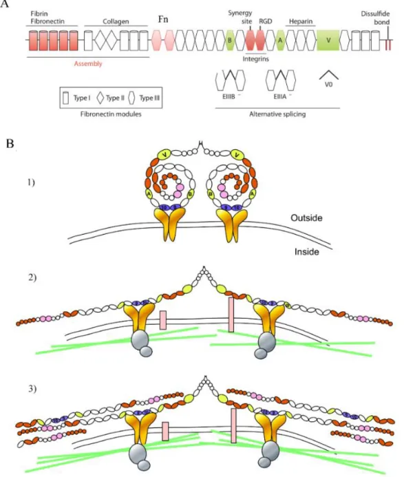
2019
Crosstalk between the myotome and muscle stem cells during the
development of the skeletal muscles of the back
“Documento Definitivo”
Doutoramento em Biologia
Especialidade de Biologia do Desenvolvimento
André Brás Gonçalves
Tese orientada por:
Professora Doutora Sólveig Thorsteinsdóttir Doutora Marianne Deries
2019
UNIVERSIDADE DE LISBOA FACULDADE DE CIÊNCIAS
Crosstalk between the myotome and muscle stem cells during the development of the skeletal muscles of the back
Doutoramento em Biologia
Especialidade de Biologia do Desenvolvimento
André Brás Gonçalves
Tese orientada por:
Professora Doutora Sólveig Thorsteinsdóttir, Doutora Marianne Deries
Júri: Presidente:
● Doutora Maria Manuela Gomes Coelho de Noronha Trancoso, Professora Catedrática e Presidente do Departamento de Biologia Animal da Faculdade de Ciências da Universidade de Lisboa.
Vogais:
● Doutora Perpétua da Conceição Pinto do Ó, Investigadora Principal, Instituto de Engenharia Biomédica da Universidade do Porto;
● Doutor Moisés Mallo Perez, Investigador Principal, Instituto Gulbenkian de Ciência;
● Doutora Ruth Cecilia Diez del Corral Baubry, Investigadora Associada, Champalimaud Centre for the
Unknown da Fundação Champalimaud;
● Doutora Maria Leonor Tavares Saúde, Professora Auxiliar Convidada, Faculdade de Medicina da Universidade de Lisboa;
● Doutor Domingos Manuel Pinto Henrique, Investigador Auxiliar, Faculdade de Medicina da Universidade de Lisboa;
● Doutora Sólveig Thorsteinsdóttir, Professora Associada com Agregação, Faculdade de Ciências da Universidade de Lisboa (orientadora).
Documento especialmente elaborado para a obtenção do grau de doutor Fundação para a Ciência e Tecnologia – SFRH/BD/90827/2012
The work presented in this dissertation was developed with the support from the Fundação para a Ciência e Tecnologia, through a PhD Fellowship (SFRH/BD/90827/2012) and the Project Grant PTDC/SAU-BID/120130/2010).
vii
Notas Prévias
1) Para a elaboração da presente tese de doutoramento foi usado integralmente como capítulo um artigo científico publicado numa revista internacional indexada. Uma vez que os trabalhos referidos na presente tese foram realizados em colaboração com outros investigadores, e de acordo com o disposto do nº1 do Artigo 45º do Regulamentos dos Estudos Pós-Graduados da Universidade de Lisboa, publicado no Diário da República, 2ª série, nº 65 de 30 de março de 2012, esclareço que participei integralmente na conceção e execução do trabalho experimental, na interpretação e discussão dos resultados, bem como na redação dos manuscritos.
2) O facto desta tese integrar um artigo científico levou a que o capítulo, cujo artigo está exposto, fosse escrito seguindo as normas da revista em que foi publicado, variando, portanto, das normas de escrita estabelecidas pela Universidade de Lisboa. O capítulo 5 consiste numa compilação de resultados considerados relevantes para esta tese, parte dos quais não foram publicados, enquanto que uma parte foi incluída num artigo já publicado (Gomes de Almeida, P. G., Pinheiro, G. G., Nunes, A. M., Gonçalves, A. B. and
Thorsteinsdóttir, S. (2016). Fibronectin assembly during early embryo development: A
versatile communication system between cells and tissues. Developmental Dynamics 245, 520-535.).
ix
Para a minha famíla,
especialmente para os meus pais
xi
Acknowledgements
Dizem que na vida devemos fazer três coisas: plantar uma árvore, ter filhos e escrever um livro. Fazendo uma introspeção ao meu trajeto penso que comecei pelo fim (e talvez pela parte mais difícil). Não que uma tese de doutoramento seja considerada um livro, mas do meu ponto de vista, todo o processo alheio à mesma, desde a escrita ao trabalho no laboratório, tem algumas parecenças relativamente. E curiosamente, em alguns casos, uma tese chega mesmo a ser considerada como um filho próprio. Naturalmente, existem imensas pessoas por trás de todo este trabalho e que contribuíram, de forma direta ou indireta, para ter eu chegado a este ponto e estar a escrever estas palavras sentidas de apreciação.
Em primeiro lugar, os meus maiores agradecimentos vão para a Sólveig e Marianne. Pela sua enorme sapiência científica e pela maneira como me orientaram ao longo destes longos anos de forma precisa, tranquila e sempre em boa disposição e em ambiente amigável. São de facto, fontes infindáveis de conhecimento e sem elas não seria capaz de dar conta do recado sozinho. Irei recordar-me das infindáveis horas de reuniões, discussões de resultados e esquemas que desenhámos para tentar perceber os meus resultados. Resumindo, aprendi muito com vocês e espero levar alguma dessa bagagem nas minhas próximas aventuras científicas. Quero também agradecer à Gabriela Rodrigues e à Rita Zilhão pela enorme camaradagem, discussões científicas e amizade destes anos. Sempre em boa disposição, aprendi também muito com vocês, desde culturas de células, boas práticas no laboratório a biologia molecular. Obrigado por puxarem pelo meu espírito crítico científico!
O meu segundo grande agradecimento vai para os meus pais e familiares. Por todo o apoio dado ao longo destes anos, mesmo não compreendendo a totalidade do meu trabalho, nunca me deixaram de me perguntar se tinha resultados novos, como iam os” meus ratos”, encorajando-me constante e diariaencorajando-mente, encorajando-mesmo quando tenha implicado ir trabalhar em horários “alternativos” pouco convencionais, nunca hesitaram em me apoiar. Muito obrigado pela educação que me deram e por me terem mostrado que o mais importante é fazermos aquilo que gostamos, mesmo por vezes quando o retorno possa parecer frustrante. Esta tese é dedicada a vocês!
De seguida não podia deixar de agradecer aos meus colegas de laboratório: Ao Pedro Rifes e à Raquel Vaz, os meus sinceros agradecimentos por ter trabalhado com vocês e aprendido imensa coisa, desde organização no trabalho, espírito crítico científico e mesmo por
xii
ter partilhado comigo um ambiente mais relaxado e corriqueiro em que senão gozássemos uns com os outros no dia a dia, nem valia a pena vir trabalhar.
Ao Luís Marques, incansável no auxílio informático e microscópico. Um sensei que sabe o que faz, desde as coisas mais fúteis às imprescindíveis no laboratório. Um grande obrigado por todas as horas de laboratório que partilhámos. Vou-me recordar dos podcasts, músicas, piadas e de nos rirmos que nem uns perdidos sobre as mais variadas temáticas discutidas, quando tínhamos o laboratório por conta própria. Como o próprio se intitula, és repositório inesgotável de informação inútil, mas muito valioso, porque assim dá para falar um pouco de tudo e parafraseando Rui Veloso sobre Carlos Tê “Quem fala assim não é gago”.
À Marta Palma, grande companheira de aventuras extra laboratoriais, nas casas assombradas, idas a jogos de futebol e entre outras. Um grande obrigado por muitas das vezes teres sido tu a organizar a maior parte delas que tanta dinâmica deram ao grupo. Não me vou esquecer daquele dia de canoagem. Obrigado, porque sem ti o trabalho no laboratório não corria de forma eficaz, nem era tão aprazível, pois a tua ajuda no laboratório foi imprescindível. Obrigado por me teres ouvido e ajudados muitas vezes sobre os mais variados temas. O teu gabinete servia como uma espécie de consultório terapêutico que me fazia bem.
Às minhas companheiras de doutoramento, Patrícia Almeida e Andreia Nunes, por todos os momentos vividos dentro e fora do laboratório. Pelas sessões musicais neste, pelos dramas científicos diários, mas também por toda a amizade destes anos vindouros que remontam ao tempo em que ainda éramos alunos de mestrado e mal sabíamos por onde iam dar os nossos caminhos. It was a hell of a ride! Entre os altos e baixos de cada um, começámos juntos isto e sendo eu o último a encerrar o departamento dos doutorados do nosso ano no laboratório penso que fizemos todos um bom trabalho. Obrigado pelo apoio e, por certas vezes, me terem compensado nalgumas tarefas que me fizeram toda a diferença na minha logística diária.
À Inês Antunes e Ana Neto um enorme obrigado pela vossa boa disposição e constante disponibilidade para ajudar.
Ao Telmo Monteiro, pela ajuda prestada na micróscopia e análise de imagens, que muitas vezes me safaram quando ninguém estava por perto.
À D. Elvira pela sua alegria e bem-estar contagiantes. É de pessoas destas que precisamos nas nossas vidas, porque nos trazem sol em dias de chuva.
xiii
Em suma, à turma da FCUL, um enorme e sentido obrigado por todas estas aventuras, desde aos almoços rotineiros sempre em ambiente (demasiado) descontraído, aos jantares, cinemas, acampamentos e noites de copos. Tudo sempre num ambiente entre amigos, cheio de estupidez natural, mas altamente gratificante. Tive muita sorte em vos ter conhecido e em ter feito parte desta equipa muito boa!
Agradeço ao Moisés Mallo, Hélia Neves e Ruth Diez del Corral pelo empréstimo dos plasmídeos para as in situs emprestadas e também à Leonor Saúde, Ana Cristovão e Domingos Henrique, não só pelo empréstimo de outros tantos, mas também por me terem deixado usufruir das suas instalações para produzir as sondas. Sem elas, não teria muitos dos resultados aqui descritos. Obrigado pela vossa simpatia e amizade!
Quero ainda agradecer à Patricia Ybot-Gonzalez e à Beatriz Lopez-Escobar por todos os ensinamentos transmitidos sobre cultura de embriões e a todos os membros do laboratório do João Relvas por me terem feito sentir como em casa durante os meus períodos de rotação laboratorial. Em especial à Joana Faria, à Ana Seixas, Nuno Dias e ao Fábio Prata, tive muita sorte por a vida me ter posto de encontro com os vossos caminhos, pois vocês são pessoas incríveis. Fábio, continuo a aguardar as mensagens quinzenais de picardia sobre quem é o melhor piloto e equipa na fórmula 1!
Ao João Brazão e à Ana Sobral, pela vindoura amizade destes longos anos. Pelas noitadas, festas académicas, almoços e jantares sempre acompanhados de grande espírito de partilha.
E finalmente aos meus amigos e treinador “da minha outra vida”, onde destaco o Francisco Quintas, Diogo Correia, Fábio Figueira, Ritinha, Jessica Vieira, Nuno Rola, João Gigante, Ricardo Lopes, Bruno e Filipa Silva, Marta Ferreira, Duarte Mourão, Daniel Viegas, Rosinhas, Simão Morgado e Fernando Couto. Para vocês não é preciso dizer muito, pois sabem o quão importante foram e são no meu dia a dia. Vivemos muito e partilhámos muito! Um grande obrigado por todo este tempo de amizade, loucura e momentos extraordinários que vivenciei convosco dentro e fora das piscinas. E espero que ainda tenhamos muitos mais pela frente. Sem dúvida contribuíram, mesmo sem se aperceberem, para a minha motivação diária e bem-estar pessoal.
Por fim, e não menos importante, um grande obrigado ao Florent Lenoir e à tia Micá, pelo incansável e preciosíssimo trabalho que tiveram em me ajudar na formatação desta tese. De longe que senão fossem vocês, eu ainda estaria aqui a desesperar para toda a eternidade.
xv
Abstract
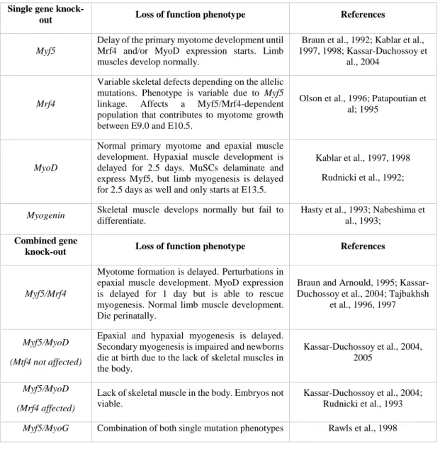
The deep back (or epaxial) muscles of amniotes derive from the transient myotomes, segmented embryonic muscles that develop from the delamination and differentiation of muscle stem cells (MuSCs) from the overlying dermomyotome. During embryonic development, myogenesis is ensured by the activation of key transcription factors: Myf5, Mrf4, MyoD and Myogenin.
The main goal of this thesis was to investigate the role of the myotome in epaxial muscle development. In Chapter 2, a technique of culturing mouse embryo explants was developed, which allowed us to study the in vivo ex utero development of the epaxial myotome and its extracellular matrix (ECM). In Chapter 3, we analysed to what extent the myotome is necessary for later epaxial muscle development using the Myf5nlacZ/nlacZ mouse line, in which the absence of Myf5 and Mrf4 results in the lack of an early myotome. We show that one specific epaxial muscle group (the transversospinalis) is able to differentiate through MyoD, while the other three epaxial muscle groups fail to form. Moreover, we show that due to the lack of myotomal factors, the maintenance of the identity of delaminating dermomyotomal MuSCs fails. In Chapter 4, we described the organisation of laminins, fibronectin and tenascin-C ECMs during myotome development showing that each one of these ECMs potentially has a specific spatial relationship with MuSCs. Finally, Chapter 5 focuses on the role of the myotome in the organisation of these same ECMs and its role in tendon development, using
Myf5nlacZ/nlacZ mouse embryos. We show that the myotome is necessary to assemble its own matrices, but these are not required for the development of the transversospinalis muscles.
The results of this thesis suggest that the transversospinalis muscles have a distinct developmental mechanism from that of the remaining epaxial muscles and we propose that they are evolutionary more recent.
Keywords: Epaxial myogenesis; Myotome; Muscle stem cell; Extracellular matrix;
xvii
Resumo
Os músculos profundos das costas (ou epaxiais) dos amniotas derivam dos miótomos transientes, músculos embrionários segmentados que se desenvolvem a partir da delaminação e diferenciação das células estaminais musculares (CEMs) do dermomiótomo sobreposto. Durante o desenvolvimento embrionário, a miogénese é assegurada pela ativação de fatores de transcrição-chave miogénicos: Myf5, Mrf4, MyoD e Miogenina.
Esta tese tem como principal objetivo perceber o papel do miótomo no desenvolvimento dos músculos epaxiais. No capítulo 2, descrevemos uma técnica de cultura de explantes de embrião de ratinho que nos permitiu estudar o desenvolvimento in vivo ex utero do miótomo epaxial e da sua matriz extracelular (MEC). No capítulo 3, analisamos até que ponto o miótomo é necessário para o desenvolvimento muscular epaxial que ocorre em fases mais tardias, recorrendo ao uso de embriões de ratinho Myf5nlacZ/nlacZ, cuja incapacidade de expressar Myf5 e
Mrf4, leva a que não formem o miótomo embrionário. Aqui, demonstramos que um grupo
muscular epaxial específico, nomeadamente o músculo transversospinalis é capaz de se diferenciar através da ativação de MyoD, ao passo que os restantes grupos musculares epaxiais não se formam. Mais ainda, demonstramos que a manutenção da identidade das CEMs do dermomiótomo que delaminam deste é comprometida devido à ausência de fatores parácrinos do miótomo. No capítulo 4, descrevemos a organização das matrizes de laminina, fibronectina e tenascina-C durante o desenvolvimento do miótomo, evidenciando que cada uma destas MECs tem uma potencial relação espacial com as CEMs. Por fim, o capítulo 5 atenta para a contribuição do miótomo na organização destas mesmas MECs e para desenvolvimento dos tendões recorrendo novamente aos embriões de ratinho Myf5nlacZ/nlacZ. Aqui, evidenciamos que o miótomoé apenas necessário para a montagem das suas próprias MECs, não sendo requerido para o desenvolvimento do músculo transversospinalis.
Os resultados desta tese sugerem que o músculo transversospinalis tenha um mecanismo de desenvolvimento distinto dos restantes músculos epaxiais, e como tal, propomos que este músculo em particular seja evolutivamente mais recente.
Palavras-chave: Miogénese epaxial; Miótomo; Célula estaminal muscular; Matriz
xix
Resumo alargado
A formação dos músculos esqueléticos, designada por miogénese, é um processo complexo que depende da combinação de uma variedade de fatores celulares autónomos e de sinais extrínsecos. Todos os músculos esqueléticos do tronco e dos membros derivam de estruturas embrionárias designadas por dermomiótomos. Estes são compostos pelas células dorsais dos sómitos epiteliais, segmentos mesodérmicos localizados de cada lado do tubo neural. Após a dispersão da parte ventral dos sómitos, que origina o esqueleto axial, as células dos dermomiótomos, adotam uma organização em camada epitelial, formando uma borda nas suas extremidades.
Os primeiros músculos esqueléticos a formar no embrião são os miótomos que se desenvolvem através da delaminação de células estaminais musculares (CEMs) das bordas dos dermomiótomos. Estas células diferenciam-se em miócitos que se dispõem paralelamente ao tubo neural, no lado ventral dos dermomiótomos, e formam assim músculos segmentados repetidos ao longo do eixo anterior-posterior do embrião. Ao entrar no miótomo, as CEMs, que se caracterizam pela expressão de Pax3 e/ou Pax7, ativam fatores de transcrição que induzem a diferenciação miogénica, nomeadamente Myf5, Mrf4, MyoD e Miogenina. A expressão destes fatores ocorre numa maneira hierárquica e específica ao longo do desenvolvimento embrionário. Myf5 é o primeiro fator de determinação a ser expresso, tanto na parte epaxial como na região hipaxial do miótomo. Seguem-se Mrf4 e MyoD que marcam os mioblastos em diferenciação no miótomo e por fim, a expressão de Miogenina que induz a diferenciação dos mioblastos em miócitos mononucleados, alongados e pós-mitóticos.
Tanto os dermomiótomos como os miótomos são estruturas transientes. Numa dada altura do desenvolvimento, as células do dermomiótomo de-epitelizam e entram nos miótomos como CEMs proliferativas que expressam Pax3 e/ou Pax7. Algumas delas diferenciam-se e fundem com os miócitos do miótomo enquanto outras mantém o estado estaminal. Simultaneamente, os miótomos transformam-se, perdendo a sua conformação segmentada embrionária e reorganizam-se na musculatura definitiva pré-adulta do tronco. Estas incluem os músculos epaxiais (ou músculos profundos das costas), que se associam à coluna vertebral contribuindo para o seu alinhamento e estabilização, e a musculatura hipaxial do tronco, onde fazem parte os músculos intercostais e abdominais que controlam os movimentos do tronco e protegem os órgãos internos.
xx
Apesar de todo o conhecimento em torno da regulação miogénica durante o desenvolvimento do miótomo, muito pouco se sabe sobre a forma como este afeta as CEMs, aquando a sua entrada para o miótomo e também como o próprio influencia o desenvolvimento da musculatura epaxial. Assim, o principal objetivo desta tese foi estudar como o miótomo embrionário influencia o comportamento das CEMs e a morfogénese dos músculos epaxiais. Usámos embriões de ratinho selvagens e embriões knock-out Myf5nlacZ/nlacZ, que não expressam nem Myf5 nem Mrf4 e consequentemente não formam os miótomos embrionários. No entanto os embriões Myf5nlacZ/nlacZ desenvolvem musculatura esquelética mais tardiamente.
O capítulo 2 atenta para o desenvolvimento de uma técnica de cultura que permite o desenvolvimento in vivo ex utero de explantes preparados a partir de embriões de ratinho de estirpe selvagem. Esta técnica foi desenvolvida e otimizada para o estudo do desenvolvimento do miótomo e do seu ambiente extracelular. Foram realizados ensaios para detetar a proliferação celular e apoptose e também foi estudada a morfologia tridimensional da musculatura esquelética epaxial, assim como a organização da matriz extracelular (MEC) dentro e à volta do miótomo. Estas características foram depois comparadas com as dos embriões que se desenvolveram no interior do útero materno. Aqui, demonstramos que dentro de uma janela temporal de 12 horas e em condições de cultura de explantes em meio desprovido de soro, a morfogénese do miótomo procede de forma semelhante em comparação com a dos embriões desenvolvidos in utero. Ao mesmo tempo verificámos também que a MEC aumentava de complexidade ao longo do tempo, acompanhado o desenvolvimento do miótomo epaxial. Apesar de um ligeiro atraso no desenvolvimento, este fenótipo foi acompanhado pela ausência de diferenças na proliferação e morte celular, atestando a validade desta técnica. Ainda neste capítulo, são discutidas as vantagens e limitações desta técnica, a qual oferece uma forma rápida, acessível em termos de equipamento e manipulação, sendo assim económica e com potencial para múltiplas aplicabilidades, uma vez que permite o uso simultâneo de uma certa quantidade de embriões e aplica-se a vários estádios de desenvolvimento. Pode também ser acrescido o uso de diferentes inibidores para o estudo de vias de sinalização num determinado contexto biológico. Não menos importante, a aplicação desta técnica não é apenas útil para o estudo do desenvolvimento do miótomo, mas poderá ser transversal à análise de outras estruturas em desenvolvimento em fases de desenvolvimento semelhantes. O trabalho inserido neste capítulo foi publicado na revista Differentiation (2016) 91, 57-67.
No capítulo 3 analisamos até que ponto o miótomo é necessário para o desenvolvimento e morfogénese dos músculos epaxiais. Para tal, usámos a linha de ratinho Myf5nlacZ/nlacZ que não
xxi
forma o miótomo e em que a miogénese se encontra atrasada por ser apenas induzida pelo fator de transcrição MyoD, uma vez que estes embriões mutantes não expressam Myf5 nem Mrf4. Neste capítulo demonstramos que apenas um grupo muscular epaxial (designado por transversospinalis) é capaz de se diferenciar através da expressão de MyoD e que se desenvolve independentemente do miótomo, enquanto que os restantes três grupos epaxiais musculares não se desenvolvem. Verificámos também que a ausência do miótomo compromete a manutenção da identidade das CEMs derivadas do dermomiótomo, aquando a sua de-epitelização e entrada no espaço onde o miótomo deveria existir, sugerindo que o miótomo emite sinais para manter estas células. Sabe-se que o miótomo normalmente expressa vários fatores parácrinos, como fatores de crescimento dos fibroblastos (Fgfs) e o fator de crescimento A derivado das plaquetas (Pdgfa), que, na ausência do miótomo, não são expressos. Colocamos assim a hipótese que a perda de identidade estaminal das CEMs poderia dever-se à ausência destes fatores parácrinos. Quando se bloqueou a via de sinalização Fgf em embriões de ratinho de estirpe selvagem (isto é, embriões que formam um miótomo normal) verificou-se uma diminuição significativa no número de CEMs. No entanto, a inibição da via de sinalização Pdgf, não teve efeitos significativos na totalidade do número destas células. Estes dados sugerem que os Fgfs do miótomo mantêm a identidade das CEMs do dermomiótomo durante e após a sua de-epitelização. Estes resultados levam ainda à hipótese de que poderão existir mecanismos de desenvolvimento divergentes entre o transversospinalis e os restantes músculos epaxiais. Este capítulo consiste num manuscrito em preparação para publicação.
No capítulo 4 descrevemos a organização dos componentes da MEC, incluindo a laminina, a fibronectina e a tenascina-C, em relação às CEMs durante o desenvolvimento do miótomo demonstrando que cada uma destas matrizes possui uma relação espacial específica relativamente às CEMs do dermomiótomo e do miótomo. Através de uma perspetiva tridimensional analisamos como as matrizes de laminina, fibronectina e tenascina-C se organizam durante a de-epitelização das CEMs do dermomiótomo central e a sua entrada no miótomo em embriões de ratinho da estirpe selvagem. Verificamos que a laminina é depositada na membrana basal do dermomiótomo e do miótomo e que as CEMs dentro do miótomo se localizam na proximidade da membrana basal deste. Verificámos ainda que as matrizes de fibronectina e tenascina-C também se encontram associadas as membranas basais do dermomiótomo e do miótomo e que são ambas depositadas no espaço entre estes dois territórios, antes de ocorrer a delaminação das CEMs do dermiótomo, levantando à hipótese de que possam ser usadas por estas células para colonizar o miótomo.
xxii
No capítulo 5, analisamos de que modo os padrões de expressão das MECs analisadas
no capítulo 4 se encontram alterados nos embriões Myf5nlacZ/nlacZ. Nestes embriões, evidenciamos que o miótomo é crucial para a formação da sua própria membrana basal composta por lamininas, uma vez que esta não se forma. Mais ainda, verificámos que o padrão típico das matrizes de fibronectina e tenascina-C do miótomo não se forma na ausência do miótomo. Contudo, quando as massas musculares do transversospinalis se desenvolvem, nos embriões Myf5nlacZ/nlacZ, estas não só conseguem produzir as suas próprias MECs, como também induzem a expressão de Scleraxis, um marcador das células precursoras de tendinócitos, no mesênquima circundante. Estes dados demonstram que a MEC do miótomo não é necessária para o desenvolvimento do transversospinalis e que o mesmo é capaz de induzir a expressão de
Scleraxis em seu redor na ausência do miótomo. Parte dos resultados deste capítulo foram
incluídos no artigo Gomes de Almeida et al. Developmental Dynamics (2016) 235, 520-535.
Finalmente no capítulo 6, são discutidos os principais resultados de todos os capítulos de acordo com a bibliografia existente, destacando o papel do miótomo durante o desenvolvimento da musculatura epaxial e as particularidades do mecanismo de desenvolvimento do transversospinalis.
Palavras-chave: Miogénese epaxial; Miótomo; Célula estaminal muscular; Matriz
xxv
Contents
Acknowledgements ... xi Abstract ... xv Resumo ... xvii Resumo alargado ... xix List of Abbreviations and Acronyms... xxxiii
Chapter 1: Introduction... 2
I. General Introduction ... 4 1. The musculoskeletal system ... 4 1.1 The anatomy of the musculoskeletal system ... 4
1.2 Skeletal muscle development at a glance ... 5
2. Cell-cell communication in development ... 8 2.1 Types of cell-cell communication ... 8
2.2 Paracrine (growth) factor signalling ... 8
2.2.1 Fgf signalling ... 8 2.2.2 Pdgf signalling ... 10 2.2.3 Wnt signalling ... 12 2.2.4 Hedgehog signalling ... 13 2.2.5 Tgfβ/Bmp signalling ... 16 2.3 Juxtacrine communication ... 17 2.3.1 Notch signalling ... 17 2.3.2 Cell adhesions ... 20
2.4 The extracellular matrix ... 22
2.4.1 A primer on extracellular molecules ... 23
3. Embryonic origin of skeletal muscle ... 28 3.1 Somitogenesis ... 31
xxvi
3.2.1 Sclerotome: the primordium of axial skeleton ... 32
3.2.2 Dermomyotome: a progenitor epithelium ... 33
3.2.3 Myotome: the first skeletal muscle ... 36
3.2.4 Syndetome: linking back muscles to the skeleton ... 38
4. Mechanisms of early stages of muscle differentiation ... 40 4.1 Transcriptional networks regulating myotomal populations ... 40
4.2 Induction of myogenesis in the trunk ... 43
4.2.1 Signalling molecules regulating epaxial myotome formation ... 43
4.2.2 Signalling molecules regulating hypaxial myotome formation ... 44
4.2.3 Activation of myogenesis in the rostral and caudal dermomyotomal lips .. 45
4.2.4 The role of the myotomal basement membrane ... 47
4.3 Signalling molecules regulating myotome growth ... 47
4.4 Myotome transformation and the formation of the trunk musculature ... 49
4.5 Limb myogenesis: migration and differentiation ... 52
4.6 Limb tendinogenesis ... 54
5. Later stages of skeletal myogenesis ... 55 5.1 Primary myogenesis ... 55
5.2 Secondary myogenesis ... 59
5.3 Neonatal and adult myogenesis ... 59
II. Aims and objectives ... 60 III. References ... 62
Chapter 2: Rapid and simple method for in vivo ex utero development of mouse embryo explants ... 80 Chapter 3: The myotome is necessary for epaxial muscle development and provides essential cues for the maintenance of muscle stem cells ... 96 Chapter 4: Extracellular matrix dynamics during axial muscle development: distinct roles for laminins, fibronectin and tenascin-C? ... 140 Chapter 5: The myotomal extracellular matrix is not required for transversospinalis and hypaxial muscle morphogenesis ... 166 Chapter 6: Discussion ... 190
xxvii
1. Role of the myotome during embryonic development ... 192 2. The myotome and its matrices ... 193 3. The myogenic pathways ... 194 4. The mechanisms of development of transversospinalis muscles share similarities with that of the hypaxial muscles ... 196 II. Final considerations ... 198 III. References ... 199
xxix
List of Figures
Figure 1.1: Anatomy of the vertebrate musculoskeletal system and development of skeletal muscles ... 6 Figure 1.2: Overview of the Fgf signalling pathway. ... 9 Figure 1.3: Pdgf ligands and Pdgfrs ... 11 Figure 1.4: Wnt signalling pathway ... 14 Figure 1.5: Generic overview of the Shh signalling pathway ... 15 Figure 1.6: The Tgfβ member superfamily and the canonical, Smad-dependent Tgfβ/Bmp signalling pathway. ... 18 Figure 1.7: Overview of the Notch signalling pathway ... 20 Figure 1.8: Extracellular matrix organisation in vertebrates ... 24 Figure 1.9: Structure of the laminin trimer. ... 26 Figure 1.10: Fibronectin structure and assembly ... 27 Figure 1.11: Structure of the tenascin-C molecule ... 28 Figure 1.12: Somitogenesis ... 29 Figure 1.13: Illustration showing somite development at distinct axial levels along the caudal to rostral axis of an E11.5 mouse embryo ... 30 Figure 1.14: Embryonic origins of the adult skeletal musculature. ... 31 Figure 1.15: Sclerotome differentiation and vertebrae formation ... 34 Figure 1.16: Dermomyotomal sources of MuSCs ... 35 Figure 1.17: Trunk and limb tendon development ... 39 Figure 1.18: Transcriptional networks regulating myogenesis in the different regions of the myotome (trunk) and in the limb. ... 41 Figure 1.19: Induction of myogenesis by signals from neighbouring tissues. ... 46 Figure 1.20: Developmental stages of the epaxial dermomyotome and myotome and segregation of the definitive axial musculature. ... 51 Figure 1.21: Specification and migration of limb MuSCs ... 54 Figure 1.22: Summary of skeletal muscle development ... 56 Figure 2.1: Dissection of an E11.5 mouse conceptus for explant culture ... 86 Figure 2.2: Whole mount immunohistochemistry protocol. ... 87 Figure 2.3: Apoptosis and proliferation in cultured explant embryos. ... 88 Figure 2.4: Morphogenesis of deep back muscles occurs normally in cultured explants ... 89
xxx
Figure 2.5: The tenascin ECM of deep back muscles grows with the muscles during the 12h culture period ... 92 Figure 3.1: Epaxial muscle morphogenesis in Myf5nlacZ/nlacZ embryos is incomplete. ... 111 Figure 3.2: MyoD expression is spatially restricted in Myf5nlacZ/nlacZ embryos ... 113 Figure 3.3: MuSCs of Myf5 nlacZ/nlacZ embryos fail to maintain Pax7 expression after central dermomyotome dissociation. ... 115 Figure 3.4: Myf5nlacZ/nlacZ embryos express Fgf6 and Pdgfa upon myogenic differentiation . 120 Figure 3.5: Comparative expression pattern of Fgfr1, Fgfr4 and Pdgfrα in Myf5+/nlacZ and
Myf5nlacZ/nlacZ embryos ... 121 Figure 3.6: Fgf signalling through Fgfr1 and Fgfr4 maintain MuSC identity after central dermomyotome dissociation. ... 124 Supplementary Figure 3.1: Differential distribution of Pax3- and Pax7-positive MuSCs in E11.5 wild type embryos ... 136 Supplementary Figure 3.2: Proliferation and apoptosis assays in Myf5+/nlacZ and Myf5nlacZ/nlacZ
embryos ... 137 Figure 4.1: Different views of 3D reconstruction of one segment ... 149 Figure 4.2: Tenascin-C appears to co-localise with laminin in the dermomyotomal and myotomal basement membranes during early myogenesis ... 151 Figure 4.3: Fibronectin accumulates between the dermomyotome and the myotome before the delamination of the dermomyotomal MuSCs ... 154 Figure 4.4: Laminin and tenascin matrices define the epaxial pouch that contains the MuSCs in the epaxial lip of the dermomyotome ... 156 Figure 4.5: Fibronectin and tenascin-C are located in the proximity of myotomal MuSCs. . 158 Figure 5.1: Deposition of laminins in the dermomyotomal basement membrane is normal, but the myotomal basement membrane does not form in Myf5nlacZ/nlacZ embryos ... 173 Figure 5.2: Normal Fn1 expression in Myf5+/nlacZ and Myf5nlacZ/nlacZ embryos but the myotomal fibronectin ECM does not form in Myf5nlacZ/nlacZ embryos ... 178 Figure 5.3: Epaxial tendinogenesis is delayed in Myf5nlacZ/nlacZ embryos and only the tendons of transversospinalis and hypaxial muscles develop ... 181 Figure 6.1: Model proposing the existence of three different myogenic transcriptional networks regulating epaxial myogenesis ... 196
xxxi
List of Tables
Table 1.1: Phenotypes of loss of function (knock-outs) of MRFs ... 42 Table 1.2: MuSC terminology ... 58 Table 2.1: Materials for mouse embryo explant culture ... 85 Table 2.2: Media for mouse embryo explant culture ... 86 Table 2.3: Description of embryo dissection and number of embryos per filter according to their age ... 87 Table 2.4: Antibodies ... 87 Table 3.1: Primary and secondary antibodies used for immunohistochemistry on sections (IHC) and in whole mount immunohistochemistry (WMIHC) ... 106 Table 3.2: Chemical inhibitors used in explant cultures of wild type embryos. ... 107 Table 41: Primary and secondary antibodies used for immunohistochemistry ... 149 Table 5.1: Primary and secondary antibodies used for immunohistochemistry on sections.. 185
xxxiii
List of Abbreviations and Acronyms
ACT - Activin
ADAM/TACE - A Disintegrin and Metalloproteinase
AKT - RAC-Alpha Serine/Threonine-Protein Kinase
AMH - Anti-Müllerian Hormone
APC - Adenomatosis Polyposis Coli
bHLH - Basic Helix-Loop-Helix
BMP - Bone Morphogenetic Protein
BMPRI/II - Bone Morphogenetic Protein Receptor Type I/II
Boc - Brother of Cdo
CAM - Cell Adhesion Molecule
CDK1 - Cyclin Dependent Kinase 1
Cdo - Cysteine Dioxygenase
CK1α- Casein Kinase I Alpha
CL - Caudal Lip of the Dermomyotome
CSL - DNA Binding Domain Protein CSL
CXCL12 - C-X-C Chemokine Ligand 12
CXCR4 - C-X-C Chemokine Receptor 4
Dhh - Desert Hedgehog
Dll - Delta-Like-Ligand
DML - Dorso-Medial Lip of the Dermomyotome
xxxiv
Dmrt2 - Doublesex and Mab-Related Transcription Factor 2
DNA - Deoxyribonucleic Acid
Dsv - Dishevelled
ECM - Extracellular Matrix
EEE - Early Epaxial Enhancer
EGF - Epidermal Growth Factor
EMT - Epithelial-to-Mesenchymal Transition
E(n) - Embryonic day (number)
En1 - Engrailed Homeobox 1
ErbB3 - Erb-B2 Receptor Tyrosine Kinase 3
ERK - Extracellular Signal - Regulated Kinase
Eya - Eyes Absent Homolog
FGF- Fibroblast Growth Factor
FGFR - Fibroblast Growth Factor Receptor
Foxc1/2 - Forkhead Box C1/2
Fu - Fused
Fzd - Frizzled
GAG - Glycosaminoglycan
Gas1 - Growth Arrest-Specific Protein 1
GDF - Growth and Differentiation Factor
GDNF - Glial-Derived Neurotrophic Factor
GFP - Green Fluorescent Protein
Gli - Gli Family Zinc Finger
xxxv
GSKβ3 - Glycogen Synthase Kinase 3 Beta Hes - Hairy/Enhancer of Split
Hey - Hes Related Family BHLH Transcription Factor with YRPW Motif 1
HGF - -Hepatocyte Growth Factor
Hhip1 - Hedgehog Interacting Protein 1
HS -Heparan Sulphate
HSPG - Heparan Sulphate Proteoglycan
Ihh - Indian Hedgehog
INH - Inhibin
Jag - Jagged
JNK - C-Jun N-terminal Kinase
Kif7 - Kinesin-Like Protein 7
Lbx1 - Ladybird Homeobox 1
TCF1 /LEF - Transcription Factor 1 / Lymphoid Enhancer Binding Factor
Lefty - Left-Right Determination Factor
LRP5/6 - Low Density Lipoprotein - Related Protein 5/6
Mam - Mastermind-Like Protein
MAPK - Mitogen-Activated Protein Kinase
MET - MET Proto-Oncogene, Receptor Tyrosine Kinase
MHC - Myosin Heavy Chain
MRF - Myogenic Regulatory Factor
mTOR - Mechanistic Target of Rapamycin Kinase
MuSC - Muscle Stem Cell
xxxvi
Myf6 /Mrf4 - Myogenic Factor 6
MyoD - Myogenic Differentiation 1
NCC - Neural Crest Cell
NECD - Notch Extracellular Domain
NFAT - Nuclear Factor of Activated T cell
Ngr -Neuregulin
NICD - Notch Intracellular Domain
Nodal - Nodal Growth Differentiation Factor
Notch -Notch Homolog 1, Translocation-Associated
NRR - Negative Regulatory Region
P (n) - Postnatal day (number)
Pax - Paired Box
PCP - Planar Cell Polarity
PDGF - Platelet-Derived Growth Factor
PDGFR - Platelet-Derived Growth Factor Receptor
PI3K - Phosphoinositide 3-Kinase
Pitx2 - Paired-like Homeodomain Transcription Factor 2
PKA - Protein Kinase A
PKC - Protein Kinase C
PLCγ - Phospholipase C, Gamma 1 PP2A - Protein Phosphatase 2
PSM - Presomitic Mesoderm
Ptch - Patched
xxxvii
RBPJk - Recombining Binding Protein Suppressor of Hairless
RGD - Arginine-Glycine-Aspartic Acid
RL - Rostral Lip of the Dermomyotome
RNA - Ribonucleic Acid
RTK - Receptor-Linked Tyrosine Kinase
SCR- SCR Proto-Oncogene, Non-Receptor Tyrosine Kinase
Scx - Scleraxis bHLH Transcription Factor
Shh - Sonic Hedgehog
Sim1 - SIM bHLH Transcription Factor 1
Six - Sine Oculis Homeobox Homolog
Slit1 - Slit Guidance Ligand 1
SMAD - Mothers Against Decapentaplegic Homolog
Smo -Smoothened
Snail - Snail Family Transcriptional Repressor
SOX - Sex Determining Region Y-box
Spry - Sprouty RTK Signalling Antagonist
STAT - Signal Transducer and Activator of Transcription
SuFu - Suppressor of Fused
TCF1 /LEF - Transcription Factor 1 / Lymphoid Enhancer Binding Factor
TFIC - Transcription Factor Inhibitory Complex
TGFβ - Transforming Growth Factor Beta
TGFRβI/II - Transforming Growth Factor Beta Receptor Type I/II VLL - Ventro-lateral Lip of the Dermomyotome
Chapter 1
4
I. General Introduction
1.
The musculoskeletal system
1.1 The anatomy of the musculoskeletal system
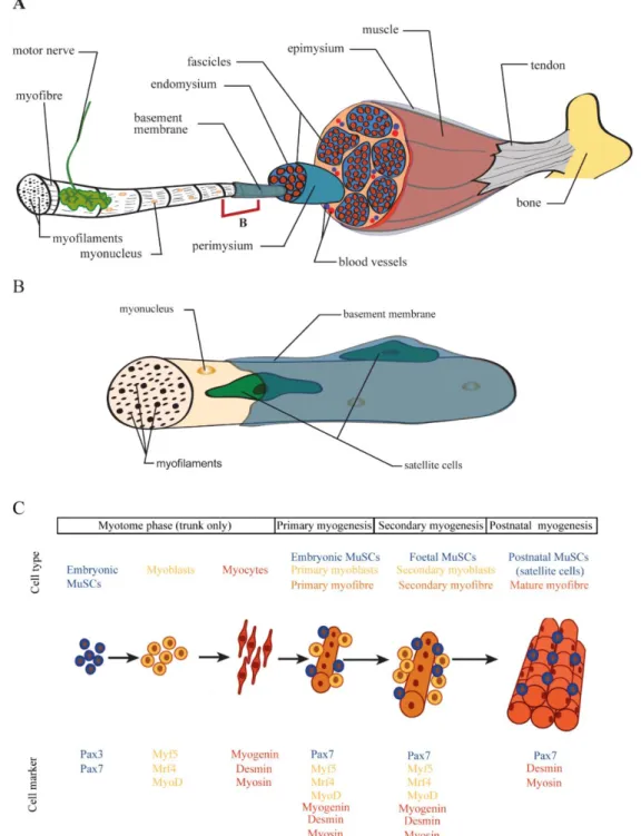
Vertebrate movement depends on the musculoskeletal system which includes skeletal muscles, bones, connective tissue (including tendons), nerves and blood vessels. Of the three types of muscles in vertebrates (skeletal, cardiac and smooth muscle), skeletal muscle is the most abundant. To be functional, skeletal muscle tissue needs to be incorporated in the whole musculoskeletal system, i.e. linked to the skeleton through tendons, become innervated by motor neurons and sufficiently vascularised to sustain muscle homeostasis (Fig. 1.1A; Deries and Thorsteinsdóttir 2016; Frontera and Ochala, 2015; Hauschka, 1994).
Skeletal muscles consist of large multinucleated, elongated and striated cells that are coated by an extracellular matrix called a basement membrane (Fig. 1.1A, B). Basement membranes are composed of a combination of collagens, non-collagenous glycoproteins and proteoglycans and play key roles in cell polarisation, differentiation and homeostasis, and, in the case of skeletal muscle, synaptogenesis, tendinogenesis and regeneration (Sanes, 2003; Thorsteinsdóttir et al., 2011). Typically, skeletal muscle cells, also named myofibres, lie parallel to each other and are arranged in multiple cylindrical bundles named fascicles (Fig. 1.1A). Connective tissue contributes to support and organise the superstructure of the muscle by providing routes for nerve fibres and blood vessels and is stratified in three main layers: the endomysium, the perimysium and the epimysium. The endomysium is the most internal layer and wraps the individual muscle fibres, the perimysium organises the muscle fibres into multiple fascicles and the epimysium, the most external layer, delimits the whole muscle apparatus (Fig. 1.1A; Frontera and Ochala, 2015; Gillies and Lieber, 2011). These three sheaths are continuous with each other and extend to attach directly to the bone or to join the tendons that connect the muscles to bone or cartilage, thus enabling force transmission generated by skeletal muscles to move the skeleton (Fig. 1.1A).
The cellular components underlying the contractile capacity of muscle fibres are revealed upon microscopic analysis. The sarcoplasm (cytoplasm of the muscle fibre) contains repeated
5
contractile elements named sarcomeres, which in turn are formed by the organisation of thick (enriched in myosin) and thin (enriched in actin) cytoskeletal filaments (Frontera and Ochala, 2015). The alternated organisation of these two types of filaments gives skeletal muscles their striation pattern and underlies their contraction capacity as they slide along each other upon the release of Ca2+ from the sarcoplasmic reticulum, a process that is triggered by motor nerves (Frontera and Ochala, 2015). Hence, skeletal muscles are responsible for crucial biological functions such as breathing, body support, locomotion and their activity also contributes to the control of body temperature.
Muscle fibres associate with and maintain adult muscle stem cells (MuSCs), also known as satellite cells (Mauro, 1961), which are located between the muscle fibres and their overlying basement membrane (Fig. 1.1B). In healthy muscle they remain quiescent, but upon muscle injury, some satellite cells become activated and proliferate, thus expanding the population. Subsequently some of these MuSCs differentiate to repair the affected muscle area, while others return to their quiescence state under the basement membrane (Motohashi and Asakura, 2012).
1.2 Skeletal muscle development at a glance
Myogenesis, the process of building skeletal muscle is very complex. It is initiated during embryogenesis and continues throughout adulthood as a mechanism to maintain a healthy tissue. Most skeletal muscles of vertebrates (see section 4 for details) derive from paraxial mesodermal cells that have activated Pax3 and/or its paralogue Pax7, both members of the paired box (Pax)/homeodomain family of transcription factors, thus marking a subpopulation of mesodermal cells that can enter the myogenic lineage (Fig. 1.1C; Buckingham, 2006; Buckingham and Relaix, 2007). During development, first embryonic and then foetal MuSCs produce a sequential series of myogenic regulatory factors (MRFs), members of the MyoD family of myogenic basic helix-loop-helix (bHLH) transcription factors, thus initiating myogenic determination and differentiation. In vertebrates there are four MRFs named the myogenic factor 5 (Myf5), the myogenic factor 6 (Myf6, also known as Mrf4 or herculin), the myogenic differentiation 1 (MyoD, also known as MyoD1) and Myogenin (Asfour et al., 2018; Buckingham and Rigby, 2014; Pownall et al., 2002; Tajbakhsh, 2009) When an embryonic MuSC turns on the expression of Myf5, MyoD or Mrf4, it becomes committed to myogenesis and is termed a myoblast. Committed myoblasts can divide a few times (Fig. 1.1C) but as they
6
progress in the myogenic differentiation programme, they upregulate Myogenin which induces their exit from the cell cycle and their terminal differentiation into postmitotic myocytes as they elongate and start the synthesis of muscle structural proteins, such as desmin and several myosin
____________________________________________________________________________________________________
Figure 1.1: Anatomy of the vertebrate musculoskeletal system and development of skeletal muscles.
A: Adult skeletal muscle movements are directed by motor nerves that transmit nerve impulses at synaptic areas triggering the contraction of the muscle. Skeletal muscle is highly vascularised and is linked to bones through the tendons to transmit the force to the skeleton. It is composed of multiple multinucleated myofibres which contain contractile myofilaments and each myofibre is wrapped by its specific basement membrane. (Continues next page).
7
isoforms (Fig. 1.1C). Finally, terminally differentiated myocytes fuse with each other and/or with myoblasts to form multinucleated myotubes, named primary myofibres (Fig. 1.1C). This first wave of myofibre formation is termed primary myogenesis and sets the muscle pattern of the embryo (Biressi et al., 2007). Some MuSCs do not differentiate during this phase but continue proliferating. Some of these differentiate during foetal development through a process called secondary myogenesis, where foetal MuSCs differentiate into myoblasts which fuse forming new myotubes around the primary myofibres. These new myotubes are called secondary myofibres and their formation around primary myofibres leads to the growth of the muscles (Fig. 1.1C). Finally, in postnatal muscle, the MuSCs that are still undifferentiated enter quiescence and are called satellite cells. These can be activated and repair the muscle upon damage or disease (Almeida et al., 2016; Kuang and Rudnicki, 2008).
In the next section (section 2), I will give a general overview of the major cell-cell communication pathways that operate during development, focussing on those that are most relevant for muscle development. Then in sections 3 and 4, I will review skeletal muscle development, from the early stages in the somite, until birth. The major focus will be on the early stages of myogenesis in vivo in the mouse embryo. However, whenever appropriate, other vertebrate model systems will also be referred. The underlying mechanisms that lead to embryonic skeletal muscle differentiation and morphogenesis of the embryonic myotome will be described together with the main signals that influence the balance between proliferation and differentiation of embryonic MuSCs, and how this balance ensures the correct development of the skeletal musculature of the body.
____________________________________________________________________________________________________
(Continued from previous page). Myofibres are organised in fascicles and are surrounded by connective tissue in distinct layers (endomysium, perimysium and epimysium). B: Adult MuSCs (satellite cells) reside underneath the basement membrane of the myofibre in a quiescent state. Myonucleus refers to the nucleus of the muscle fibre. C: Simplified scheme illustrating the progression of myogenesis. The major cell types and respective myogenic markers involved in skeletal muscle differentiation are depicted. MuSCs: muscle stem cells. Adapted from Deries et al. (in press) (A, B) and Deries and Thorsteinsdóttir, 2016 (C).
8
2.
Cell-cell communication in development
2.1 Types of cell-cell communication
During the development of multicellular organisms, cell behaviour is tightly regulated to assure an appropriate cellular response for successful morphogenesis of tissues and organs.
Cells communicate with each other (both within the same tissue and between distinct tissues) and this cell-cell communication is critical for the development of organisms, since it allows cellular coordination, cooperation, and organisation.
Cell-cell communications during the early stages of development can be divided into paracrine and juxtacrine communication events (Gilbert and Barresi, 2016). Paracrine communication events are usually mediated by paracrine factors, also called growth factors (Gilbert and Barresi, 2016). Paracrine factors are secreted extracellular-signalling proteins which act on neighbouring target cells by binding to specific high affinity plasma membrane receptors on these cells (Gilbert and Barresi, 2016; Sherbet, 2011). Upon binding, they usually trigger signal transduction pathways leading to the activation of effector mechanisms within the responding cell. Paracrine factors can be grouped into families or super families according to their structural characteristics (Gilbert and Barresi, 2016; Sherbet, 2011). Juxtacrine cell-cell communication events on the other hand depend on cell contact (Gilbert and Barresi, 2016). They include cell-cell adhesion processes (i.e. when a cell adheres to another cell), binding of the ligand on one cell to a receptor on the neighbouring cells (e.g. Notch signalling) or indirect cell-cell interactions mediated by the extracellular matrix (ECM).
2.2 Paracrine (growth) factor signalling
2.2.1 Fgf signalling
Fibroblast growth factors (Fgfs) form a big family of secreted proteins that are expressed in almost all tissues of our body and participate in a wide variety of crucial biological functions, such as proliferation, survival, migration and differentiation (Ornitz and Itoh, 2015).
9
Phylogenetic analysis led to the identification of a total of twenty-two structural related Fgf polypeptides and four Fgf receptors (Fgfrs) in humans and mice. This large Fgf family is further divided into subfamilies based on their structure and function which are conserved between vertebrates and invertebrates (Goetz and Mohammadi, 2013; Ornitz and Itoh, 2015). The seven subfamilies are grouped into three different groups, namely the canonical, endocrine and intracellular Fgfs (Fig. 1.2A; Goetz and Mohammadi, 2013; Ornitz and Itoh, 2015). Here, I will focus on the canonical Fgfs, their receptors and their downstream signalling pathways.
Canonical Fgfs are secreted molecules that can act in both an autocrine and paracrine manner. These Fgfs bind to Fgfrs on the cell surface and are very important during development. They synergise with heparin/heparan sulphate (HS) and heparan sulphate proteoglycans (HSPGs), which serve as co-factors that can modulate their binding to Fgfrs (Fig. 1.2B; Ornitz and Itoh, 2015; Yayon et al 1991).
____________________________________________________________________________________________________
Figure 1.2: Overview of the Fgf signalling pathway.
A: Phylogenetic fibroblast growth factor (Fgf) tree including the twenty-two Fgfs that are grouped into seven distinct subfamilies according to their action mechanisms. Canonical Fgfs associate with heparin and heparan sulphate to bind to Fgf receptors (Fgfrs), while endocrine Fgfs bind to Fgfrs as well, but use α- and β-Klotho as co-factors. Intracellular Fgf are non-signalling proteins serving as co-factors for voltage gated sodium channels (Nav channels) and other molecules. Branch lengths are proportional to the evolutionary distance between each Fgf gene. B: A simplified perspective of the Fgf signalling pathway. Fgf-Fgfr association can trigger several signalling cascades including the PLCγ, STAT, PI3K/AKT/mTOR and the most common signalling cascade activated by Fgfs, the RAS/MAPK/ERK pathway. All these signalling cascades lead to the expression of target genes that codify proteins affecting cell behaviour. Some of these can act as activators or repressors of the Fgf signalling pathway. Based on Ornitz and Itoh, 2015.
____________________________________________________________________________________________________
Fgfrs are receptor-linked tyrosine kinases (RTKs) that possess three immunoglobulin-like domains. After binding to a specific Fgfr with its co-factors, canonical Fgfs induce the
10
phosphorylation of a subset of tyrosine kinase residues, which are coupled to and can activate intracellular signalling cascades (Fig. 1.2B; Goetz and Mohammadi, 2013; Ornitz and Itoh, 2015).
The binding of Fgfs to Fgfrs activates signalling platforms such as the RAS/MAPK/ERK (Ras/Mitogen-activated protein kinase/ Extracellular signal-regulated kinase) pathway involved in cell proliferation and differentiation, the PI3K/AKT/mTOR (phosphoinositide 3-kinase/ RAC-Alpha serine/threonine-protein 3-kinase/ mechanistic target of rapamycin kinase) pathway involved in cell survival, the PLCγ (phospholipase C, gamma 1) signalling involved in cell adhesion and epithelial-to-mesenchymal transitions (EMTs) and the STAT (signal transducer and activator of transcription) pathway that controls for example cell motility (Fig. 1.2B; Ornitz and Itoh, 2015). These intracellular signalling cascades can be regulated by specific inhibitors such as those codified by the Sprouty (Spry) family of genes (Fig. 1.2B; Yang et al., 2013). Hence, the activation of Spry genes in a cell receiving Fgf signalling through the RAS/MAPK/ERK or PI3K/AKT/mTOR pathway dampens these signalling cascades and the respective cellular response (Fig. 1.2B; Yang et al., 2013). Phosphorylation of specific Fgfr residues recruit STAT proteins and PLCγ enzymes that initiate the STAT and PLCγ signalling cascades, respectively (Ornitz and Itoh, 2015). Thus, Fgf signalling activation can induce a variety of downstream effects in cells and the nature of these effects depends on the tissue type and on the biological context.
2.2.2 Pdgf signalling
Mammalian platelet-derived growth factors (Pdgfs) are a family of secreted proteins that encode four ligands (Pdgfa, Pdgfb, Pdgfc and Pdgfd) which bind to two receptors (Pdgfrα and Pdgfrβ). All Pdgf ligands consist of di-sulphide homodimers, but Pdgfa and Pdgfb types can also form functional heterodimers (Pdgfab) and the same happens with both Pdgfrs (Pdgfrαβ; Heldin and Westermark, 1999; Hoch and Soriano, 2003; Kazlauskas, 2017). Pdgfs are divided into two subfamilies according to structural differences and proteolytic processing, which determine ligand-receptor binding affinity (Fig. 1.3). The first subgroup includes Pdgfa and Pdgfb, which are cleaved and secreted in their active forms, while Pdgfc and Pdgfd are both activated after being secreted, therefore constituting another subgroup (Heldin and Westermark, 1999; LaRochelle et al., 2001; Li et al., 2000; Kazlauskas, 2017).
11
Like Fgfrs, Pdgfrs are RTKs exhibiting five immunoglobulin repeats and a split tyrosine domain in the cytoplasm (Fig. 1.3). Pdgf ligand binding induces the dimerization of Pdgfrs activating the cytoplasmic tyrosine kinase domains, which promotes the recruitment of signalling and adaptor proteins that consequently trigger downstream signalling pathways. These pathways are similar to the ones triggered by Fgf signalling as Pdgf ligands are able to trigger the PI3K/AKT/mTOR, STAT, RAS/MAPK/ERK and the PLCγ pathways (Fig. 1.2B) but they also are able to activate Scr family members (SRC proto-oncogene, non-receptor tyrosine kinase), the JNK (Jun N-terminal kinases) pathway and the N-Wasp proteins (Wiskott-Aldrich syndrome proteins) involved in the regulation of embryo development and cell growth, pro-inflammatory responses and filipodia formation, respectively (Claesson-Welsh, 1994). Structural differences between Pdgfrα and Pdgfrβ lead to the recruitment of distinct proteins that induce the activation of different signalling pathways. Thus, Pdgfrα and Pdgfrβ play distinct biological functions in vivo (Klinghoffer et al., 2001; Rosenkranz and Kazlauskas, 1999; Kazlauskas, 2017).
____________________________________________________________________________________________________
Figure 1.3: Pdgf ligands and Pdgfrs.
Platelet-derived growth factor (Pdgf) ligands form homo- or heterodimers that have distinct affinities for the different Pdgf receptors (Pdgfrs). Pdgf dimers are represented by the letters that compose their subunits. The arrows between ligands and receptors indicate the Pdgf/Pdgfr binding interactions. Based on Hoch and Soriano, 2003.
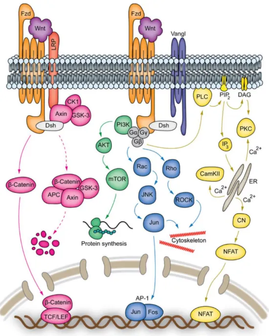
12 2.2.3 Wnt signalling
Wnt signalling is a well conserved pathway in Metazoans and plays a role in numerous important biological events, including cell proliferation, motility, polarisation, migration and determination, in the context of the establishment of the body plan as well as in the patterning of tissues and in organogenesis (Komiya and Habas, 2008; Teo and Kahn, 2010). The name Wnt derives from the fusion between the name of the Drosophila segment polarity gene
wingless with the vertebrate homologue integrated or int-1. Wnts are secreted glycoproteins,
which in humans and mice include a total of nineteen distinct Wnt proteins that share a core homology but exhibit distinct biological functions (Clevers and Nusse, 2012). Wnt ligands bind to Frizzled (Fzd) receptors, which are G protein-coupled receptor (GPCR)-like proteins that span the cell membrane seven times (Clevers and Nusse, 2012).
The best known Wnt signalling pathway operates through the activity of β-catenin and is termed the canonical Wnt pathway (Fig. 1.4). In the absence of Wnt ligands, cytoplasmic β-catenin is degraded through the activity of a degradation complex composed of Axin, APC (adenomatosis polyposis coli), PP2A (protein phosphatase 2A), CK1α (casein kinase 1α) and GSK3β (glycogen synthase kinase 3β). When Wnt ligands are present, they bind to the low-density lipoprotein-related protein 5/6 (LRP5/6) which acts as a co-factor to Fzd receptors and this coupling leads to the activation of Fzd (He et al., 2004; Fig. 1.4). The activation of Fzd is transduced by the phosphoprotein Dishevelled (Dsh) which directly interacts with Fzd (Wallingford and Habas, 2005). Activated Dsh recruits GSK3β and Axin to the membrane, thus dissociating the degradation complex, permitting the accumulation of β-catenin and its translocation to the nucleus, thereby inducing specific transcriptional gene expression (Fig. 1.4; Komiya and Habas, 2008). Wnt signalling can also occur through at least three so called non-canonical pathways (Fig. 1.4). These pathways are overall less well understood. Importantly, they are independent of LRP5/6 proteins and do not involve β-catenin activity. Rather they act through the PI3K/AKT/mTOR pathway, through G-protein mediated planar cell polarity (PCP) pathway or through the so called Wnt/Ca+2 pathway (Fig. 1.4) and are involved in a plethora of cellular responses crucial for tissue morphogenesis and organogenesis (Dale et al., 2009; De, 2011; Komiya and Habas, 2008; von Maltzahn et al., 2012a). Importantly, Wnt signalling can crosstalk with elements of other major signalling networks including Notch, Fgf and Shh pathways (Sethi and Vidal-Puig, 2010).
13 2.2.4 Hedgehog signalling
Hedgehog (Hh) signalling intervenes in a variety of important biological events such as cell differentiation, morphogenesis, tissue homeostasis and regeneration (Carballo et al., 2018; Lee et al., 2016). Hh proteins are found in a wide range of invertebrates and vertebrates and each Hh molecule is composed of a N-terminal domain, named “the Hedge” and a C-terminal domain termed “the Hog”. The Hedge domain is responsible for the signalling activity, while the latter domain undergoes autoproteolytic cleavage and is lipidated before Hh is secreted (Briscoe and Thérond, 2013; Ingham et al., 2011). In mammals there are three different types of Hhs: Sonic-Hedgehog (Shh), Indian-Hedgehog (Ihh) and Desert-Hedgehog (Dhh). Shh is the the most common Hh protein in our body and is crucial for a myriad of developmental decisions, including the patterning of the neural tube and the limbs, whereas Ihh is more specific, playing a role in skeletogenesis for example (Cai and Liu, 2016; Dessaud et al., 2008; Tickle and Towers, 2017). Dhh is the most restricted of the Hhs in terms of expression, being important for the development of the gonads (Bitgood et al., 1996; Yao et al., 2002). In this section, I will review the Shh signalling pathway (Carballo et al., 2018).
Canonical Shh signalling occurs in a ligand-dependent manner (Fig. 1.5A). First, Shh ligand binds to its co-factors Cdo (cysteine dioxygenase), Boc (brother of Cdo) and Gas1 (growth arrest-specific protein 1) which promote its binding to the transmembrane receptor protein Patched1 (Ptch1; Allen et al., 2011). Ptch proteins are also involved in limiting Shh signalling by sequestering and endocytosing Shh ligands through Hhip1 (Hh interacting protein 1; Holtz et al., 2015). In the absence of Shh ligands, Ptch1 inhibits the G-protein coupled receptor Smoothened (Smo). When Shh binds to Ptch1, Ptch1 and its Shh ligand are internalised for degradation, lifting the Ptch1-induced repression on Smo that enters the membrane. When in the membrane, Smo initiates the Shh transduction signalling pathway (Fig. 1.5A; Carballo et al., 2018; Incardona et al., 2002; Lee et al., 2016).
In the absence of ligands, Smo is not detected in the membrane and the transcription factor inhibitory complex (TFIC) is active, which leads to the transformation of Gli family zinc finger (Gli) transcription factors from activator into repressors by inducing their proteolytic cleavage (Fig. 1.5B; Carballo et al., 2018; Lee et al., 2016). Repressor forms of Gli2 and Gli3 translocate into the nucleus and block Shh target genes, leading to processes such as differentiation, pro-
14
____________________________________________________________________________________________________
Figure 1.4: Wnt signalling pathway.
A: Wnt signalling can be transduced into a canonical pathway (pink signalling cascade), which leads to the intracellular accumulation of β-catenin, which is translocated to the nucleus where it acts as a transcription factor and associates with TCF/LEF (transcription factor 1 / lymphoid enhancer binding factor) proteins inducing the expression of target genes. Binding of certain Wnt ligands to Frizzled (Fzd) receptors can also trigger non-canonical Wnt pathways that are independent of LRP5/6 (low-density lipoprotein-related protein 5/6) proteins and β-catenin activity. These pathways are (1) the PI3K/AKT/mTOR (green signalling cascade), which is directly activated upon Wnt-Fzd binding and is involved in cellular proliferation, growth, survival and migration. (2) the G-protein mediated PCP (planar-cell-polarity) pathway, which leads to activation of several small-GTPases and activates a Jun kinases (JNK) cascade (blue signalling cascade) that causes cytoskeletal remodelling and cell migration. (3) the Wnt/Ca2+ pathway (yellow signalling cascade), which involves the activation of the PLC/PKC
(phospholipase C/protein kinase C) signalling cascade leading to an increase of intracellular Ca2+ levels, which in turn
culminate with the entry of NFAT (nuclear factor of activated T cell) into the nucleus where it drives the expression of genes involved in cell adhesion, motility, polarity and proliferation. Based on Dale et al., 2009; De, 2011; Komiya and Habas, 2008; von Maltzahn et al., 2012a, b.
15
apoptotic and anti-proliferative cellular responses (Fig. 1.5B; Carballo et al., 2018; Lee et al., 2016). When a Shh ligand is present, on the other hand, the activation of Smo induces the repression of the TFIC culminating in the inhibition of Gli2 and Gli3 protein cleavage, and their entry into the nucleus as transcriptional activators which promote processes such as cell proliferation, de-differentiation and survival (Fig. 1.5A; Carballo et al., 2018; Lee et al., 2016).
Patched2 (Ptch2) is another transmembrane receptor for Hh proteins. It shares more than 50% homology with Ptch1 and is also able to trigger the Shh signalling pathway. However, Ptch2 exhibits a lower efficiency in the inhibition of Smo and thus does not have the same effect as Ptch1 (Rahnama et al., 2004). Shh signalling can also be triggered in a non-canonical way, which is a process independent of Smo or Gli activity and can result in the modulation of the cytoskeleton and regulation of the cell cycle, respectively (Brennan et al., 2012).
____________________________________________________________________________________________________
Figure 1.5: Generic overview of the Shh signalling pathway.
A: Secreted Sonic hedgehog (Shh) glycoproteins interact with transmembrane Patched (Ptch1) receptors. From these interactions Ptch1 and the Shh binding to it are internalised releasing the inhibition that Ptch1 exerted on Smoothened (Smo). Smo is stabilised and activated in the membrane, which results in the dissociation of the transcription factor inhibitory complex (TFIC) composed of Fu (Fused), SuFu (suppressor of Fused) and Kif7 (kinesin-like protein 7). proteins. This dissociation leads to the translocation of activated forms of Gli proteins (Gli1/2/3 A) leading to the activation of Shh target genes. B: In the absence of the ligand, Ptch1 remains inactive and inhibits Smo. With Smo inhibited the TFIC complex plus signalling cascades such as PKA (protein kinase A), CDK1 (cyclin dependent kinase 1) and GSK3β (glycogen synthase kinase 3β) promote the proteolytic change of Gli proteins, making them act as transcriptional repressors (Gli1/2/3 R). These forms of Gli proteins enter the nucleus and repress Shh target genes. Based on Carballo et al., 2018 and Lee et al., 2016.
16 2.2.5 Tgfβ/Bmp signalling
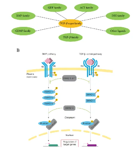
The transforming growth factor β (Tgfβ) superfamily is a conserved family of paracrine factors which include the Tgfβs, bone morphogenetic proteins (Bmps), growth and differentiation factors (Gdfs), glial-derived neurotrophic factors (Gdnfs) activins (Acts), inhibins (Inhs) and other ligands, such as anti-Müllerian hormone (Amh), left-right determination factor (Lefty) and nodal growth differentiation factor (Nodal) (Fig. 1.6A; Poniatowski et al., 2015). These different Tgfβ superfamily ligands induce multiple effects in the organism, being involved in a variety of critical cell decisions such as growth inhibition, cell differentiation, EMTs, ECM remodelling and immune suppression (Vortkamp et al., 1998; Zelzer et al., 2014). For the purposes of this thesis, I will focus on Tgfβs and Bmps.
Tgfβs are produced in a wide variety of cell types, including bone cells and the entire white blood cell lineage, in three major isoforms (Tgfβ1-3). In humans, they are encoded by genes located in distinct chromosomes, but the three proteins are highly homologous (Barton et al., 1988). More than a dozen of Bmp molecules are found in vertebrates and the Bmp family is divided into six main subgroups according to their structural characteristics and biological function (Katagiri and Watabe, 2016).
Tgfβs and Bmps family members are synthetised as inactive large homodimers in association with latent proteins. When these latent proteins are dissociated, Tgfβ and Bmp ligands become active (Schultz-Cherry et al., 1994). Other factors, such as changes in the pH environment or interactions with integrins (see section 2.3.2.) can also activate Tgfβ/Bmp ligands. After activation, Tgfβ ligands interact with a receptor complex forming a heterotetrameric combination containing two type I (TgfβrI) and two type II (TgfβrII) serine-threonine kinase receptors (Poniatowski et al., 2015). Bmps bind to Bmp type I (BmprI) receptors and type II (BmprII) serine-threonine kinase receptors but can also bind with less affinity to activin type II receptors (Katagiri and Watabe, 2016). Type II receptors for Bmps and Tgfβs ligands are constitutively active and control the binding of these ligands to type I receptors by inducing their phosphorylation. The specificities of the binding of Tgfβ /Bmps to their type I receptors depend on the identities of the interacting type II receptors and the cell type (Katagiri and Watabe, 2016; Poniatowski et al., 2015). After this, transduction of the Tgfβ or Bmp signalling pathways is initiated with the activation of Smad (mothers against decapentaplegic homolog) proteins. Smads can be classified as receptor (R-Smads), inhibitory