Universidade do Minho
Escola de Ciências
Ana Sofia Pereira de Freitas
outubro de 2015
Evaluation of bioactivities of a propolis sample
(Gerês) of Portuguese origin
Ana Sof ia P er eira de F reitas Ev
aluation of bioactivities of a propolis sample (Gerês) of P
or
tuguese origin
UMinho|20
Universidade do Minho
Escola de Ciências
Ana Sofia Pereira de Freitas
outubro de 2015
Evaluation of bioactivities of a propolis sample
(Gerês) of Portuguese origin
Trabalho realizado sob orientação do
Professor Doutor Rui Pedro Soares Oliveira
e da
Professora Doutora Cristina Alexandra de Almeida Aguiar
Dissertação de Mestrado
iii
A
CKNOWLEDGEMENTS
Firstly, I would like to thank my family: my parents, my brothers and my sisters for being my best friends and for supporting me in every way they could. More important, for always believing in me and for never let me give up of anything.
To Rui Silva, a special thank for all the love, care and patience and for always be there for me, in the best and worst moments.
I would like to express my sincere gratitude to my supervisors, Cristina Aguiar and Rui Oliveira, for all their tireless help, patience and friendship during these last years, and for giving me the opportunity to work with them. Without them this would not be possible.
Besides my supervisors, I would like to thank professors Ana Cunha and Dulce Geraldo for being part of this work and for sharing their knowledge with me and to all the technical and auxiliary staff of the Biology Department for all the help that made much easier the realization of this work, especially Cristina Ribeiro and Luís Correia.
I could not forget to thank my lab mates, Paulinha Vilela, Renata Moreira, Adriana Novais, Ilisa Antunes, Manuel Oliveira, Cláudio Oliveira, Adriana Gomes, Hugo Ribeiro, Marek Puškár, Nikola Grčić and Luis Giraldo for all the support and for all the fun we have had.
I would also like to thank to the beekeeper, Amadeu Fortunas, for gently provided the propolis samples used in this work and to Paulo Antunes for his help and contribution to this work by performing the chemical analysis of some of the samples.
v
Avaliação de bioatividades numa amostra de própolis (Gerês) de origem
Portuguesa
RESUMO
Própolis é uma mistura complexa formada por material resinoso e balsâmico, produzida pelas abelhas a partir de ramos, flores, pólen, brotos e exsudados de árvores, a qual é misturada com secreções salivares das abelhas. As abelhas utilizam o própolis na defesa contra invasores, protegendo a colmeia de infeções resultantes da putrefação. A composição química do própolis pode variar geograficamente, com a flora disponível, o clima, com a altura da colheita e a espécie de abelha. Diferentes grupos de compostos têm sido identificados em própolis, tais como flavonóides, ácidos fenólicos e os seus ésteres. Estes compostos têm sido associados com diversas atividades biológicas, nomeadamente: antimicrobiana; anti tumoral; antioxidante e quelante de radicais livres; anti-genotóxica e genotóxica; e antimutagénica.
O objetivo deste trabalho prende-se com o estudo de amostras de própolis português, particularmente no que diz respeito à sua caracterização química e à avaliação das suas bioatividades, visando a possibilidade da sua utilização/ exploração em aplicações médicas, cosmecêuticas e nutracêuticas. O própolis selecionado para este estudo foi colhido num apiário no Gerês (G), em quatro anos consecutivos, e foi utilizado para preparar extratos etanólicos (EE), que por sua vez foram testados em diferentes
ensaios, usando o eucariota simples Saccharomyces cerevisiae como modelo biológico.
O ensaio cometa foi realizado para analisar a genotoxicidade/ antigenotoxicidade e os resultados evidenciam que o EE preparado com própolis do Gerês recolhido em 2012 (G12.EE) não apresenta efeito genotóxico significativo. Por outro lado, o própolis do Gerês também não protege as células contra os
danos de DNA causados por peróxido de hidrogénio (H2O2), um comportamento exibido por qualquer um
dos extratos testados (G11.EE, G12.EE, G13.EE e G14.EE. Contudo, células co- ou pré-incubadas com
G.EEs e H2O2 10 mM exibiram maior viabilidade do que células incubadas apenas com H2O2, sugerindo
que o propolis protege as células de levedura contra o stresse oxidativo. Esta atividade antioxidante foi também demonstrada por citometria de fluxo - a oxidação do fluorocromo intracelular diacetato de
diclorofluoresceina (H2DCFDA) foi menor em células co- ou pré-incubadas com G.EEs e H2O2 do que em
células incubadas apenas com H2O2 - e corroborada por outros ensaios in vitro que demonstraram um
efeito quelante de radicais livres por parte dos G.EEs. Foi ainda constatado que os G.EEs, embora revelem baixa citotoxicidade para as células eucariotas testadas, têm atividade antimicrobiana particularmente expressiva contra bactérias Gram-positivas produtoras de esporos, tendo sido igualmente
observado um efeito sinérgico com o antibiótico gentamicina.A análise de células tratadas com os vários
G.EEs, na presença do fluorocromo rodamina 123, mostrou que o própolis do Gerês exerce influência sobre o potencial da membrana mitocondrial interna.
Todas as amostras de própolis estudadas exibiram um comportamento muito semelhante nas diversas bioatividades avaliadas, o que de um modo geral contraria a variabilidade atribuída a este produto natural quando colhido em diferentes anos, mesmo que proveniente de um só local. Para este perfil de bioatividades mais constante contribui possivelmente o tipo de produção padronizada de própolis usada pelo apicultor responsável, ao contrário do que faz a grande maioria de outros apicultores, particularmente os portugueses. Uma análise química preliminar de G11.EE e G12.EE, releva não haver diferenças significativas em termos do seu perfil em compostos fenólicos, aos quais se atribuem as bioatividades de propolis, justificando assim o comportamento mais constante evidenciado pelos quatro extratos estudados.
vii
Evaluation of bioactivities of a propolis sample (Gerês) of Portuguese origin
ABSTRACT
Propolis is a complex mixture composed by resinous and balsamic material, produced by bees from branches, flowers, pollen, buds and exudates of trees and mixed with bees’ salivary secretions. Bees use propolis in the defense against invaders, protecting the hive from infections resulting from putrefaction. The chemical composition of propolis varies geographically, with the available flora, the climate, the harvesting time and the bee species. Different groups of compounds can be found in propolis extracts, such as flavonoids, phenolic acids and their esters. These compounds have been associated with different biological activities such as antimicrobial; antitumor; antioxidant and free radical scavenger; antigenotoxic and genotoxic; and antimutagenic.
The aim of this work relates to the investigation on Portuguese propolis, particularly with regard to its chemical characterization and the evaluation of biological activities of this product in order to assess the possibility of its use/ exploitation in medical applications, cosmeticeutics and nutraceutics. The propolis samples selected for this study were collected in an apiary from Gerês, over four consecutive years and were used to prepare ethanol extracts (EE) which were tested in different assays, using the simple
eukaryote S. cerevisiae as biological model.
The comet assay was performed to analyze the genotoxicity/ antigenotoxicity and the results suggest that the EE prepared with propolis from Gerês harvested in 2012 (G12.EE) do not display significant genotoxic effect. On the other hand, propolis from Gerês does not protect cells against DNA damages caused by
H2O2 either, a behavior displayed by any of the tested extracts (G11.EE, G12.EE, G13.EE e G14.EE).
However, cells co- and pre-incubated with G.EE and 10 mM H2O2 displayed higher viability than cells
incubated only with H2O2, suggesting that G.EEs protect yeast cells against oxidative stress. The same
antioxidant activity was demonstrated by flow cytometry – a lower fluorescence of the intracellular
fluorochrome dichlorofluorescein diacetate (H2DCFDA) was detected in cells co- and pre-incubated with
G.EE and 5 mM H2O2 as compared with cells incubated only with H2O2 - and corroborated by several other
assays in vitro that show the free radical scavenging activityof G.EEs. Antimicrobial activity was evaluated
by the agar dilution method and the results suggest that G.EEs have antimicrobial activity, especially against Gram-positive spore forming bacteria. A synergistic effect of G.EEs when mixed with gentamicin was also demonstrated in the present work. The analysis of cells treated with G.EEs in the presence of the fluorochrome rhodamine 123 showed that propolis from Gerês has an influence on the inner mitochondrial membrane potential, decreasing the emitted fluorescence.
All the studied propolis samples exhibited a very similar behavior in the different evaluated bioactivities, which in generally is contrary to the variability attributed to this natural product when harvested in different years, even from a single location. For this more constant bioactivities profile possibly contributes the type of standardized production of propolis used by the beekeeper in charge, unlike what makes the great majority of other beekeepers, particularly the Portuguese. A preliminary chemical analysis of G11.EE and G12.EE reveals no significant differences in terms of phenolics profiles, compounds to which the bioactivities of propolis are attributed, thus justifying the more constant behavior evidenced by the four studied extracts.
ix
I
NDEX
ACKNOWLEDGEMENTS ... iii RESUMO ... v ABSTRACT ... vii INDEX ... ix ABBREVIATION LIST ... xiINDEX FIGURES ... xiii
INDEX TABLES ... xvii
1. INTRODUCTION ... 19
1.1. Propolis ... 21
1.2. Chemical composition and biological activities ... 22
1.2.1. Antibacterial activity ... 24
1.2.2. Antifungal activity ... 26
1.2.3. Antiviral activity ... 27
1.2.4. Antioxidant activity ... 28
1.2.5. Other biological activities ... 32
1.3. Biological problem and aim of this work ... 33
2. MATERIALS AND METHODS ... 37
2.1. Propolis samples and preparation of propolis extracts ... 39
2.2. Chemical analysis of G.EE ... 39
2.3. Yeast strains, media and growth conditions ... 40
2.4. Genotoxic and antigenotoxic properties of propolis ... 41
2.5. Assessment of propolis cytotoxicity ... 42
2.5.1. Evaluation of propolis protective effects against oxidative stress ... 42
2.6. Evaluation of the antimicrobial properties of propolis from Gerês ... 42
x
2.7. In vitro evaluation of the antioxidant activity of G.EEs ... 44
2.7.1. DPPH scavenging activity ... 44
2.7.2. Superoxide anion scavenging activity ... 45
2.7.3. Iron chelating activity ... 45
2.8. Evaluation of the antioxidant activity of G.EE by flow cytometry ... 46
2.9. Evaluation of the G.EEs influence on inner mitochondrial membrane potential by flow cytometry ... 47
2.10. Statistical analysis ... 48
3. RESULTS AND DISCUSSION... 49
3.1. Effects of propolis on S. cerevisiae DNA ... 51
3.1.1. G.EEs do not protect yeast cells from DNA damage caused by H2O2 ... 52
3.2. Effects of G.EE on cell viability ... 54
3.2.1. G.EE protects yeast cells under stress conditions caused by H2O2 ... 55
3.3. Antimicrobial activity of G.EE ... 57
3.3.1. Synergistic effect between G.EE and gentamicin ... 59
3.4. G.EE has significant DPPH radical scavenging activity ... 62
3.5. G.EE has superoxide anion scavenging activity ... 64
3.6. G.EE has iron chelating activity ... 65
3.7. G.EEs decreases intracellular oxidation ... 66
3.8. Influence of G.EE on inner mitochondrial membrane potential ... 69
3.9. Chemical composition of G.EE ... 71
4. CONCLUSIONS ... 73
REFERENCES... 77
xi
A
BBREVIATION LIST
BHA – butylated hydroxyl anisole BHT – butylated hydroxyl toluene CAPE – caffeic acid phenyl ester CAT – catalase
CPE – cytopathogenic effect DNA – deoxyribonucleic acid
DPPH – 2,2-diphenyl-1-picryl-hydrazyl EDTA – ethylenediamine tetraacetic acid EE – ethanol extract
G – Gerês
G11.EE – ethanol extract of propolis from Gerês harvested in 2011 G12.EE – ethanol extract of propolis from Gerês harvested in 2012 G13.EE – ethanol extract of propolis from Gerês harvested in 2013 G14.EE – ethanol extract of propolis from Gerês harvested in 2014 GA – gallic acid
GSH – glutathione
GSH-Px – glutathione peroxidase GSH-R – glutathione reductase H2DCF – dichlorofluorescein
H2DCFDA – dichlorofluorescein diacetate
HCL – human lung carcinoma HIV – human immunodeficiency virus HSV – human simplex virus
LC-MS – liquid chromatography-mass spectrometry LMA – low melting agarose
MIC – minimum inhibitory concentration
NADH – nicotinamide adenine dinucleotide, reduced form
NADPH – nicotinamide adenine dinucleotide phosphate, reduced form NBT – nitroblue tetrazolium
xii
OD – optical density
P.EE – ethanol extract of propolis PBS – phosphate buffered saline PMS – phenazine methosufate RNA – ribonucleic acid
ROS – reactive oxygen species RPM – revolutions per minute SOD – superoxide dismutase UV – ultra-violet
xiii
I
NDEX FIGURES
Figure 1 - Cellular reactions which result in the production of HO· - the Fenton reaction. Adapted from Valle et al., 2010. ... 29 Figure 2 - Main endogenous antioxidant defences of the cell. SOD: superoxide dismutase; CAT: catalysis; GSH: glutathione; GSH-Px: glutathione peroxidase; GSH-R: glutathione reductase; Vit. C: ascorbic acid; Vit. E: α-tocopherol. Adapted from Ferreira et al., 2007. ... 30 Figure 3 - Examples of comets observed with a fluorescence microscope after DNA labeling with GelRed. (A) Saccharomyces cerevisiae cells untreated (C-) or (B) treated with 10 mM H2O2 (C+).
... 51 Figure 4 - Assessment of propolis genotoxic effects by the comet assay. Incubation of S. cerevisiae with 5, 10, 25, 50, 100 or 200 µgmL-1 G12.EE. (C-) cells treated with ethanol in the
same volume as the extracts. (C+) cells treated with 5 mM H2O2. Mean±SD values of comet tail
length are from three independent experiments (* means p < 0.05, ** means p < 0.01 and *** means p < 0.001). ... 52 Figure 5 - Comet assay to evaluate propolis protective effects on DNA damage induced by oxidative stress. Co-incubation of S. cerevisiae with 5, 10, 25, 50 or 100 µgmL-1 of G11.EE (A),
G12.EE (B), G13.EE (C) and G14.EE (D). Propolis from Gerês does not seem to protect cells from DNA damage under oxidative stress caused by H2O2 (10 mM). (C-) cells treated with ethanol, the
solvent used for extraction. (C+) cells treated with H2O2. Mean±SD values are from three
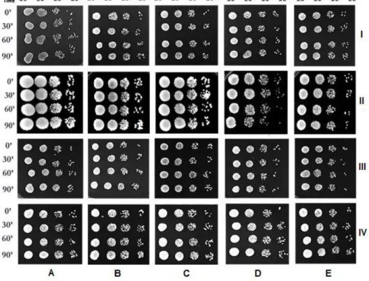
independent experiments (* means p < 0.05, ** means p < 0.01 and *** means p < 0.001). .. 53 Figure 6 - Viability of S. cerevisiae cells after 30, 60 and 90 min of incubation with (A) ethanol alone (control) or 100 µgmL-1 (B); 200 µgmL-1 (C); 500 µgmL-1 (D) and 750 µgmL-1 (E) of G11.EE
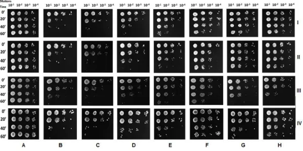
(I), G12.EE (II), G13.EE (III) and G14.EE (IV).. Data are from a representative experiment from three independent experiments. ... 54 Figure 7 - Viability of S. cerevisiae cells after 20, 40 and 60 min of co-incubation with H2O2 (5
mM) and (C) 25 µgmL-1; (D) 50 µgmL-1; (E) 100 µgmL-1; (F) 200 µgmL-1; (G) 500 µgmL-1 or (H)
750 µgmL-1 of G11.EE (I), G12.EE (II), G13.EE (III) and G14.EE (IV). (A) Cells treated with
ethanol alone; (B) cell treated with 5 mM H2O2 to assess the damage caused by the oxidizing
agent alone. Data are from a representative experiment from three independent experiments. . 56 Figure 8 - Viability of S. cerevisiae cells previously incubated with (C) 25 µgmL-1; (D) 50 µgmL-1;
xiv
G13.EE (III) or G14.EE (IV) for 20 min, washed with deionized water and suspended in YPD medium, and subsequently, incubated with H2O2 (5 mM), for 20, 40 and 60 min. (A) Cells treated
with ethanol alone; (B) cell treated with 5 mM H2O2 to assess the damage caused by this oxidizing
agent alone. Data are from a representative experiment from three independent experiments. . 56 Figure 9 - Detail of the assays to detect synergism between propolis and gentamicin. Drops of a Staphylococcus aureus prepared as described in section 2.3 were placed on top of LBA plates supplemented with 100 µgmL-1 or 200 µgmL-1 G.EE (A), gentamicin (B) or G.EE and gentamicin
(C) (100 µgmL-1 G.EE and 0.01 µgmL-1 gentamicin or 25 µgmL-1 G.EE and 0.75 µgmL -1gentamicin). (C-) LBA plate supplemented with ethanol. Data are from a representative
experiment from three independent experiments. ... 61 Figure 10 - Intracellular oxidation of S. cerevisiae cells loaded with H2DCFDA, incubated for 1 h
with different concentrations (50. 100 or 200 µgmL-1) of G11.EE (A), G12.EE (B), G13.EE (C) or
G14.EE (D) and analysed for fluorescence by flow cytometry. The control, (C-), representative of the extract solvent and the cells used in the experiment, only had ethanol. Data are from a representative experiment from three independent experiments. ... 67 Figure 11- Intracellular oxidation of S. cerevisiae cells loaded with H2DCFDA and analyzed for
fluorescence by flow cytometry, after co-incubation with 5 mM H2O2 and different concentrations
(50. 100 or 200 µgmL-1) of G11.EE (A), G12.EE (B), G13.EE (C) or G14.EE (D) for 20 min. Two
controls, one with ethanol (C-), representative of the extract solvent and the cells used in the experiment, and another with H2O2 5 mM (C+), to assess the damage caused by H2O2 alone, were
included. Data are from a representative experiment from three independent experiments. ... 68 Figure 12 - Intracellular oxidation of S. cerevisiae cells loaded with H2DCFDA, previously
incubated with different concentrations (50. 100 or 200 µgmL-1) of G11.EE (A), G12.EE (B),
G13.EE (C) or G14.EE (D) for 20 min, washed once with PBS and subsequently, incubated with 5 mM H2O2 for 20 min and analyses for fluorescence by flow cytometry. Two controls, one with
ethanol (C-), representative of the extract solvent and the cells used in the experiment, and another with H2O2 5 mM (C+), to assess the damage caused by H2O2 alone, were included. Data
are from a representative experiment from three independent experiments. ... 69 Figure 13 - Inner mitochondrial membrane potential state of S. cerevisiae cells loaded with rhodamine 123 after treatment with different concentrations (50, 100 or 200 µgmL-1) of G11.EE
xv
negative control, of cells treated with ethanol was included. Data are from a representative experiment from three independent experiments. ... 70 Figure 14 - Microphotographs of fluorescence microscopy of S. cerevisiae loaded with rhodamine 123 after incubation with G12.EE 100 µgmL-1 (A) and 200 µgmL-1 (B). ... 71
Figure 15 - Chromatographic profile of G11.EE. Each peak in the figure represents a different compound, corresponding to the compounds showed in Table 11. ... 72 Figure 16 - Percentage of reduction in absorbance of DPPH (517 nm) by adding increasing concentrations of gallic acid. ... 95
xvii
I
NDEX TABLES
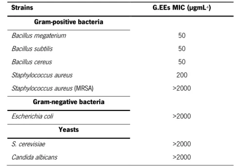
Table 1 - Strains used in this work as indicator strains in the antimicrobial assay ... 43 Table 2 - MIC values (µgmL-1) of G.EEs against the panel of susceptibility indicator strains. Results
are the same for G11.EE, G12.EE, G13.EE and G14.EE. ... 58 Table 3 - MIC values (µgmL-1) of gentamicin against the panel of tested bacteria. ... 59
Table 4 - Bacillus megaterium growth in the presence of sub-MIC concentrations of G.EEs and gentamicin. Results were the same for all the four studied G.EEs. ... 60 Table 5 - Bacillus subtilis and Bacillus cereus growth in the presence of sub-MIC concentrations of G.EEs and gentamicin. Results were the same for all the four studied G.EEs. ... 60 Table 6 - Staphylococcus aureus growth in the presence of sub-MIC concentrations of G.EEs and gentamicin. Results were the same for all the four studied G.EEs. ... 61 Table 7 - Escherichia coli and Staphylococcus aureus (MRSA) growth in the presence of sub-MIC concentrations of G.EEs and gentamicin... 62 Table 8 - Percentage of reduction of DPPH free radical by the four G.EEs tested. ... 63 Table 9 - Total antioxidant activity of G.EEs expressed as GA equivalents, in µgmL-1
. ... 64
Table 10 - Superoxide anion scavenging activity displayed by the four G.EEs studied in the present work... 65 Table 11 - Iron chelating activity displayed by the four G.EEs studied. ... 66 Table 12 - Chemical composition of G11.EE obtained by LC-MS. ... 72 Table 13 - Antioxidant activity (%) by the free radical DPPH scavenging by gallic acid concentration (µgmL-1). ... 95
19
21
1.1. Propolis
Throughout history, man has learned to use natural products in medicine. Propolis has been used in traditional medicine since the primordial times of humanity, having acquired popularity among Egyptians, Arabs, Greeks, and many other civilizations (Moreira et al., 2008). For Egyptians, propolis was well known due to its anti-putrefactive properties and its capability to embalm cadavers. Incas used propolis as an anti-pyretic agent and Greek and Roman physicians used it as an oral disinfectant, as antiseptic and to heal wounds, being prescribed for topical treatment of skin and mucosa (Burdock, 1998). Balkans used propolis to treat wounds and burns, sore throat and stomach ulcers. In the Second World War, the Soviets used propolis to treat tuberculosis due to the observed decrease of lung problems (Wollenweber et al., 1990). Registered as an official drug in the pharmacopoeia of London in the 17th century, propolis has
become very popular in Europe during the following years in particular due to its antibacterial activity (Fokt et al., 2010).
Propolis, or bee glue as it is also called, is a complex mixture composed by resinous and balsamic material, produced by honeybees (mainly Apis mellifera L.) from branches, flowers, pollen, buds and exudates of trees and mixed with salivary enzymes, waxes and other compounds resulting from the metabolism of bees (Fokt et al., 2010). Etymologically the word propolis is derived from the Greek pro (for ‘in front of’) and polis (for ‘community’), meaning that this natural product contributes to the defence of the hive (Sforcin, 2007). In the hive, propolis is used against invaders to immobilize their carcasses, protecting the hive from pests resulting from putrefaction. Another function of propolis is the mechanical and thermal insulation of the hive, being used to fill cracks or openings (Moreira et al., 2008).
The chemical composition and biological activities of raw propolis vary with the source plant species that exist around the hive where propolis is collected (Katalinic et al., 2004; Falcão et al., 2010), the climate characteristics (Falcão et al., 2010), the time of harvest, the technique used to harvest, the species of bee (Pereira et al., 2002) as well as the extraction method (Sheng et al., 2006). Even though, and in general, this complex mixture is composed of around 50 % resins and vegetable balsams, 30 % wax, 10 % essential oils, 5 % pollen and 5 % of other substances, including organic compounds (Fokt et al., 2010).
22
Propolis has a characteristic and pleasant aromatic odour and its colour can vary from yellow-green, red and dark brown, depending on their origin and age (Bankova et al., 2000). It is hard and brittle when cold, but becomes soft and very sticky when warm (Loutfy, 2006). It is difficult to remove from human skin since it appears to interact strongly with the proteins and oils of the skin (Burdock, 1998).
1.2. Chemical composition and biological activities
There are many compounds that have been identified in different samples of propolis (Marcucci et al., 1995; Bankova et al., 2000; de Castro et al., 2001), and new compounds are yet to be acknowledged during the chemical characterization of new samples. From all the identified compounds, phenolics are the most important. The most common phenolic compounds in propolis from temperate zones are flavonoids like pinocembrin, galangin and chrysin and phenolic acids such as caffeic acid, ferulic acid and cinnamic acid (Marcucci et al, 1995; Bankova et al., 2000; Huang et al., 2014). Propolis from tropical areas, especially in South-eastern Brazil, proved to be rich in prenylated phenylpropanoids, and some non-typical compounds such as kaempferide and isosakuranetin have also been found (Bankova et al., 2000).
In European propolis, the main bioactive compounds are flavonoids (flavones, flavonols and flavonones), phenolic acids and their esters (Huang et al., 2014). In a study concerning the phenolic compounds of an ethanol extract of propolis from Northeast Portugal, made by Falcão et al. (2010), it was shown that chemical compounds such as flavonoids were also found in the Portuguese sample, side by side with rare pinocembrin or pinobanksin derivatives that contain basic structures of phenolic acids, as well as p-coumaric ester derivative dimer (Fokt et al., 2010). Upon analysis by liquid chromatography-mass spectrometry (LC-MS) of the ethanol extract (EE) of propolis from Gerês collected in 2012 (G12.EE), the main compounds found were p-coumaric acid, pinocembrin, caffeic acid, quercetin, pinobanksin, chrysin and ferulic acid, among many others and similarly to other chemical profiles of European propolis described (Freitas, 2013).
Certain biological activities are always present in propolis and they can be associated with completely different chemical profiles in samples from diverse geographic and climate areas. Different chemical compositions of propolis from different origins led to the explanation that their
23
biological properties would be dissimilar, but this is amazingly untrue as samples of different origins can display identical biological activities. The main compounds responsible for propolis biological activities are the flavonoids, aromatic acids, diterpenic acids, phenolic compounds and cinnamic acid derivatives including caffeic acid esters but, very often, different propolis types have distinct main bioactive compounds (Borreli et al., 2002).
Propolis is commercialized in different parts of the world and it is recognized as an important source of compounds with properties for several applications (Moreira et al., 2008). There is a long history of propolis use, that continues today in home remedies and personal products, and that happens because propolis has an endless list of preparations and uses. The demand for this substance is becoming larger due to the growing consumers´ preference for natural products. Propolis can be found in pharmaceutical and cosmetic products such as face creams (vanishing creams and beauty creams), ointments, lotions and solutions. It is also found in dermatological items, useful in wound healing, tissue regeneration, treatment of burns, neurodermatitis, leg ulcers, psoriasis, morphoea, herpes simplex and genitalis and active against dermatophytes (Burdock, 1998). Propolis is commercially available and can be found in the form of capsules (pure or combined with aloe gel, Rosa canina or pollen), extracts (hydroalcoholic or glycolic), mouth wash solutions, creams, and many others (Fokt et al., 2010).
It is well known that ethanol and non-ethanol extracts of propolis have different chemical compositions and display diverse biological activities (Majiene et al., 2010; Ramanauskiené and Inkéniené, 2011; Mavri et al., 2012; Kubiliene et al., 2015). In recent years, this product has been the subject of intensive studies, highlighting their biological and pharmacological properties (Falcão et al., 2010; Piccinelli et al., 2013; Kurek-Górecka et al., 2014; Silva-Carvalho et al., 2014, 2015; Boisard et al., 2015; Szweda et al., 2015). Regardless of the plant source (species or geographical origin) and the composition, the biological activity of propolis, particularly the antimicrobial activity, has always been reported. Due to the plant diversity, there are different types of propolis, which contain numerous chemical constituents responsible not only for antimicrobial activity, but also for other valuable bioactivities (Bankova, 2005).
Propolis biological properties include antibacterial activity against various pathogenic bacteria (Burdock, 1998; Kujumgiev et al., 1999; Koo et al., 2000; Borreli et al., 2002; Uzel et al., 2005 Falcão et al., 2010; Castro et al., 2011, 2012), antifungal (Burdock, 1998; Koo et al.,
24
2000; Borreli et al., 2002; Moreira et al., 2008; Falcão et al., 2010; Castro et al., 2011, 2012), anti-protozoan (Castro et al., 2011, 2012), anti-viral (Borreli et al., 2002; Sheng et al., 2006; Moreira et al., 2008) as anti-HIV (Falcão et al., 2010; Castro et al., 2011, 2012), antioxidant (Banskota et al., 2001b; Borreli et al., 2002; Sheng et al., 2006; Falcão et al., 2010), anti-inflammatory (Borreli et al., 2002; Sheng et al., 2006; Lofty et al., 2006; Sforcin, 2007, Moreira et al., 2008; Falcão et al., 2010), anti-tumor (Grunberger et al., 1998; Sforcin, 2007; Moreira et al., 2008; Valença et al., 2013; Silva-Carvalho et al., 2014), hepato-protective (Sheng et al., 2006), anti-neurodegenerative (Chen et al., 2008; Falcão et al., 2010), local-anaesthetic (Moreira et al., 2008), anti-tuberculosis (Falcão et al., 2010), free-radical-scavenging (Sheng et al., 2006; Castro et al., 2012), immunostimulating (Borreli et al., 2002; Lofty et al., 2006) and cytotoxic (Matsuno et al., 1997). Propolis extracts were also tested as a food preservative due to its bacteriostatic and bactericidal properties (Tosi et al., 2007). Furthermore, Gregoris et al. (2011) showed that propolis protects against UV radiation and could be used in the formulation of sunscreens. Propolis is also capable of inhibiting the action of the enzyme hyaluronidase, allowing to retard cell aging (Kim et al., 2005) and, more recently, experimental data showed that propolis can be used to treat fungal infections of Candida (Castro et al., 2012).
For all these reasons, this natural product has sparked interest in the pharmaceutical and food industries, being introduced in different products for human consumption as drinks, food and cosmetics, though mostly because of its antioxidant and antimicrobial properties.
1.2.1. Antibacterial activity
Many researchers have studied the antibacterial activity of propolis to evaluate this property against a large panel of Gram-positive and Gram-negative bacteria, normally using one or two of the most popular methods used to evaluate this activity - the disc diffusion method and the broth or agar dilution method (Fokt et al., 2010). Several studies demonstrated that propolis has activity against a wide range of Gram-positive bacteria but had a limited or even no activity against Gram-negative ones (Bankova et al., 2000; Uzel et al., 2005; Lofty, 2006; Jorge et al., 2008; Ramanauskiené and Inkéniené, 2013).
Although the propolis mechanism of action for its antibacterial activity is not yet clearly understood, some studies suggest that propolis and some of its cinnamic and flavonoid components were able to uncouple the energy-transducing cytoplasmatic membrane, to inhibit
25
bacterial motility (Mirzoeva et al., 1997). It was also suggested that propolis inhibits bacterial growth by preventing cell division, resulting in the formation of pseudo-multicellular forms. In addition, propolis also disorganized the cytoplasm, the cytoplasmic membrane and the cell wall, which led to a partial bacteriolysis, and inhibited protein synthesis (Takasi et al., 1994). Other study (Uzel et al., 2005) suggests that the mechanism of action may be related to the inhibition of RNA-polymerase from bacteria.
The antimicrobial activity of propolis may be linked with its complex composition involving a complex mechanism putatively attributed to the synergistic effect of phenolic compounds such as cinnamic acid and ester derivatives including caffeic acid and acid phenyl ester (CAPE). Other compounds such as flavonoids - including quercetin, naringenin (Santos et al., 2002; Boisard et al., 2015), galangin, pinostrobin, and pinocembrin, ferulic acid, hydroquinones - and terpenic acids such as isopimaric, abietic and dehydroabetic acid (Patel et al., 2014) are also suspected to be responsible for this biological activity.
Park et al. (1998) reported that an ethanol extract of propolis (P.EE) from various regions of Brazil inhibited the growth of Streptococcus, an oral pathogen. Other studies made with periodontitis-causing bacteria, such as Peptostreptococcus anaerobius (Santos et al., 2002) Porphyromonas gingivalis, Prevotella intermedia (Santos et al., 2002; Gebara et al., 2002), Prevotella melaninogenica, Actinobacillus actnomycetemcomitans, Fusobacterium nuclatum and Capnocytophaga gingivalis (Gebara et al., 2002) showed the susceptibility of these strains to the EE. Several studies showed the antimicrobial action of P.EE against Staphylococcus aureus, a pathogen reported to produce food poisoning (Hegazi et al., 2000; Lu et al., 2005; Ramanauskiené et al., 2009; Ramanauskiené and Inkéniené 2013). Ramanauskiené et al. (2009) and Ramanauskiené and Inkéniené (2013) not only showed the antibacterial activity of Lithuanian P.EE against Staphylococcus aureus, but also against Enterococcus faecalis, Escherichia coli, Klebsiella pneumonia, Pseudomonas aeruginosa, Proteus mirabilis, Bacillus subtilis and Bacillus cereus. Wojtyczka et al. (2013a) demonstrated the antibacterial activity of Polish P.EE against the methicillin-sensitive (MSSA) and the methicillin-resistant (MRSA) Staphylococcus aureus, both clinical isolates. Other study showed the antibacterial activity of a propolis samples from Lebanon against Staphylococcus aureus (MRSA) (Chamandi et al., 2015). Antimicrobial activity of Korean propolis was showed by Kim and Chung (2011) against various foodborne pathogens such as Bacillus cereus, Staphylococcus aureus and Listeria onocytogenes.
26
Polish P.EE showed antibacterial activity against Staphylococcus epidermidis reducing the biofilm formation and bacterial growth (Wojtyczka et al., 2013b).
Bianchini and Benedo, (1998) demonstrated the inhibitory effect of aqueous extracts of propolis against some phytophatogenic bacteria such Agrobacterium tumefaciens, Clavibacter michiganensis subsp. michiganensis and Xanthomonas axonopodis. Piermann et al. (2007) tested an extract of propolis from a commercial product 10% concentrated and showed its antimicrobial activity against eight phytopathogenic bacteria (Pseudomonas syringae pv. tomato, Pseudomonas corrugata, Clavibacter michiganensis subsp. michiganensis, Erwinia carotovora subsp. Carotovora and several species of the genus Xanthomonas. Other study with phytopathogenic bacteria showed the susceptibility of Pseudocercospora vitis, Elsinoe ampelina and Phakopsora euvitis to alcoholic extracts of propolis (Marini et al., 2012).
A synergistic effect of P.EE with bactericidal anti-tuberculosis drugs, including streptomycin, rifamycin and isoniazide was reported. In the same study, two of the tuberculosis bacilli strains tested, found to be resistant to some of the drugs, lost part of their resistance when treated with P.EE in combination with the drug (Scheller et al., 1999). The synergistic effect between propolis and the antimicrobial drugs ampicillin, ceftriaxone and doxycycline against Staphylococcus aureus and with nystatin against Candida albicans was noticed by Stepanović et al. (2003), authors that also proved that bacterial resistance to antibiotics had no influence on the susceptibility to propolis extracts. Fernandes et al. (2005) found a synergistic effect between propolis and antimicrobial drugs against Staphylococcus aureus, especially for those agents that interfere on bacterial protein synthesis.
1.2.2. Antifungal activity
The antifungal activity is normally evaluated using the disc diffusion method and/ or the dilution method, as for the estimation of antibacterial activity. For antifungal activity, as well as for antibacterial activity, the effect is associated with the presence of flavonoids and other phenolic components (Farnesi et al., 2009). The propolis mechanism against fungal strains may be related to genes involved in the mitochondrial electron transport chain, vacuole acidification, negative regulation of transcription from RNA polymerase II promoter, regulation of macroautophagy associated with protein targeting to vacuoles, and cellular response to starvation (Castro et al., 2011).
27
Samples from European propolis demonstrated a fungicidal effect against species belonging to the genera Candida, Microsporum, Mycobacteria, Trichophyton, Fusarium and other dermatophytes (de Castro et al., 2001). Several other studies demonstrated also the susceptibility of clinical yeasts belonging to Candida genus such as Candida albicans, (Hegazi et al., 2000; Trusheva et al., 2006; Ramanauskiené et al., 2009; Noori et al., 2012; Ramanauskiené and Inkéniené 2013; Chamandi et al., 2015) as well as of some filamentous fungi, mainly dermatophytes. S. cerevisiae and Trichosporon sp. showed to be susceptible to propolis as well (Oliveira et al., 2006). The susceptibility of S. cerevisiae was also found in a study with Spanish P.EE made by Banvehí and Gutiérrez (2012). Al-Daamy et al. (2015) assessed the antifungal activity of propolis from Iraq against the two dermatophytes: Trischophyton mentagrophytes and Trichophyton tonsurans and on five clinical isolates of Candida albicans isolated from oral cavities of different patients, and showed the susceptibility of all the strains. Recently, Szweda et al. (2015) studied a sample of P.EE from Poland and showed its fungicidal activity against Candida albicans, Candida glabrata and Candida krusei. D’auria et al. (2003) not only showed the antifungal activity of propolis against Candida albicans strains but described additionally its inhibitory effect on yeast-mycelial conversion and a reduction on hyphal length. Other study, made by de Castro et al. (2013) demonstrated that propolis inhibited the transition from yeast-like to hyphal growth on Candida albicans mutants’ strains.
1.2.3. Antiviral activity
The methodology normally used to evaluate the antiviral activity is the cytopathogenic effect (CPE) reduction assay (de Castro et al, 2001). The data about the antiviral effect of propolis are very few but the studies performed have shown that propolis displays significant antiviral activity at different levels, interfering with the replication of some viruses (de Castro et al., 2001) like herpes simplex types 1 and 2, adenovirus type 2, influenza virus, or human immunodeficiency virus (HIV), among others (Schinitzler et al., 2010; Sartori et al., 2012). Indeed, it was also found that propolis suppressed HIV-1 replication (Hadi and Hedazi, 2002) and inhibited its variants expression (Gekker et al., 2005) too. According to Tait et al. (2006), natural and synthetic flavonoids may interfere with picornavirus replication by preventing the decapsidation of viral particles and RNA release within cells or blocking viral RNA synthesis. In fact, apigenin, luteolin, naringenin and quercetin showed to be active aginst enterovirus 71
28
infections (Ji et al., 2015).Schinitzler et al. (2010) analysed the antiviral effect of P.EE and some of the constituents against herpes simplex virus type 1 (HSV-1) and proved that P.EE exhibited high anti-HSV-1 activity and that galangin and chrysin were the main bioactive compounds. Other study made by Shvarzbeyn and Huleihel (2011), who tried to determine which step of Tax oncoprotein-induced NF-kB activation is blocked by propolis and CAPE, showed that both inhibited substantially the activation of NF-kB-dependent promoter by Tax and also that both prevented Tax binding to IkBα and its degradation.
1.2.4. Antioxidant activity
Reactive oxygen species (ROS) such as hydroxyl (HO·), superoxide anion (O2·), nitric oxide
(NO) and hydrogen peroxide (H2O2) are continuously generated in the cell due to aerobic
metabolism. A free radical can be defined as any molecular species that contains an unpaired electron in an atomic orbital (Lobo et al., 2010). O2·, normally considered a primary ROS, can be
formed by the addiction of an electron to the molecular oxygen (Cadenas and Sies, 1998). Despite not being a very active radical, it is able to interact with other molecules to form other radicals, usually called secondary ROS, such as H2O2 and HO· (Ferreira et al., 2007). H2O2 can
yield HO· when with metal ions, being the ROS that causes more cellular damage due to its strong reactivity (Ferreira et al, 2007). In Fenton reaction (Figure 1), iron reacts with H2O2,
leading to the formation of HO· radicals which have a high redox potential, attacking all the species present in the reaction medium. The high reactivity of HO· results in rapid and non-specific reactions with different substrates, implying that the reaction rate can be limited by the diffusion rate. When Fe3+ is used instead of Fe2+, in combination with excess of H
2O2, other
radicals of lower oxidation potential such as hydroperoxyl (HO2·) and O2·, are also formed. The
proportion in which these radicals are produced is determined by the pH, due to the protonation of the O2· that occurs in acid medium. The HO· radical can act like an electrophile or like a
common nucleophile, attacking organic molecules by the rejection of hydrogen ions or engaging in double bonds and aromatic rings (hydroxylation). The decomposition of H2O2 by Fe3+ can
generate the reduced species Fe2+ which also reacts with H
2O2 and HO· (Aguiar et al., 2007).
ROS can also be generated due to exogenous agents such as heat shock, dehydration, toxic chemicals, ultraviolet and ionizing radiation (Nakajima et al., 2009; Sá et al., 2013; Mitra and Uddin, 2014). At low or moderate concentrations, ROS can be beneficial to the cell, being
29
involved in several physiological processes such as signalling and redox regulation and defence against infections (Fridovich, 1999). Once produced, most free radicals are removed by the cell antioxidant defences including enzymes and non-enzymatic molecules. Maintaining the balance between free radical production and antioxidant defences is a prerequisite for the normal functioning of the body. However, this balance can be destroyed when generation of ROS overwhelms the cellular antioxidant components or because there is a deficiency in the antioxidant defences of the cell, causing a drastic oxidative stress (Ferreira et al., 2007; Sá et al., 2013). When ROS production exceeds cellular antioxidant capacity, the consequences are oxidative damage of membrane lipids, proteins and nucleic acids, which can lead to cell death or to acceleration in aging and to a number of diseases such as cancer (prostate and colon) (Karamian and Ghasemlou, 2013), Alzheimer, Parkinson or multiple sclerosis (Wilms et al., 2007; Weiner, 2009; Politis et al., 2011).
Figure 1 - Cellular reactions which result in the production of HO· - the Fenton reaction. Adapted from Valle et al., 2010.
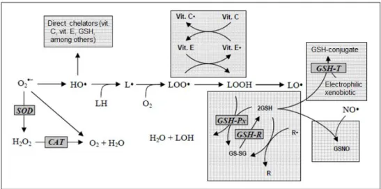
The exposure of organisms to free radicals led to the selection of those who have developed a number of defence mechanisms. Examples of these defences are superoxide dismutase (SOD), catalase (CAT), glutathione peroxidase (GSH-Px) and glutathione reductase (GSH-R). Between the non-enzymatic antioxidant defences are compounds such as glutathione (GSH), α-tocopherol (vitamin E), ascorbic acid (vitamin C), flavonoids and carotenoids (Ferreira et al, 2007) (Figure 2) that are mainly related to the elimination and detoxification of the components that can be damaged by ROS (Sá et al., 2013).
30
Figure 2 - Main endogenous antioxidant defences of the cell. SOD: superoxide dismutase; CAT: catalysis; GSH: glutathione; GSH-Px: glutathione peroxidase; GSH-R: glutathione reductase; Vit. C: ascorbic acid; Vit. E: α-tocopherol. Adapted from Ferreira et al., 2007.
There are also synthetic antioxidants like butylated hydroxyl anisole (BHA), butylated hydroxyl toluene (BHT), tertiary butylated hydroquinone and gallic acid esters (Talla et al., 2014), used by food industries to prevent lipid peroxidation and oxidation of food constituents. However, these compounds are suspected to have some negative health effects as liver damage and carcinogenesis. These problems increased the research and the demand for safer natural antioxidants in many applications, renewing the interest on natural products that have been used for centuries for a variety of reasons (Geckil et al., 2005).
Propolis is recognized for being a natural antioxidant agent. The antioxidant potential of propolis is important for immunomodulatory properties because it increases the cellular immune response through the increase of mRNA for interferon-γ and activates the production of cytokines (Fischer et al., 2007). The propolis main mechanisms of action for its antioxidant activity involve the inhibition of the activity of some enzymes which are involved in ROS generation, inhibiting that way the production of ROS; the scavenging activity, interrupting the reactions that result in lipid peroxidation; by chelating metal ions, generally iron and cooper; or potentiating the action of other antioxidants (Kurek-Górecka et al., 2013; Silva-Carvalho et al., 2015).
Polyphenols and flavonoid compounds were the main bioactives reported to be responsible for antioxidant potential in different propolis samples (Kumazawa et al., 2004). According to
Orsolić et al. (2006), ferulic acid, quercetin, prenylated compounds, apigenin, galangin and
p-coumaric acid are involved with the antioxidant potential of propolis samples. A study with samples of P.EE from Transylvania showed a positive correlation between high polyphenolic
31
composition and high antioxidant activity (Mihai et al., 2011). Chen et al. (2009) found also CAPE as a component which plays an important role in the antioxidant activity. Other study made with a ethyl acetate fraction of propolis collected in Anhui, China, showed strong scavenging activity and ferric reducing activity and related these activities with the presence of caffeic acid, phenethyl caffeate, cinnamyl caffeate and benzyl caffeate (Yang et al., 2011).
Propolis extracts have been reported to possess a potent antioxidant activity (Viuda-Martos et al., 2008). This activity can be evaluated by various methodologies. By flow cytometry it was possible to observe a decrease in the fluorescence intensity of dichlorofluorescein (H2DCF) in S.
cerevisiae propolis-treated cells, which correlated to propolis ability to decrease intracellular oxidation (Cigut et al., 2011). This antioxidant ability of a propolis sample from China was also demonstrated for RAW264.7 cells (Zhang et al., 2015). Sá et al. (2013) reported also a potent antioxidant activity of propolis in yeast cells: a reduction of the level of ROS produced when cells were treated with propolis after treatment with H2O2.
Propolis also showed strong reducing power and the ability to chelate metal ions (Miguel et al., 2010; Mavri et al., 2012; Talla et al., 2014) and scavenges free radicals (Moreira et al., 2008; Nakajima et al., 2009; Miguel et al., 2010; Mavri et al., 2012; Campos et al., 2014; Zhang et al., 2015). Geckil et al. (2005) reported high metal chelating capacity of propolis and comparable antioxidant activity to the two most widely used synthetic antioxidants, BHA and BHT. Sheng et al. (2007) and Talla et al. (2014) indicated propolis as a potential natural antioxidant by DPPH free-radical-scavenging activity. Other antioxidant activities of different extracts of propolis were found such as ferric reducing activity (Yang et al., 2011; Piccinelli et al., 2013) and reduction of lipid peroxidation (Valente et al., 2011; Silva et al., 2011; Campos et al., 2014).
A study with the same samples used in this work (G11.EE, G12.EE, G13.EE and G14.EE) using cyclic and differential pulse voltammetry methods, showed significant antioxidant capacity for all the G.EEs in a concentration dependent-manner. The highest antioxidant capacity, in cyclic voltammetry, was found in G12.EE, while G11.EE possesses the lowest antioxidant capacity values, at both concentrations tested. The highest antioxidant capacity, in differential pulse voltammetry, was found in G14.EE, while G11.EE possesses the lowest antioxidant capacity values, at both concentrations tested. The sequence orders of antioxidant capacity on cyclic
voltammetry and on differential pulse voltammetry were respectively
32
1.2.5. Other biological activities
Propolis showed antitumor activity, including cytotoxicity (Valença et al., 2013; Silva-Carvalho et al., 2014; Silva-Carvalho et al., 2015) against several cancer cell lines. Its main mechanism of action involves apoptosis, cell cycle arrest and interference on metabolic pathways (Watanabe et al., 2011; Valença et al., 2013; Silva-Carvalho et al., 2014). The cytotoxic effect of different flavonoids such as quercetin, chrysin and caffeic acid was shown to be strong against five leukaemia cell lines (MOLT, JURKAT, HL-60, RAJI and U937) (Watanabe et al., 2011). Some diterpenic acids, isolated from propolis, exhibited cytotoxicity towards human lung carcinoma HLC-2 and human carcinoma HeLa cells (Banskota et al., 2001a). Other component of propolis, CAPE, was investigated for its effect on the angiogenesis, tumor invasion, and metastasis and the results showed inhibition of the angiogenesis and of cell proliferation concluding that CAPE has potential as an anti-metastatic agent (Liao et al., 2003). It was also reported that CAPE showed a dose-dependent decrease in cell viability of CT26 colon adenocarcinoma cells (Lofty, 2006). Also, Hernandez et al. (2007) showed that CAPE, galangin, xanthomicrol and chrysin had a significant antiproliferative activity on several cancer cells. A study made with a fractionated Portuguese propolis sample from Angra do Heroísmo, Azores Archipelago, on HCT-15 colon cancer cell line showed the ability of propolis to decrease cell viability of colon tumor cells and also a disturbance of cancer cell glycolytic metabolism (Valença et al., 2013). Other study, made by Silva-Carvalho, (2014), with a Portuguese propolis sample, from Pereiro, showed a decrease on cell viability of different tumour cells. The same study showed a decrease on MDA-MB-231 and DU145 cells proliferation and migration, with cell cycle changes.
There are several studies describing the anti-inflamatory activity of propolis (Naito et al., 2007; Funakoshi-Tago et al., 2015; Teles et al., 2015; Valenzuela-Barra et al., 2015; Silva-Carvalho et al, 2015). A study showed that P.EE from Croatia suppressed functional activity of macrophages improving psoriatic-like skin lesions on male albino mice (Oršolić et al., 2014). Another studies made on male mice showed that P.EE, from Chile and from Brazil, inhibited the NO release by the macrophages (Valenzuela-Barra et al., 2015) and decreased renal macrophages infiltration (Teles et al., 2015), respectively.
The immunomodulatory effect of propolis was evaluated by Da Silva et al. (2013) in Leishmania (Viannia) braziliensis infection, using a propolis sample from Brazil. Propolis was able to increase the interiorization of macrophages and further killing the parasites. The same study
33
also demonstrated an increase of TNF-α production while IL-12 was downregulated during the infection.
1.3. Biological problem and aim of this work
Over the years, the interest in natural products has been surprisingly increasing because of its potential for the development of new drugs. Also, the industries have revealed an increased interest in these natural products because of its diversified bioactive profiles that can be used in food, in order to replace some usual synthetic compounds that can be toxic, as well as in cosmetic and pharmaceutical industries (Silva-Carvalho et al., 2015). Propolis biological and pharmacological actions have been reported (Silva-Carvalho et al., 2015) and its biological properties have been used by several industries (Marcucci, 1995; Pereira et al., 2002). On the other hand, and unlike the products derived from medicinal plants, propolis has great variability in terms of chemical composition, which is a major problem to its standardization and consequently to its use and acceptance by the medical community (Bankova et al., 2000; Falcão et al., 2010). Propolis purity, regarding the percentage of beeswax or insoluble residues, among others, must be considered as well (Bankova et al., 2000). Propolis composition depends on a variety of factors like the source of plant species, environmental factors (Valença et al., 2013) such climate characteristics (Marcucci, 1995), the time and the technique of harvest, the species of bee (Pereira et al., 2002), as well as the extraction method (Sheng et al., 2006). These factors account for the wide range of compounds found in propolis samples from different regions (Mărghitaş et al., 2013), which in turn constitute a problem for the medical use of propolis and its quality control (Bankova et al., 2000). There is considerable information about the chemical composition and the biological activities of propolis but, in order to be accepted into the health care system, propolis needs chemical analysis performed by some standardized methods (Mărghitaş et al., 2013) and requires systematic investigations of the chemical composition and the biological action, particularly the antimicrobial action (Bankova et al., 2000).
The aim of this work relates to the investigation on Portuguese propolis, particularly with regard to its chemical characterization and the evaluation of biological activities in order to assess the possibility of their use/ exploitation in medical, cosmeticeutics and nutraceutics applications. The propolis samples selected for this study were collected in an apiary from Gerês (G), harvested over four consecutive years (2011, 2012, 2013 and 2014), and used to prepare EE
34
which were tested in different assays, using the simple eukaryote S. cerevisiae as biological model. There are several characteristics that contribute to the choice of the yeast S. cerevisiae as the experimental biological model, including the easy handling, non-pathogenicity, and the well-known biology, genetics and metabolism (Guthrie and Fink, 2004). When in rich medium, cultures of S. cerevisiae display several distinct stages of growth. After a short period of adjustment, the lag phase, the culture grows exponentially using the energy derived mainly from the fermentation of hexoses such as glucose, with simultaneous repression of genes required for the respiratory metabolism. S. cerevisiae is a facultative anaerobe and in the presence of glucose does not perform oxidative phosphorylation. When glucose is exhausted, the repressed genes cease to be, and the cells adjust to the respiratory metabolism. After this phase, cell division is resumed at a reduced rate, via the respiratory metabolism using the final fermentation products like ethanol, acetate, glycerol, and other sources of carbon and energy, thereby performing the aerobic energy metabolism. When these carbon sources are exhausted, the cells stop dividing, thus entering the stationary phase (Winde et al., 1997).
Considering the relative few studies concerning the effects of propolis on yeast DNA, it seemed relevant to evaluate the genotoxic/ antigenotoxic effect of G.EEs in the present work. DNA damages caused by G.EEs alone or in co-incubation conditions with 5 mM H2O2 were
investigated in S. cerevisiae cells by the comet assay. The antimicrobial activity is one of the most important biological activities found on propolis samples and to evaluate the antimicrobial potential of propolis from Gerês, the antimicrobial activity of G.EEs was determined against a panel of bacteria and yeast strains, being expressed by the minimum inhibitory concentration (MIC) of propolis for which no microbial growth was detected. With microbial antibiotic resistance becoming a significant world health problem in recent years, strategies to overcome microbial resistance as well as new antimicrobial drugs are urgently needed. Being the synergistic effect of propolis with several antibiotics already reported, a possible synergistic effect of G.EEs and the antibiotic gentamicin was also studied in this work, being evaluated against the bacteria Bacillus megaterium, Bacillus subtilis, Bacillus cereus, Escherichia coli, Staphylococcus aureus and methicillin resistant Staphylococcus aureus (MRSA).
The antioxidant activity is another important activity found in propolis, and naturally it was important to evaluate if propolis samples from Gerês also have potential to be used as an antioxidants. In vitro antioxidant assays were carried out to measure the G.EEs ability to: scavenge DPPH free radical (by the DPPH assay); to quench the superoxide anions (O2•-) (by the
35
superoxide anion scavenging activity) and chelate Iron (Fe2+) (by iron chelation activity). The
intracellular oxidation and the antioxidant activity were also analysed in vivo by flow cytometry with the intracellular redox-sensitive fluorochrome dichlorofluorescein diacetate in yeast cells incubated with G.EE alone and in co- and pre-incubation conditions with 5 mM H2O2.
Propolis influence on the inner mitochondrial membrane potential was also investigated as such effect was described for other propolis samples. The influence of G.EE on the inner mitochondrial membrane potential was evaluated by flow cytometry and using rhodamine 123 as probe, which stains the mitochondria directly and distributes electrophoretically into the mitochondrial matrix in response to the mitochondrial electrical potential, making possible to detect alterations in mitochondrial distribution (Ludovico et al., 2001). In parallel, in order to overcome the problem of propolis standardization and given the need to chemically characterize and compare propolis samples from Gerês, G1.EE was analyzed by LC-MS to characterize its profile in phenolic compounds.
37
39
2.1. Propolis samples and preparation of propolis extracts
The propolis samples used in this work are from Gerês (G) and were kindly provided by the beekeeper Amadeu Fortunas whose apiary is located near the Cávado River, between the villages of Paradela and Sirvozelo, in Montalegre, Gerês, Portugal (41⁰45’41.62’’ N; 7⁰58’03.34’’ W). Four samples of propolis were collected in different years - 2011 (G11), 2012 (G12), 2013 (G13) and 2014 (G14) – and were used to prepare ethanol extracts (EE).
For alcoholic extraction, approximately 15 g of propolis were incubated with 80 mL of absolute ethanol in an orbital shaker at 25 ⁰C, 100 revolutions per minute (rpm), for 24 h in the dark. The resulting solution was filtered with Macherey-Nagel filter papers, using a Buchner funnel and a Kitasato system attached to a vacuum pump. The residues were collected and extracted again, with 50 mL of absolute ethanol. The resulting filtrates were polled and dried in a Büchi Rotavapor RE 121 with a water bath (Büchi 461), at 40 ⁰C, 40 rpm, yielding the ethanol extracts of propolis – G11.EE (Carvalho, 2012; Pereira, 2013), G12.EE (Pereira, 2013), G13.EE (Araújo, 2014) and G14.EE (this work) - which were stored at 4 ⁰C, in the dark, until further use. The stock solutions prepared for the following described assays were performed by diluting the propolis extracts in the same solvent used for the extraction.
2.2. Chemical analysis of G.EE
Propolis chemical analysis was performed by Paulo Antunes at Centro de Apoio Tecnológico Agro-Alimentar (CATAA), Castelo Branco. Briefly, samples were homogenized and diluted with 80 % ethanol, at 70 ⁰C for 1 h. The resulting mixture was filtered directly to a vial, through a nylon 0.22 µm filter. Standards for gallic acid, siriginc acid, ferulic acid, p-coumaric acid, apigenin and kaempferol were acquired from Sigma-Aldrich Co. Luteolin and gentisic acid standards were acquired from Extrasynthese, France. The chromatographic system consists of an Agilent 1200 series equipped with a model of a triple quadrupole mass spectrometer Agilent 6400. A Sorbax SB-C18 (50 mm x 4.6 mm i.d. x 1.8 µm particle diameter – Agilent technologies) column was used for the separation of the components of a flow rate of 0.7 mL/ min, at 30 ⁰C. Elution was performed using a gradient of 0.1 % formic acid (eluent A) and acetonitrile (eluent B). The gradient was as follows: started at 10 % of B, then 20 % of B in 10
40
min, 40 % of B in 40 min, 60 % of B in 60 min, 90 % of B in 80 min and at 81 min return to the initial conditions, stabilizing for 9 min. ESI operated with a nitrogen flow of 10 L/ min, at 300 ⁰C. MS detector operated in MS2-Scan, scan type in the range 80-1000 Da, and negative mode was selected. The capillary voltage was set to 4.0 kV, the quadrupole temperatures were 100 ⁰C, the fragmentation energy was 145 Kjmol-1 and the cell accelerator voltage was 7 kV. Data were
acquired and analysed using Masshunter Workstation Software (version B.04.00) from Agilent technologies.
For MS/ MS confirmation, the same equipment and chromatographic conditions were used. MS detector operated in Product Ion scan type, selecting the precursor ions and performing a scan of the fragments in the range 80-500 Da, and the negative mode was selected. The capillary voltage was set to 4.0 kV, the quadrupole temperatures were 100 ⁰C, the fragmentation energy was 135 Kjmol-1, cell accelerator voltage was 7 kV and the collision energy
was 15 eV. Compounds were identified (Antunes, P., personal communication) based in standards retention times and by comparison of the ESI-MS/MS with the data from MS/MS published in the literature, such as in Falcão et al. (2010).
2.3.
Yeast strains, media and growth conditions
In all experiments the haploid yeast strain S. cerevisiae BY4741 (MATa his3Δ1 leu2Δ0 met15Δ0 ura3Δ0) was used. Yeast cell cultures were prepared on liquid YPD medium (1 % (w/ v) yeast extract DB BactoTM, 2 % (w/ v) peptone DB BactoTM, 2 % (w/ v) glucose) or YPE (the same
as liquid YPD with 2 % (v/ v) of ethanol instead of 2 % glucose) depending on the experiments, while bacterial cultures were grown on liquid LB medium (Luria-Bertani – 0.5 % (w/v) yeast extract DB BactoTM, 1 % (w/ v) tryptone BactoTM, 1 % (w/ v) NaCl). For solid media (YPDA and LBA)
2 % agar (w/ v) was added to the same recipes. For liquid cultures a ratio flask/medium volume of 5/1 was used and incubation was performed at 30 ⁰C, for yeasts, or at 37 ⁰C, for bacteria, both at 200 rpm. The cultures growth was monitored by optical density at 600 nm (OD600).
To prepare the cells to the experiments an overnight microbial culture was diluted in fresh medium to an OD600 = 0.1 (10 mL final volume) and incubated at 30 ⁰C for 4 h, for yeasts, or at
37 ⁰C for 2 h, for bacteria, and 200 rpm until OD600 reached 0.4-0.8 (at least two generation
41
2.4. Genotoxic and antigenotoxic properties of propolis
A volume of 1 mL of a yeast culture obtained as described in the above section was harvested by centrifugation at 14500 rpm, 4 ⁰C for 2 min and washed twice with the same volume of deionized water at 4 ⁰C. The resulting pellet was suspended in S buffer (sorbitol 1 M, KH2PO4 25 mM, pH 6.5) with 6600 U lyticase (66 Uµl-1) and 50 mM β-mercaptoethanol, being
incubated at 30 ⁰C, 200 rpm for 30 min in order to obtain spheroplasts. The spheroplasts were collected by centrifugation at 14500 rpm, 4 ⁰C for 2 min, washed twice with the same volume of S buffer and resuspended in 1 ml S buffer. The suspension was divided by 100 µL aliquots and centrifuged under the same conditions. Supernatants were discarded and spheroplasts were treated with 5, 10, 25, 50 or 100 µgmL-1 of G12.EE to evaluate the genotoxic effects of propolis.
For negative and positive controls similar cell suspensions were treated with ethanol or ethanol and H2O2 (10 mM), respectively. All aliquots were incubated at 30 ⁰C, 200 rpm, during 20 min.
After incubation, spheroplasts were collected from each sample by centrifugation, at 14500 rpm for 2 min, washed with S buffer and each pellet was resuspended in 40 µL of low melting agarose (LMA) 1.5 % (w/ v in S buffer) at 35 ⁰C. The mixture was spread onto glass slides pre-coated with normal melting agarose (NMA) 0.5% (w/ v) and covered with cover slides. Glass slides were placed on ice for 5 min to solidify the agarose. The cover slips were removed and the glass slides were submerged in ice-cold lysing buffer (30 mM NaOH, 1 M NaCl, 50 mM EDTA, 10 mM Tris-HCl, 0.05 % (w/ v) laurylsarcosine, pH 10) for 20 min, followed by 20 min immersion in ice-cold electrophoresis buffer (30 mM NaOH, 10 mM EDTA, 10 mM Tris-HCl, pH 10). The slides were then placed in the electrophoresis tank filled with electrophoresis buffer and an electric field of 0.7 V/ cm was applied for 10 min. Samples were fixed with 76 % (v/ v) ice-cold ethanol and subsequently with 96 % (v/ v) ice-cold ethanol, both for 10 min. The gels were dried at room temperature and stored at 4 ⁰C until observation. Slides were analysed by fluorescence microscopy (Leica DM5000B+CTR5000+ebq100) after staining with 10 µL of GelRedTM (3.300x
diluted; Biotium), with 400x magnification. Comets´ tail length was measured by informatic analysis of the images.
To assess the antigenotoxicity of propolis, the procedure was the described above with the exception of the incubation step, being the cells incubated with each of the four G.EEs simultaneously with H2O2 (10 mM).
42
2.5. Assessment of propolis cytotoxicity
To assess propolis cytotoxicity a yeast culture (see 2.3.) was divided into 1 mL aliquots and treated with 25, 50, 100, 200, 500 or 750 µgmL-1 of each of the four G.EEs. The same
volume of ethanol was added to a similar cell suspension for the negative control. Immediately after G.EE or ethanol addition, 100 µL of each suspension were removed and serially diluted to 10-4. This procedure was repeated after 30, 60 and 90 min incubation at 30 ⁰C, 200 rpm. After
dilutions, drops of 5 µL of each sample were transferred to YPDA plates and then incubated at 30 ⁰C for 48 hours.
2.5.1. Evaluation of propolis protective effects against oxidative
stress
A possible protective effect of propolis against oxidative stress was evaluated in similar viability assays but in the presence of hydrogen peroxide. In co-incubation conditions, the procedure was the described above with the exception of a simultaneous incubation with 5 mM H2O2 and a particular concentration of each G.EE. In pre-incubation conditions, the procedure
was also basically the same but a previous incubation with G.EE for 20 min was followed by incubation with 5 mM H2O2, both at 30 ⁰C and 200 rpm. A positive control for both co- and
pre-incubation experiments was prepared adding 5 mM H2O2 to a similar cell suspension, which
followed the experimental procedure herein described.
2.6. Evaluation of the antimicrobial properties of propolis from
Gerês
To evaluate the antimicrobial properties of propolis from Gerês, the MIC (minimum inhibitory concentration) values of all the G.EEs studied in the present work were determined against two yeast strains and six bacterial strains, one gram-negative and five gram-positive (Table 1) using an adaptation of the agar dilution method (Sforcin et al., 2000). The cultures of yeasts and bacteria were grown on YPD and LB medium, respectively (see 2.3.). Overnight cultures were diluted with fresh medium to OD600 = 0.1 and incubated until OD600 = 0.4-0.8, to