ABSTRACT
Hospital Universitário, Department of Surgery, Faculdade de Medicina da
Universidade de São Paulo (FMUSP), São Paulo, Brazil
INTRODUCTION
Mediastinitis can be defi ned as an infl am-mation of connective tissue that involves medi-astinal structures and fi lls interpleural spaces.1,2 It can be secondary to infectious or non-infec-tious causes and, depending on the etiology, it can be acute or chronic. In its acute form, it is a life-threatening condition if not diagnosed early or if treated inadequately.1 The major-ity of cases are associated with cardiovascular operations (affecting 1 to 2% of heart surgery patients).3,4 However, other etiologies such as esophageal perforation, tracheobronchial per-foration, mediastinal extension of pulmonary infections or mediastinal extension of head and neck infections are also possible.5,6 When the condition has an infectious origin located in the cervical or oral region, the mediastinal infl ammation is called “descending mediasti-nitis” (DM). In this case, it is characterized by acute polymicrobial infection with extensive fascial necrosis that may spread toward the skin and underlying muscles.2 One peculiarity of this type of infection is its capacity to affect several anatomical zones, thereby provoking muscle and fascia necrosis, cellulitis, abscess formation and systemic toxicity induction.
The conditions that can lead to de-scending mediastinitis include, for example, retropharyngeal abscess, Ludwig’s angina and odontogenic infection.7,8 Since descen-ding mediastinitis is a lethal condition if not promptly treated, it must be considered to represent an emergency situation.
Data sources
Data sources
The subject was examined in the light of the authors’ own experiences and by reviewing the literature available on the subject. The Medline, Lilacs and Cochrane databases were searched for articles, without time limits, screening for the term “descending mediastinitis”. From this, 160 articles in all
languages were found, of which 94 in English and Spanish were selected. Among these, there were 54 case reports (58%), 28 original articles (30%, including 11 case-series studies and one clinical trial), 10 letters to editors (11%) and one review (1%). No meta-analyses on descending mediastinitis were found. The articles were then read, and all the informa-tion we considered relevant for accomplishing the aim of this review was used. Articles with repeated information were rejected.
Anatomical considerations
Anatomical considerations During the fetal period, the neck, chest and abdomen communicate with each other and the mediastinum is the anatomical pas-sageway. The communicating pathways between these anatomical spaces progressively close as the cervicothoracic organs form and as the body elongates cephalocaudally. Neverthe-less, at adult ages, vestiges of these pathways can, under special conditions, allow orocervi-cal infection to reach the thoracic cavity and go as far as the mediastinum.8
The mediastinum is bordered by the tho-racic inlet superiorly, the diaphragm inferiorly, the sternum anteriorly, the vertebral column posteriorly, and the parietal pleura laterally. The three major divisions of the mediasti-num are the anterior mediastimediasti-num, middle mediastinum and posterior mediastinum. The structures in the anterior mediastinum include the aortic arch and branches, the great veins, the lymphatics and the thymus gland. The middle mediastinum contains the bronchi, the heart and pericardium, the hila of both lungs, lymph nodes, phrenic nerves and the trachea. In turn, the posterior mediastinum includes the azygos vein, the descending aorta, the esophagus, lymph nodes, the thoracic duct and the vagus and sympathetic nerves.
The deep fascia of the neck is divided into three layers,9 which in turn divide the
CONTEXT: Mediastinitis is an inflammation of connective tissue that involves mediastinal structures. When the condition has an infectious origin located in the cervical or oral region, it is termed “descending mediastinitis” (DM). DATA SOURCES:The subject was examined in the light of the authors’ own experiences and by reviewing the literature available on the subject. The Medline, Lilacs and Cochrane databases were searched for articles, without time limits, screening for the term “descending mediastinitis”. The languages used were English and Spanish. DATA SYNTHESIS: There are three main fascial pathways by which oral or cervical infections can reach the mediastinum: pretracheal, lateropha-ryngeal and retrophalateropha-ryngeal. About 70% of DM cases occur via the retropharyngeal pathway. The mortality rate is about 50%. According to infection extent, as seen using computed tomog-raphy, DM can be classifi ed as focal (type I) or diffuse (type II). The clinical manifestations are nonspecifi c and resemble other systemic infec-tions or septic condiinfec-tions. The primary treatment for DM consists of antibiotics and surgical drain-age. There are several approaches to treating DM; the choice of approach depends on the DM type and the surgeon’s experience.In spite of all the improvements in knowledge of the microbiol-ogy and physiopatholmicrobiol-ogy of the disease, contro-versies still exist regarding the ideal duration of antibiotic therapy and whether tracheostomy is really a necessary procedure.
CONCLUSION: Since DM is a lethal condition if not promptly treated, it must always be conside-red to represent an emergency situation. KEY WORDS:Mediastinitis. Infection. Mediasti-num. Dental focal infection. Sepsis.
REVIEW AND
UPD
A
deep neck into three major fascial pathways by which oropharyngeal infections can spread towards the mediastinum (Table 1). The three layers are the pretracheal or superfi cial, visceral and prevertebral layers. In turn, there are three main fascial pathways. The fi rst of these is the pretracheal pathway, which is anterior to the trachea and ends in the anterior mediastinum at the level of the carina. This space is limited superiorly by the thyroid cartilage and is the most superfi cial of these spaces. The second is the lateropharyngeal pathway, which extends from the base of the skull to the aortic arch and drains into the middle mediastinum. This is formed by fusion of the major layers of the cervical fascia, and it has communication with all the cervicofascial spaces. It is also called the “perivascular space”, because it is surrounded by the carotid sheath and thus contains the carotid artery, internal jugular vein and vagus nerve. Finally, the retropharyngeal pathway is located between the esophagus and spine and is also called the “prevertebral” or “retrovisceral” space.1,10 This interfascial space starts at the C6 level of the spine and continues as far as the T1 level (where the alar fascia joins the inferior constrictor muscles of the pharynx); from that
point onwards, the so-called “danger space” starts. This name is given because this space is patent from the skull base to the diaphragm, thereby allowing the spread of infection to the mediastinum. When infection reaches this level, the prognosis is usually poor.
About 70% of the cases of DM occur through the retropharyngeal pathway9-11 and 8% occur via the pretracheal route.9 The lat-ter is more common in infections originating from thyroid gland.9 The remainder of the cases occur via perivascular spreading and, in these cases, the process is frequently complicated with arterial hemorrhage. In general, pharyngeal abscesses spread into the retropharyngeal space to reach the posterior mediastinum, whereas submental and submaxillary abscesses spread towards the anterior mediastinum.12 It is im-portant to remember that transdiaphragmatic spread via either the esophageal hiatus or the vena cava foramen may also occur, especially in immunocompromised patients.2
Epidemiology and
Epidemiology and classifi cation classifi cation
DM mainly affects young adults. The median age is 36 years and 86% of the patients are men.13 Odontogenic infection is the most common cause of descending mediastinitis,8,14 especially when the second and third lower molars are involved. It accounts for 40-60% of the cases. The second most common cause is retropharyngeal abscess (14%). Peritonsil-lar abscesses makes up 11% of the etiologies. Either retropharyngeal or peritonsillar abscess may cause violation of the lateropharyngeal spaces and downward spread of the infection to the mediastinum.12 Less common causes in-clude cervical lymphadenitis (7%), traumatic endotracheal intubation (7%), clavicular os-teomyelitis (7%), external trauma (5%),
intra-venous drug abuse, parotitis and thyroiditis13 (Table 2). It is believed that conditions like diabetes, alcoholism, neoplasms and radione-crosis are risk factors for the development of DM.15,16 In addition, age greater than 70 years and diabetes have been associated with worse prognosis. Poor oral hygiene, malnutrition and long-term corticotherapy are also factors that might worsen the outcomes and hinder the treatment.17
Endo et al.18 classifi ed descending me-diastinitis as focal and diffuse types (Table 3), according to the degree of dissemination revealed by computed tomography scan (CT scan). In type I (focal type), the infection is located in the superior mediastinal space above the tracheal bifurcation. In type II (diffuse type), there are two subtypes. In subtype IIA, infection is still located in the inferior anterior mediastinum; in subtype II, the infectious pro-cess has already reached the inferior posterior mediastinum.
Physiopathology and
Physiopathology and microbiology
microbiology
Oral infections are common in popula-tions and only rarely do they lead to serious complications. Among them, DM can be considered the most severe, with a mortality rate ranging between 25% and 50%.1,19,20 The main causes of poor prognosis in DM cases are the diffi culty in making an early diagnosis, inadequate debridement and drainage of the cervicomediastinal spaces, the patient’s clinical state (which is often poor) and the rarity of this disturbance, which hinders accumula-tion of experience by surgeons and general practioners.21
The microorganisms involved may be aerobes or anaerobes. The type most commonly isolated is beta-hemolytic oral Streptococcus (a consequence of the fact that most DM cases are caused by odon-togenic infections).2,8 Other organisms commonly found include Prevotella, Pep-tostreptococcus, Fusobacterium, Veillonella, Actinomyces, Bacterioides, Staphylococcus and also alpha-hemolytic Streptococcus.22 The anaerobic germs have a high affi nity for the
Table 1. Main fascial pathways of the neck
Pretracheal Lateropharyngeal Retropharyngeal
anterior to the trachea and limited superiorly by the thyroid cartilage
the most superfi cial
ends in the anterior mediastinum at the level of the carina
•
• •
also called “perivascular space” formed by fusion of the major layers of the cervical fascia
extends from the base of the skull to the aortic arch drains into the middle mediastinum
• •
• •
also called the “prevertebral” or “retrovisceral space”
located between the esophagus and spine starts at the C6 level of spine and continues as far as the T1 level
•
• •
Table 2. Causes of descending
medias-tinitis12,13
Odontogenic infection (40-60%) Retropharyngeal abscess (14%) Peritonsillar abscess (11%) Cervical lymphadenitis (7%) Clavicular osteomyelitis (7%) Traumatic endotracheal intubation (7%) External trauma (5%)
Intravenous drug abuse, parotitis and thyroiditis
Table 3. Classifi cation of descending mediastinitis based on radiological fi ndings18
Type I (focal type) Type II (diffuse type)
Type IIa Type IIb infection is located in the superior
mediastinal space above the tracheal bifurcation
• infection is still located in the inferior anterior mediastinum
lipidic constituents of cell membranes, thus leading to lysis of muscle cells, erythrocytes and platelet cells. Pathological states that lower tissue oxygenation (diabetes or im-munodefi ciency) favor the spread of infection caused by anaerobic organisms. Infection by these pathogens can involve any of the medi-astinal structures, causing physiological com-promise by compression, bleeding, systemic sepsis, or a combination of these.23
Proteasis produced by Streptococcus and Gram-negatives anaerobes is probably related to tissue destruction.22 Enzymes such as fi brinolysin and coagulase lead to ischemia and favor bacterial proliferation. In turn, hyaluronidase and collagenases distort the tissue elements and supporting structures, thereby facilitating the spread of the infection through fascial spaces. The tissue destruction is explained by multiple small-vessel thromboses that cause hypoxia and extensive edema.23 Helped by gravity, respiration and the negative pressure in the mediastinum, pus from the orocervical spaces rapidly reaches the mediastinal area. Sometimes, gas production also occurs, which is named “non-clostridium gaseous gangrene”. Both Gram-positive cocci and Gram-negative bacilli can cause tissue lesion by gas release.23
Deficient vascularization and rarity of cell defenses are features of cervicomediastinal spaces.23 Over the course of mediastinitis, a thick layer of fi brin is formed, causing decreased mobility of the mediastinal structures. With the spread of infection, an increasing area of dead space is formed under the sternum bone.23
Clinical manifestations
Clinical manifestations
Since clinical features depend upon the lo-cation of the infection, descending mediastinitis presents as a wide clinical spectrum, ranging from subacute forms to devastating forms that require immediate intensive care12 (Table 4). Chills, high-fever, tachycardia, dyspnea and non-productive cough are the main symptoms and the most common ones associated with mediastinitis.1,8 When the upper mediastinum is involved, retrosternal pain that radiates up-wards into the neck may be present. On the other hand, when the posterior compartment of the inferior mediastinum is affected, pain originates between the scapulae and radiates around the chest.1 These symptoms usually appear 24-48 hours after the stimulation pro-cess. At more advanced stages, the patient may present with sepsis and hypotension, which may require large volumes of crystalloids and vasopressor medication.14 Decreased urine
output and signs of tissue malperfusion may also be present in septic cases. The Hamman sign (crunching sound heard with a stethoscope over the precordium during systole) is usually present, although its absence does not change the likelihood of correct diagnosis. Cervical abscess can also occur; in these cases, symptoms like dysphagia, odynophagia, dysphonia, regur-gitation and cervical skin edema also appear. Cranial nerve defi cits are common and are usually manifested by trismus.9
The diagnostic criteria for descending mediastinitis, according to Estrera et al. are: 1) evidence of oropharyngeal infection; 2) radiographic characteristics of mediastinitis; 3) intraoperative or postmortem documentation of infection; and 4) establishment of a rela-tionship between the oropharyngeal process and mediastinitis.24
Diagnosis
Diagnosis
Delay in treating mediastinitis can in-crease morbidity and mortality. Therefore, early diagnosis is very important. Blood cells counts usually present leukocytosis with shift to the left. Hematocrit and hemoglobin may be decreased in cases of active bleeding. In pa-tients with sepsis, the platelet count increases in the early stages and decreases in the late stages because of disseminated intravascular coagulation. The main fi nding from chest X-ray is pneumomediastinum with an air-fl uid level, which is best viewed in lateral projection. Widening of the mediastinal shadow, pleural and pericardial effusions and lung abscess also may be found.1,12 However, conventional chest X-ray may mislead the diagnosing of DM. Therefore, computed tomography (CT) scan is a better and more commonly used test (Figures 1-3). Contrast-enhanced cervicotho-racic CT imaging can identify DM in its early course. CT usually shows varying degrees of tissue necrosis, soft tissue infi ltration, localized collections, subcutaneous emphysema and gas
bands (Figure 4). CT scans make it possible to assess the spread of the infection and decide on the best surgical approach,8,15 and also to recognize pleural involvement and vascular complications (internal jugular vein throm-bosis and carotid pseudoaneurysm). They are also useful for monitoring outcomes following surgery, and should always be repeated if there is any sign of clinical deterioration.15
Magnetic resonance imaging (MRI) has poor use in diagnosing mediastinitis, except in cases when the fi rst two tests (CT scan and chest X-ray) still leave doubts regarding the patient’s condition.25 Cultures and bac-terioscopy with Gram staining are defi nitive diagnostic procedures. Biological material can be obtained by mediastinoscopy or subxiphoid aspiration. Several authors have pointed out that the sensitivity rate for subxiphoid aspira-tion ranges from 60 to 70%.23
Treatment
Treatment
The primary treatment for DM consists of antibiotics and surgical drainage. Correct and well-indicated antibiotic therapy is essential
Table 4. Clinical manifestations of descen-ding mediastinitis
Chills High fever Tachycardia Dyspnea
Non-productive cough Retrosternal pain Hypotension Hamman sign Dysphagia Odynophagia Dysphonia Regurgitation Edema of cervical skin Trismus
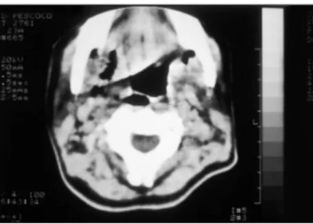
Figure 2. Chest computed tomography
scan showing widening of mediastinal region and pleural effusion.
Figure 1. Computed tomography scan of
for effective treatment of mediastinitis. Broad-spectrum antibiotics and good coverage against anaerobes should be the fi rst choice for descend-ing mediastinitis.8,9,26 Piperacillin-tazobactam and vancomycin are a good choice for empirical treatment while the culturing results are still ongoing. Another option is clindamycin plus ceftriaxone or ceftazidime. When the results are
negative, empirical treatment is indicated, based on clinical signs and the existence of purulent material with fetid smell. Patients who are allergic to penicillin can receive quinolone plus clinda-mycin. An association of beta-lactamic plus aminoglycoside plus imidazole may be utilized after microbiological diagnosis and antibiogram (considering the great number of microorgan-isms that produce beta-lactamase in DM). Che-motherapy with carbapenem and metronidazole is also suggested. Hyperbaric oxygen may also be useful as adjuvant therapy.13,23,27
When the clinical course appears to be resistant to treatment, mycotic infection needs to be considered, as already described.28
However, intravenous broad-spectrum antibiotic therapy alone is not curative without surgical drainage of the cervical and mediastinal collections, with debridement and excision of all the necrotic tissue located in the affected region. Continued irrigation of the mediastinal and pleural space should be performed as well.
Four different types of surgical approach have been described (Table 5).15,21,23,29,30 The decision on which one to choose depends upon the stage of the infection. In the early stages, when the collection is located above the carina anteriorly, and above the fourth vertebra posteriorly (type I mediastinitis), a transcervical approach is suffi cient. The incision must be wide and lateral along the sternocleidomastoid muscle.23 It is less in-vasive, but may not reach deep regions, and many procedures may be needed in order to achieve complete debridement. Nevertheless, in order to maximize the chances of obtaining a cure in more advances cases, posterolateral thoracotomy (PLT) may be performed.29 A focus located at the middle or inferior medi-astinum, below the fourth thoracic vertebra (descending mediastinitis types IIA and IIB) should be drained by PLT, because this allows easier access to all the mediastinal structures, complete debridement and excision of ne-crotic tissue, and also adequate drainage of the pleural and pericardial cavities.31 The PLT approach is the one most utilized, although this should not be used for collections in the superior mediastinum, because of the risk of spreading the infection to the inferior spaces; this approach also allows pulmonary decortication, if necessary.11
Other options include median sternotomy and the transthoracic approach, using either a clamshell incision or a subxiphoid approach (also called anterior mediastinotomy). Me-dian sternotomy allows a good view of the operative fi eld and can also be performed in cases of bilateral collections. However, since descending dissemination occurs mainly through the prevertebral pathway, it hinders the drainage of collections by the anterior approach. Moreover, there is a high risk of sternal osteomyelitis and subsequent dehis-cence of the bone.21,29 In addition, access to the posterobasal compartments of the chest is diffi cult when this approach is used. On the other hand, a subxiphoid transthoracic approach may be useful in cases of anterior
Figure 3. Chest computed tomography scan demonstrating mediastinal collection.
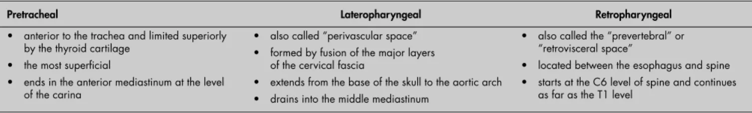
Figure 4. Cervical computed tomography
scan showing gas in lateropharyngeal space, and also parapharyngeal and retropharyngeal bilateral abscess.
Table 5. Surgical approaches to descending mediastinitis (DM)21
Transcervical Posterolateral thoracotomy Median sternotomy Transthoracic
indicated for DM type I less invasive
may not reach deep regions •
• •
indicated for DM types IIA and IIB
allows easily access to all mediastinal structures allows pulmonary decortication •
•
•
allows a good view of the operative fi eld
can also be performed in cases of bilateral collections risk of sternal osteomyelitis and dehiscence
•
•
•
can use either a clamshell incision or a subxiphoid approach
clamshell incision yields excellent exposure of all the mediastinal struc-tures, but is very invasive and there is a risk of damaging the phrenic nerve subxiphoid access is useful in cases of anterior mediastinal collections •
•
1. Akman C, Kantarci F, Cetinkaya S. Imaging in mediasti-nitis: a systematic review based on aetiology. Clin Radiol. 2004;59(7):573-85.
2. Mihos P, Potaris K, Gakidis I, Papadakis D, Rallis G. Manage-ment of descending necrotizing mediastinitis. J Oral Maxillofac Surg. 2004;62(8):966-72.
3. El Oakley RM, Wright JE. Postoperative mediastini-tis: classification and management. Ann Thorac Surg. 1996;61(3):1030-6.
4. Callister ME, Wall RA. Descending necrotizing mediastinitis caused by group A streptococcus (serotype M1T1). Scand J Infect Dis. 2001;33(10):771-2.
5. von Rahden BH, Feith M, Dittler HJ, Stein HJ. Cervical esopha-geal perforation with severe mediastinitis due to an impacted dental prosthesis. Dis Esophagus. 2002;15(4):340-4. 6. Sato S, Kajiyama Y, Kuniyasu T, et al. Successfully treated
case of cervical abscess and mediastinitis due to esophageal perforation after gastrointestinal endoscopy. Dis Esophagus. 2002;15(3):250-2.
7. Jiménez Y, Bagán JV, Murillo J, Poveda R. Infecciones odontogénicas. Complicaciones. Manifestaciones sistémicas. [Odontogenic infection. Complications. Systemic manifes-tations]. Med Oral Patol Oral Cir Bucal. 2004;(9 Suppl): S139-47. Available from: http://www.medicinaoral.com/me-doralfree01/v9Suppli/medoralv9supplip143.pdf. Accessed in 2006 (Aug 8).
8. Mitjans MS, Sanchís JB, Padro XB, Garcia I, Garolera JM, Garay MR. Descending necrotizing mediastinitis. Int Surg. 2000;85(4):331-5.
9. Kiernan PD, Hernandez A, Byrne WD, et al. Descending cervical mediastinitis. Ann Thorac Surg. 1998;65(5):1483-8. 10. Moncada R, Warpeha R, Pickleman J, et al. Mediastinitis from
odontogenic and deep cervical infection. Anatomic pathways of propagation. Chest. 1978;73(4):497-500.
11. Moriwaki Y, Sugiyama M, Matsuda G, et al. Approach for drain-age of descending necrotizing mediastinitis on the basis of the extending progression from deep neck infection to mediastinitis. J Trauma. 2002;53(1):112-6.
12. Novellas S, Kechabtia K, Chevallier P, Sedat J, Bruneton JN. Descending necrotizing mediastinitis: a rare pathology to keep in mind. Clin Imaging. 2005;29(2):138-40.
13. Harar RP, Cranston C, Warwick-Brown N. Descending nec-rotizing mediastinitis: report of a case following steroid neck injection. J Laryngol Otol. 2002;116(10):862-4. 14. Sancho LM, Minamoto H, Fernandez A, Sennes LU, Jatene FB.
Descending necrotizing mediastinitis: a retrospective surgical experience. Eur J Cardiothorac Surg. 1999;16(2):200-5. 15. Balkan ME, Oktar GL, Oktar MA. Descending necrotizing
mediastinitis: a case report and review of the literature. Int Surg. 2001;86(1):62-6.
16. Figueroa-Damián R. Manifestaciones clínicas y letalidad de la mediastinitis necrosante descendente. [Clinical manifestations and lethality of descending necrotizing mediastinitis]. Rev Invest Clin. 2001;53(1):35-40.
17. Basa S, Arslan A, Metin M, Sayar A, Sayan MA. Mediastinitis caused by an infected mandibular cyst. Int J Oral Maxillofac Surg. 2004;33(6):618-20.
REFERENCES
mediastinal collections, in combination with the transcervical approach (although this is less frequent).8,11 In some cases, a combination of transcervical and subxiphoid transthoracic approaches can be also utilized.32
Clamshell incision (bilateral thoracotomy plus median sternotomy) yields excellent exposure of the mediastinal structures, but it is very invasive and the risk of damaging the phrenic nerve is high.21 Comparing patients who underwent transcervical and transtho-racic drainage, Corsten et al.33 demonstrated statistically signifi cant greater survival for the transthoracic group (53% versus 81%).
Furthermore, the use of video-assisted thoracoscopy has been reported by one author,34 but its indication is still limited to the early stages of infection or to posterior mediastinal collections. In any of these ap-proaches, abundant irrigation and continu-ous drainage with straight tube drains must be performed.11 Through the drains, it is also possible to carry out continuous wash-ing with antiseptic solutions. The drains must also be constantly monitored, because they are liable to become obstructed by necrotic tissue particles. The median length of time for keeping the drain in position is around three weeks.23 In spite of all this care, abscesses tend to form. This is why
it is recommended that control CT scans should be performed regularly, following the intervention. In cases of persistent abscesses or non-drained collections, a second surgical approach is essential in order to avoid clini-cal deterioration. Some authors also believe that the use of tracheostomy in order to avoid airway obstruction due to pharyngeal abscess or infl ammation of the tracheal wall is necessary.9,32,35 Nevertheless, others believe that tracheostomy can spread the infection to non-involvedareas and hinder subsequent surgical treatment.8,35 However, tracheostomy should be considered in cases of prolonged mechanic ventilation.
Complications and prognosis
Complications and prognosis The main complication of descending me-diastinitis is sepsis (see the signs and symptoms above). Other complications include pneu-moperitoneum, pneumothorax and pleural effusions (which can lead to empyema) and pericarditis. According to Hirai et al.,31 the most frequent complication other than sepsis is thoracic empyema. Hemorrhage may occur following debridement procedures due to ves-sel erosion. The main vascular complications are thrombosis of the internal jugular veins and carotid pseudoaneurysm.12 Moreover, aor-topulmonary fi stula, aspiration pneumonia,
epidural abscess and adult respiratory distress syndrome have also been reported.10,15,32
DM is a severe condition. The mortality rate (about 50%) is still high and few changes have been observed over the past few years. The outcome depends upon the degree of infection and the patient’s underlying disease, and also on the comorbidities (such as diabe-tes, HIV infection etc.). The causes of death are multiple, ranging from septic shock and respiratory insuffi ciency to gastrointestinal hemorrhages.12 The most important factor for improving the clinical course, and the course that must be pursued by the healthcare team, is early detection and readily available aggres-sive treatment.
Controversies and
Controversies and perspectives
perspectives
AUTHOR INFORMATION Luis Marcelo Inaco Cirino, MD, PhD. Associate
professor, Hospital Universitário, Department of Surgery, Faculdade de Medicina da Universidade de São Paulo (FMUSP), São Paulo, Brazil.
Fernando Melhem Elias, MD. Oral surgeon, Hospital Universitário, Department of Surgery, Faculdade de Medicina da Universidade de São Paulo (FMUSP), São Paulo, Brazil.
José Luiz Jesus de Almeida. Medical student, Hospital Universitário, Department of Surgery, Faculdade de Medicina da Universidade de São Paulo (FMUSP), São Paulo, Brazil.
Address for correspondence:
José Luiz Jesus de Almeida
Rua Ernesto Paglia, 29 — Butantã São Paulo (SP) — Brasil — CEP 05547020 Tel. (+55 11) 3782-3313
E-mail: josluizalmeida@yahoo.com.br
Copyright © 2006, Associação Paulista de Medicina
Resumo Mediastinite descendente: uma revisão
CONTEXTO: Mediastinite é um processo infl amatório do tecido conectivo que envolve as estruturas medias-tinais. Quando essa condição é causada por uma infecção em sítio cérvico-oral, a infl amação mediastinal é dita mediastinite descendente (MD).
FONTE DE DADOS:O assunto foi examinado através de revisão da literatura disponível e à luz da expe-riência dos autores. Os bancos de dados Medline, Lilacs e Cochrane foram pesquisados, sem limite de tempo, através do termo “descending mediastinitis”. As línguas utilizadas foram inglês e espanhol. SÍNTESE DOS DADOS: Existem três vias fasciais principais pelas quais um foco infecioso em região cérvico-oral pode se espalhar para o mediastino: pré-traqueal, latero-faríngeo e retrofaríngeo. Cerca de 70% dos casos de MD ocorrem através da via retrofaríngea. O índice de mortalidade situa-se ao redor de 50%. De acordo com a extensão da infecção e baseado nos achados de tomografi a computadorizada (TC), MD pode ser classifi cada como focal (tipo I) e difusa (tipo II). As manifestações clínicas são inespecífi cas e semelhantes às de outras infecções sistêmicas. O tratamento primário da MD consiste em antibióticos e drenagem cirúrgica. Existem diversas formas de abordagem no tratamento cirúrgico da MD; a escolha de qual via será utilizada depende do tipo de MD e da experiência do cirurgião. Apesar de todo o avanço no conhecimento da microbiologia e fi siopatologia da doença, ainda há controvérsias quanto à duração ideal da antibioticoterapia e à necessidade de se realizar traqueostomia nos pacientes portadores de MD. CONCLUSÃO: Como a MD é uma condição rapidamente fatal se não diagnosticada e tratada a tempo, ela deve ser sempre considerada uma emergência médica.
PALAVRAS-CHAVE: Mediastinite. Infecção. Mediastino. Infecção focal dentária. Sepse.
18. Endo S, Murayama F, Hasegawa T, et al. Guideline of surgical management based on diffusion of descending necrotizing me-diastinitis. Jpn J Thorac Cardiovasc Surg. 1999;47(1):14-9. 19. Pérez A, Cueto G, de la Escosura G, Cícero R. Mediastinitis
necrosante descendente. Resultados del tratamiento médico-quirúrgico en 17 casos. [Descending Necrotizing Mediastinitis.
Results of Medical-Surgical Treatment in 17 Cases].Gac Med
Mex 2003;139(3):199-204.
20. Bulut M, Balci V, Askkose S, Armagan E. Fatal descending necrotising mediastinitis. Emerg Med J. 2004;21(1):122-3. 21. Lavini C, Natali P, Morandi U, Dallari S, Bergamini G.
Descen-ding necrotizing mediastinitis. Diagnosis and surgical treatment. J Cardiovasc Surg (Torino). 2003;44(5):655-60. 22. Sakamoto H, Aoki T, Kise Y, Watanabe D, Sasaki J.
Descend-ing necrotizDescend-ing mediastinitis due to odontogenic infections. Oral Surg Oral Med Oral Pathol Oral Radiol Endod. 2000;89(4):412-9.
23. Makeieff M, Gresillon N, Berthet JP, et al. Management of descending necrotizing mediastinitis. Laryngoscope. 2004;114(4):772-5.
24. Estrera AS, Landay MJ, Grisham JM, Sinn DP, Platt MR. Descending necrotizing mediastinitis. Surg Gynecol Obstet. 1983;157(6):545-52.
25. Scaglione M, Pinto A, Romano S, Giovine S, Sparano A, Romano L. Determining optimum management of descending necrotizing mediastinitis with CT; experience with 32 cases. Emerg Radiol. 2005;11(5):275-80.
26. Marty-Ane CH, Berthet JP, Alric P, Pegis JD, Rouviere P, Mary H. Management of descending necrotizing mediastinitis: an aggressive treatment for an aggressive disease. Ann Thorac Surg. 1999;68(1):212-7.
27. Watanabe S, Kariatsumari K, Sakasegawa K, Nakamura Y, Sakata R. A new combined surgical procedure for severe descending necrotizing mediastinitis with bilateral empyema. Thorac Cardiovasc Surg. 2002;50(5):308-10.
28. Jahnke K. Mediastinitis mit Pilznachweis. [Mediastini-tis with detection of Candida albicans]. HNO. 1982; 30(8):299-300.
29. Papalia E, Rena O, Oliaro A, et al. Descending necrotizing mediastinitis: surgical management. Eur J Cardiothorac Surg. 2001;20(4):739-42.
30. Macrí P, Jiménez MF, Novoa N, Varela G. Análisis des-criptivo de una serie de casos diagnosticados de mediasti-nitis aguda. [A descriptive analysis of a series of patients diagnosed with acute mediastinitis]. Arch Bronconeumol. 2003;39(9):428-30.
31. Hirai S, Hamanaka Y, Mitsui N, Isaka M, Mizukami T. Surgical treatment of virulent descending necrotizing mediastinitis. Ann Thorac Cardiovasc Surg. 2004;10(1):34-8.
32. Wheatley MJ, Stirling MC, Kirsh MM, Gago O, Orringer MB. Descending necrotizing mediastinitis: transcervical drainage is not enough. Ann Thorac Surg. 1990;49(5):780-4. 33. Corsten MJ, Shamji F, Odell PF, et al. Optimal treatment of
descending necrotising mediastinitis. Thorax. 1997;52(8):702-8. 34. Min HK, Choi YS, Shim YM, Sohn YI, Kim J. Descending
necrotizing mediastinitis: a minimally invasive approach us-ing video-assisted thoracoscopic surgery. Ann Thorac Surg. 2004;77(1):306-10.
35. Brunelli A, Sabbatini A, Catalini G, Fianchini A. Descending necrotizing mediastinitis. Surgical drainage and tracheostomy. Arch Otolaryngol Head Neck Surg. 1996;122(12):1326-9. 36. Oczenski W, Waldenberger F, Nehrer G, et al. Vacuum-assisted
closure for the treatment of cervical and mediastinal necrotizing fasciitis. J Cardiothorac Vasc Anesth. 2004;18(3):336-8.
Sources of funding:None Confl ict of interest: None
Date of fi rst submission: September 26, 2005 Last received: August 17, 2006