https://doi.org/10.1177/0022034517709464
Journal of Dental Research 2017, Vol. 96(9) 999 –1005
© International & American Associations for Dental Research 2017
Reprints and permissions: sagepub.com/journalsPermissions.nav DOI: 10.1177/0022034517709464 journals.sagepub.com/home/jdr
Research Reports: Biomaterials & Bioengineering
Introduction
Enzymatic degradation of dentin collagen fibrils and hydroly-sis of resin polymer matrix represent the main mechanisms responsible for bonding failure over time (Pashley et al. 2011). Preservation of the structural integrity of collagen fibrils is essential for the longevity of the resin-dentin interface, particu-larly when restorations are performed after selective caries removal (Aguiar et al. 2014; Abuna et al. 2016). Inactive and/ or proform of proteolytic enzymes such as matrix metallopro-teinases (MMPs) exist within mineralized dentin (Palosaari et al. 2003). MMPs are exposed and activated when bacteria-derived acids demineralize dentin, as well as when using phos-phoric acid etchants or self-etch adhesives (Lehmann et al. 2009). Although a small fraction of such acids (De Munck et al. 2009) may extract these proteases, most remain bonded to the matrix in active forms and hydrolyze C-terminal cross-linked telopeptide (ICTP) (Pashley et al. 2004; DeVito-Moraes et al. 2016). Cathepsin K (CTP) is a further proteolytic enzyme found in active form in demineralized dentin, which hydro-lyzes the C-terminal telopeptide (CTX) (Nascimento et al. 2011; Tezvergil-Mutluay et al. 2014).
It has been demonstrated that dentin remineralization may reduce enzymatic-mediated matrix degradation (Osorio et al. 2012; Tezvergil-Mutluay et al. 2014). The rationale is that calcium
(Ca2+) and phosphate (PO
4 3–
) ions released by bioactive mate-rials may precipitate onto demineralized dentin collagen fibrils and fossilize active endogenous proteases (Tjäderhane et al. 2013). Bioactive glasses are known to form a biomimetic non-stoichiometric nano-crystalline apatite when immersed into physiological solutions (O’Donnell et al. 2009).
1
Department of Restorative Dentistry and Cariology, Institute of Dentistry, University of Turku, Turku, Finland, and Turku University Hospital, TYKS, Turku, Finland
2
Research Division, Paulo Picanço School of Dentistry, Fortaleza, Brazil
3
Polymer Chemistry & Biomaterials Group, University of Ghent, Ghent, Belgium
4
Otto Schott Institute of Materials Research, Friedrich Schiller University Jena, Jena, Germany
5
Dental Biomaterials, Departamento de Odontología, Facultad de Ciencias de la Salud, University CEU-Cardenal Herrera, Valencia, Spain
6
Biomaterials, Biophotonics and Tissue Engineering, King’s College London Dental Institute (KCLDI), London, UK
Corresponding Author:
S. Sauro, Dental Biomaterials, Preventive and Minimally Invasive Dentistry, Departamento de Odontología, Facultad de Ciencias de la Salud, Universidad CEU-Cardenal Herrera. C/Del Pozo s/n, Alfara del Patriarca, 46115 Valencia, Spain.
Email: [email protected]
Effects of Composites Containing Bioactive
Glasses on Demineralized Dentin
A. Tezvergil-Mutluay
1, R. Seseogullari-Dirihan
1, V.P. Feitosa
2,
G. Cama
3, D.S. Brauer
4, and S. Sauro
5,6Abstract
The aim of this study was to evaluate the degradation of completely demineralized dentin specimens in contact with a filler-free or 2 ion-releasing resins containing micrometer-sized particles of Bioglass 45S5 (BAG) or fluoride-containing phosphate-rich bioactive glass (BAG-F). Fourier transform infrared spectroscopy (FTIR) and scanning electron microscopy (SEM) were also used to evaluate the remineralization induced by the experimental ion-releasing resin-based materials. Dentin beams were totally demineralized in H
3PO4
(10%) and placed in direct contact with a filler-free (RESIN) or 2 experimental ion-releasing resins (BAG or BAG-F) and immersed in artificial saliva (AS) up to 30 d. Further specimens were also processed and submitted to FTIR and SEM analysis to evaluate the remineralization induced by such ion-releasing resins before and after AS immersion. BAG and BAG-F alkalinized the incubation media. A significant decrease of the dry mass was observed between the specimens of all groups stored for 3 and 30 d in AS. However, the fluoride-containing phosphate-rich bioactive glass incorporated into a resin-based material (BAG-F) showed greater ability in reducing the solubilization of C-terminal cross-linked telopeptide (ICTP) and C-terminal telopeptide (CTX) after prolonged AS storage. Moreover, after 30 d of AS storage, BAG-F showed the greatest remineralizing effect on the stiffness of the completely demineralized dentin matrices. In conclusion, fluoride-containing phosphate-rich bioactive glass incorporated as micrometer-sized filler in dental composites may offer greater beneficial effects than Bioglass 45S5 in reducing the enzyme-mediated degradation and remineralization of demineralized dentin.
Moreover, fluoride has been advocated as a potential inhibi-tor of cathepsin K and matrix-bound MMPs (Mei et al. 2014), as it was shown to reduce the degradation of dentin matrix (Altinci et al. 2016). However, it is not known whether fluoride-containing bioactive glass (Groh et al. 2014) can promote den-tin remineralization and inhibition of the denden-tin-matrix degradation.
Thus, this study aimed at evaluating the amount of solubi-lized CTX and ICTP, as well as the modulus of elasticity of completely demineralized dentin specimens in contact for 3 or 30 d with a filler-free or 2 ion-releasing resin composites filled with micrometer-sized particles of Bioglass 45S5 or a fluoride-containing phosphate-rich bioactive glass. Fourier transform infrared spectroscopy (FTIR) and scanning electron micros-copy (SEM) were also employed to evaluate the dentin remin-eralization induced by such ion-releasing resin composites. The null hypotheses tested were that there would be no differ-ence between the 2 ion-releasing resins: 1) in the release of ICTP and CTX collagen fragments at 3 and 30 d and 2) in the remineralization of completely demineralized dentin after 30 d of artificial saliva (AS) storage.
Materials and Methods
Preparation of Bioactive Glasses
and Ion-Releasing Composites
A control resin co-monomer blend (RESIN) was formulated using 35 vol% urethane-dimethacrylate, 6.6 vol% bisphenol-A-dimethacrylate, 30.4 vol% triethylene-glycol-dimethacry-late (Esstech Essington), 15 vol% 2-hydroxyethyl-methacrytriethylene-glycol-dimethacry-late (HEMA; Sigma-Aldrich), 10 vol% absolute ethanol (Sigma-Aldrich), 0.5 wt% camphorquinone, and 1.0 wt% ethyl 4-dimethylami-nobenzoate (Sigma-Aldrich).
Bioglass 45S5 was prepared by a high-temperature melting process as described by Sauro et al. (2013) (nominal composi-tion: 46.1 mol% SiO
2, 26.9 mol% CaO, 24.4 mol% Na2O, and
2.5 mol% P
2O5). The experimental fluoride-containing
bioac-tive glass (nominal composition: 42.7 mol% SiO
2, 26.2 mol%
CaO, 26.1 mol% Na
2O, 4.0 mol% P2O5, and 1 mol% CaF2) was
prepared as described previously (Groh et al. 2014). Glasses were milled and finally sieved (<30 µm). They were incorpo-rated with the resin control (filler/resin: 50/50 wt%) to create an ion-releasing fluoride-free (BAG) and a fluoride-containing composite (BAG-F).
Dentin Specimen Preparation
Ten unerupted human third molars were used in accordance with the ethical guidelines of the research ethics committee and stored in 0.5 wt% chloramine-T/water solution at 4°C. Four beams (5 mm × 1 mm × 1 mm) were obtained from the mid-coronal dentin (total = 40) and randomly distributed into 4 different groups (n = 10/group) to reduce the tooth-effect variability: 1) DENTIN, 2) RESIN, 3) BAG, and 4) BAG-F. All dentin beams were completely demineralized in 10%
phosphoric acid for 12 h at 25°C (10 rpm). Absence of resid-ual minerals was confirmed using digital radiography. The beams in group 1 were left completely demineralized and served as control, while the beams in groups 2, 3, and 4 were placed in direct contact (only on one side) with prepolymer-ized (40 s, 1,200 mW/cm2 ; Radii Plus; SDI) resin beams (6 mm × 3 mm × 2 mm) using orthodontics rubber bands.
Assessment of Solubilized Telopeptides
The dentin beam-resin assembly was immersed in individual propylene tubes containing 0.5 mL AS (pH 7.4) at 37°C for 3 or 30 d. The AS consisted of 5 mm HEPES, 2.5 mm CaCl
2,
0.05 mm ZnCl
2, 0.68 mmol/L KH2PO4, 30 mmol/L KCl, and
120 mm NaCl. After each incubation period, the medium of each specimen was collected and used to quantitate the solubi-lized collagen fragments.
MMP-mediated degradation was assessed by measuring the amount of solubilized ICTP using the UniQ EIA ELISA kit (Orion Diagnostica). Cathepsin K–mediated collagen degrada-tion was evaluated by measuring the amount of solubilized CTX using the Serum Cross Laps ELISA (Immunodiagnostic System).
ICTP and CTX release was calculated in each assay from a standard curve created using standards of known concentra-tions provided in the kit. Triplicate analysis was performed from each sample for each incubation period. Dentin beams were dried over anhydrous calcium sulfate for 8 h, and the dry mass was then measured using a digital microbalance (Model MT5; Mettler Toledo) with a readability of 0.001 mg and repeatability of 0.0008 mg. The pH of the incubation medium (AS) was also evaluated at 3 and 30 d of storage (Fig. 1).
The normality and equality of variance assumptions were statistically analyzed by Komolgorov-Smirnov normality test and modified Levine’s test. Since the normality assumptions were not violated, the data were analyzed by 1-way analysis of variance (ANOVA) and post hoc multiple-comparisons Tukey’s test (α = 0.05) (Sigma-Plot 11.5; Systat).
FTIR Microspectroscopic Imaging and SEM Analysis
The chemical mapping was performed on the specimens’ sur-faces before and after AS storage in contact with the composite blocks using a FTIR microspectroscopic system (Spectrum Spotlight FTIR imaging system; Perkin Elmer) with both sin-gle-point and imaging mapping mode. Specimens were scanned (4,000 and 650 cm–1) at a 4-cm–1 spectral resolution, with 2 scans per pixel. Image size was 80 to 40 µm, using a 6.25-µm pixel resolution. An atmosphere correction was applied to the raw image to subtract the absorbance of water vapor and carbon dioxide. Images were elaborated using the “Spectrum Spotlight” software (Perkin Elmer). The amide I band and the v
1/v3 PO domain between 1,200 and 900 cm –1
Three-Point Bending of
Demineralized Dentin Beams
A further 40 beams (5 mm × 1 mm × 1 mm) were prepared and distrib-uted into different groups (n = 10/ group) as previously described. The stiffness of the wet specimens was assessed (0.5 mm min–1) using a 3-point bending testing device (Instron 3345; Instron Corp.) at time 0 and after 30 d of AS storage. The compressive force necessary to induce strain (10%) in dentin beams was measured in a 1N load cell; load was returned immediately to 0% stress to prevent creep of the demin-eralized collagen matrix. The modu-lus of elasticity of each specimen (E′) was calculated using the follow-ing equation: E = mL3/4bh3, where m is the steepest slope along the lin-ear portion of the load-displacement curve (N/mm), L is the span length (2.5 mm), b is the width of test spec-imens (1 mm), and h is the thickness (1). E′ was expressed in MPa. Since the distributions of data were not normally distributed, nonparametric tests were selected to analyze these variables. All data sets were submit-ted to Wilcoxon nonparametric and Mann-Whitney tests (α = 0.05).
Results
The experimental BAG as well as
BAG-F resin showed an important alkalization potential at both 3 and 30 d. By contrast, a neutral pH was observed for the incubation media of the groups RESIN and DENTIN (Fig. 1A). The specimens in contact with BAG showed a slight E′ reduction (–3.7%), while the demineralized speci-mens stored alone (DENTIN: –31.5%) and those in contact with filler-free resin (RESIN: –31.7%) showed the lowest moduli (P < 0.05) at 30 d (Fig. 1B). Conversely, the speci-mens in contact with BAG-F showed the highest (P < 0.05) increase in modulus of elasticity (13.0%) at 30 d. The release of CTX and ICTP telopeptide fragments at 3 and 30 d of AS incubation is shown in Figure 1-C1 and 1-C2, respectively. The demineralized specimens in contact with BAG showed CTX release both at 3 d (2.6 ± 0.5 ng/mg-dry-dentin) and 30 d of AS storage (0.9 ± 0.2 ng/mg-dry-dentin) significantly lower (P < 0.05) than the CTX fragments in groups DENTIN and RESIN (Fig. 1-C1). However, the specimens in contact with BAG-F showed the lowest (P < 0.05) CTX release both at 3 d (0.7 ± 0.3 ng/mg-dry-dentin) and 30 d (0.6 ± 0.1 ng/
mg-dry-dentin); no significant difference was observed at 30 d between BAG-F and BAG.
The demineralized specimens with the highest (P < 0.05) ICTP release after 3 d of storage were those in group DENTIN (36.5 ± 4.5 ng/mg-dry-dentin), but there was no significant dif-ference (P > 0.05) between the groups BAG, BAG-F, and RESIN (Fig. 1-C2). Conversely, the lowest (P < 0.05) ICTP release at 30 d was observed in group BAG-F (39.6 ± 5.7 ng/ mg-dry-dentin), followed (P < 0.05) by those in group BAG (48.6 ± 4.8 ng/mg-dry-dentin). No significant difference (P > 0.05) was found between the groups RESIN and DENTIN.
Specimens from all groups showed similar (P > 0.05) loss of dry mass (range, –2% and –4%) at 3 d storage. After 30 d of incubation, there was a significant loss of dry mass (P < 0.05) in all groups compared to the results observed at 3 d of AS storage (range, –12% and –16%), but again no significant dif-ference (P > 0.05) was observed among the groups at 30 d of AS storage (Fig. 1C3).
The FTIR spectra showed that the specimens in contact with the filler-free resin showed no sign of remineralization,
Figure 1. Alkalinization activity, elastic modulus, ICTP-CTX release, and dry mass loss.(A) Alkalinizing activity/pH values of the materials tested in this study at different storage periods in artificial saliva
(AS) evaluated by means of a professional pH electrode (Mettler-Toledo). (B) Elastic modulus (MPa)
of completely demineralized dentin specimens before and after storage. Values are expressed as MPa. Within each treatment (initial vs. storage E-modulus), values identified by the same letter are not
statistically different (Wilcoxon, P > 0.05). Change in elastic modulus (E%). Within each application time
(columns), groups identified by the same letter are not statistically different (Mann-Whitney, P > 0.05).
(C1) Means and standard deviations of C-terminal cross-linked telopeptide (ICTP) released from dentin
specimens at 3 and 30 d of storage. Within each period of storage (3 or 30 d), means designated with
the same letters indicate no significant differences (P > 0.05). (C2) Means and standard deviations of
C-terminal telopeptide (CTX) released from dentin specimens at 3 and 30 d of storage. Within each period of storage (3 or 30 d), means designated with the same letters indicate no significant differences
(P > 0.05). (C3) Means and standard of the loss of dry mass (%) at 3 and 30 d of AS storage. Same letters
indicate no significant differences (P > 0.05) between groups at the same storage period (30 d, uppercase;
3 d, lowercase). Symbol (*) indicates significant differences (p < 0.05) for the same group but at different
but only a noncrystallinity phase in the PO region at 1,014 cm−1
(Fig. 2A). Conversely, the specimens in contact with BAG or BAG-F for 30 d in AS showed precipitation of calcium phos-phate (1,100 cm−1). This was also confirmed with the FTIR mapping-images; the 2 bioactive glass–doped resins were able to remineralize the dentin surface (Fig. 2B). Indeed, along with calcium phosphate (1,100 cm−1), it was also possible to detect orthophosphate (1,038 cm−1) and pyrophosphate (746 cm−1). Carbonate precipitation (range between, 1,120 cm−1 and 1,140
cm−1) was also detected on the surface of the specimens in con-tract with the bioactive composites. The SEM results (Fig. 3) showed that the resin BAG induced the precipitation of min-eral crystals onto the surface and inside the dentin tubules (Fig. 3A, B). Similar mineralization was observed in the specimens in contact with BAG-F for 30 d in AS (Fig. 3C, D). Conversely, the specimens in contact with RESIN presented no clear rem-ineralization but several patent dentin tubules with very few crystals deposited onto the dentin surface (Fig. 3E–H).
Discussion
Active endogenous proteases such as MMPs and cysteine cathepsins (e.g., cathepsin K) in demineralized dentin matrix (DM) are able to cleave the helical and telopeptide segments of the collagen matrix, resulting in a decrease of mechanical properties of dentin and longevity of restorations (Tjäderhane et al. 2013). The rationale for analyzing the specific C-terminal telopeptide fragments (e.g., ICTP and CTX) is to evaluate MMP- and cathepsin K–mediated degradation separately (Garnero et al. 2003; Mutluay et al. 2014). Tezvergil-Mutluay et al. (2013) reported that the release of ICTP frag-ments from DM is higher than CTX fragfrag-ments; a similar trend was observed in the present study. This difference may be asso-ciated with the pH of the incubation media, as well as with the optimum pH activity of MMPs and cathepsin K (Tezvergil-Mutluay et al. 2014). The pH of the incubation media where the DM specimens were stored alone or in contact with the filler-free resin was neutral (Fig. 1A); these groups showed the greatest CTX and ICTP release after 30 d of AS storage. Holman et al. (1999) reported that the optimum pH for several MMPs to function at near-maximum rates is between 7.2 and 7.5. To degrade telopeptides at same rate of MMPs, cathepsin K should work at pH 5.5 (Marini et al. 2000). However, at neutral pH, cathepsin K is still able to accomplish its catalytic activity at lower rates (Kometani et al. 2010).
The experimental bioactive composites tested in this study (BAG and BAG-F) were able to alkalinize the incubation media (Fig. 1A). The DM specimens in these 2 groups presented CTX and ICTP release significantly lower compared to RESIN and DENTIN. BAG-F induced greater CTP inhibition at 3 d and MMP inhibition at 30 d of AS storage compared to BAG (P < 0.05). This is somewhat in contrast with the previous findings, which showed a collagen degradation zone in contact with strong alkaline mate-rials such as calcium silicate cements (Atmeh et al. 2012; Leiendecker et al. 2012). Conversely, resin-based materials doped with bioactive fillers such as Bioglass 45S5 have been shown to
preserve the integrity of collagen fibrils within the hybrid layer even after prolonged storage in simulated body fluids (Profeta et al. 2012).
The ICTP results obtained with the DM specimens stored in contact with BAG are in accordance with previous studies (Osorio et al. 2012; Tezvergil-Mutluay et al. 2014), with the only difference that the specimens in this study were not infiltrated with the tested resins, but only one side of the DM specimens was placed in direct contact with prepolymerized resin beams. The rationale behind employing such an approach was to avoid possible interference with the diffusion of ICTP and CTX telo-peptide fragments by the resin monomers that infiltrated the demineralized collagen matrices. Comparing the results attained in this study to those presented by Osorio et al. (2012) and Tezvergil-Mutluay et al. (2014), it is possible to affirm that both experimental methods may be considered valid for the assess-ment of ICTP and CTX. Conversely, the results of dry mass loss obtained in this study were not in accordance with those reported by Tezvergil-Mutluay et al. (2014). Such a difference may be attributed to the fact that the DM specimens in the present study were not totally resin infiltrated. Indeed, we hypothesize that such an experimental design may have been responsible for the lack of significant differences among the groups, as only one side of the DM specimens was protected by the bioactivity of the experimental BAG-F and BAG, which may have inhibited MMPs and CTPs at the resin-dentin interface. Moreover, it is well known that bioactive resin-based bioactive glass-contain-ing materials are able to release (Ca2+) and (PO
4 3–
) ions, which reprecipitate as apatite-like crystallites (Sauro et al. 2013). Such mineral precipitation may have occurred within collagen fibrils without forming excessive mineral clusters and thus without increasing the dry mass of the specimens after prolonged incu-bation. This latter hypothesis about remineralization of collagen fibrils requires further investigation.
However, it is also important to consider that the presence of bioactive glasses within resin-dentin interfaces may induce the release of a hydrated silica Si(OH)4, which then binds nonspe-cifically to demineralized collagen fibrils and polymerizes into a porous SiO
2-rich layer. Such a layer might serve as a template
for precipitation of amorphous calcium phosphate (Hench and Wilson 1993; Sauro and Pashley 2016), which subsequently converts into biomimetic nonstoichiometric apatite in an alka-line environment (Hoppe et al. 2011). This latter environment is attained through a rapid exchange of sodium (Na+) and hydro-gen ions (H+) or hydronium ion (H
3O
–); such an alkaline pH,
along with Si(OH)
4 condensation and precipitation of Ca 2+
and PO
4 3–
ions, contributes to fossilize proteolytic enzymes, thereby reducing their degradation activity (Tjäderhane et al. 2013). Makowski and Ramsby (2004) showed that mineral precipita-tion and apatite crystallizaprecipita-tion might immobilize proteases through the formation of [Ca/PO-MMP] complexes.
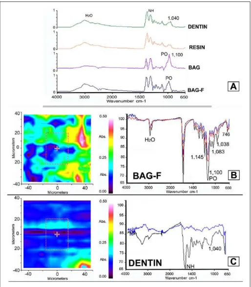
Figure 2. Fourier transform infrared spectroscopy (FTIR) of dentine remineralization induced by
the 2 bioactive composites. (A) FTIR specta (650 to 4,000 cm−1
) of the demineralized dentin matrix (DENTIN) and dentin matrices in contact with the filler-free resin (RESIN) after 30 d of storage in artificial saliva (AS). These spectra represented the average of 3 single scanning analyses that were then processed by smoothing, baseline correction, and normalization to the amide I peak. FTIR spectra
showed mainly the absorption band of collagen at 1,650 cm−1
(amide I), 1,550 cm−1
(amide II), 1,450 cm−1
,
and water (O–H: 2,800 to 3,100 cm−1). The absorption band at 1,014 cm−1 indicates a noncrystallinity
phase in the PO region. The FTIR spectra of the demineralized dentin matrix in contact for 30 d in
AS with the BAG resin or BAG-F resin show an intense band at 1,100 cm−1, which is associated with
the inorganic PO component. (B) Representative FTIR image (80 × 80 µm) and relative spectra of a
specimen of demineralized dentin matrix in contact for 30 d in AS with the resin containing the fluoride-doped bioglass (BAG-F). It is possible to note the remineralization effect of such a bioactive resin on
the entire dentin surface. The most prominent absorption band at 1,100 cm−1 is associated with the
inorganic PO component, along with an intense band at 1,650 cm−1
from the collagen. Orthophosphate
optical absorption band (1,038 cm−1) and absorption bands at 1,083 cm−1 (orthophosphate stretching
vibrations—ν3) are also clearly visible. It is also possible to observe carbonate stretching vibrations
between 1,120 cm−1 and 1,140 cm−1 and OPO stretching of pyrophosphates at 746 cm−1. (C) Representative
FTIR image and relative spectra of a specimen of demineralized dentin matrix immersed in AS for 30 d. It is possible to note no clear dentin remineralization as the main absorption bands are those of demineralized
collagen (1,650 cm−1
and 1,550 cm−1
) and 1,014 cm−1
, which indicates very little phosphate uptake (mineral precipitation) in dentin.
Moreover, the fluoride-containing bioactive glass used in this study has higher phosphate content com-pared with Bioglass 45S5. Both elements are known to accelerate apatite formation maintaining the same network connectivity (i.e., the same polymerization of the sil-icate network) of Bioglass 45S5 (Mneimne et al. 2011). Therefore, we assume that the release of fluo-ride ions and the greater bioactiv-ity of such an innovative bioglass incorporated in BAG-F resin may have contributed to attaining such better results.
It has been advocated that fluo-ride might inhibit both pro- and active forms of MMP-2 and MMP-9 from human saliva Kato et al. (2014). Brackett et al. (2015) showed that 150 ppm NaF could inhibit both soluble and matrix-bound MMP-2, MMP-8, and MMP-9. Moreover, these authors hypothesized that 75 to 600 ppm fluoride could complex not only with ionized calcium bound to the enzyme but also with Ca2+ in the incubation medium to form tightly complexed CaF
2. Therefore,
fluo-ride may have chelated Ca2+ and Zn2+ to inactivate MMPs; MMPs
depend on calcium to maintain their 3-dimensional configuration for enzymatic activity (Thompson et al. 2012).
Furthermore, fluoride has been demonstrated to be effective in inhibiting human recombinant cathepsin K and B (Mei et al. 2014). Brackett et al. (2015) stated that since EDTA does not inhibit cathepsin activity, fluoride ions may not inhibit dentin matrix cathepsin activity via a chelating approach. We believe that there may be a chemical interaction between cathepsin K and fluoride, which caused alteration of the 3-dimensional configuration as well as its ability to bind and cleave telopeptides in a specific zone of
the collagen fibril. However, the mechanism of inhibition of fluoride on cathepsin K is still unclear, and this should be fur-ther investigated in future studies.
resin-based dental composite showed greater impairment of the degradation activity of CTP at 3 d and MMPs at 30 d of AS storage. Conversely, the second null hypothesis must be rejected as the resin containing the fluoride-containing phos-phate-rich bioactive glass had greater ability than the resin containing Bioglass 45S5 in remineralizing the demineralized dentin matrices after prolonged AS storage.
In conclusion, we canassume the fluoride-containing phosphate-rich bioactive micro-filler incorporated into resin-based composites is more bioactive than BAG 45S5. This may be due to the release of fluoride ions, as well as to the greater amount of phosphates, which accelerate dentin remineralization as well as fossilization/inhibition of both MMPs and CTPs. Moreover, since Altinci et al. (2016) dem-onstrated that it was possible to inhibit matrix-bound cathep-sin-mediated dentin matrix degradation by using a minimum concentration of NaF (≥120 mm), we hypothesize that the fluoride/phosphate-rich bioactive glass used in this study might also have a direct inhibition effect on CTP-mediated degradation related to its ability to release fluoride ions dur-ing early contact with biological fluids (e.g., 3 d in AS) (Brauer et al. 2010).
Author Contributions
A. Tezvergil-Mutluay, D.S. Brauer, S. Sauro, contributed to conception, design, data acquisition, analysis, and interpretation, drafted and critically revised the manuscript; R. Seseogullari-Dirihan, contributed to conception, design, data analysis, and interpreta-tion, drafted and critically revised the manuscript; V.P. Feitosa, contributed to conception, design, data acquisition, and analysis, drafted and critically revised the manuscript; G. Cama, con-tributed to design and data acquisition, drafted and critically revised the manu-script. All authors gave final approval and agree to be accountable for all aspects of the work.
Acknowledgments
This work was supported by the research grant INDI16/34 and INDI1527B, Programa de Consolidación de Indica- dores: Fomento Plan Estatal CEU-UCH 2014-2017 to S.S. (principal investigator [PI]). This study was also supported by grant 8126472 from the Academy of Finland to A.T.-M. (PI) and EVO funding of Turku University Hospital to A.T.-M. (PI). The authors declare no potential con-flicts of interest with respect to the author-ship and/or publication of this article.
References
Abuna G, Feitosa VP, Correr AB, Cama G, Giannini M, Sinhoreti MA, Pashley DH, Sauro S. 2016. Bonding performance of experimental bioactive/metic self-etch adhesives doped with calcium-phosphate fillers and biomi-metic analogs of phosphoproteins. J Dent. 52:79–86.
Aguiar TR, Vidal CM, Phansalkar RS, Todorova I, Napolitano JG, McAlpine JB, Chen SN, Pauli GF, Bedran-Russo AK. 2014. Dentin biomodification potential depends on polyphenol source. J Dent Res. 93(4):417–422. Altinci P, Mutluay M, Seseogullari-Dirihan R, Pashley D, Tjäderhane L,
Tezvergil-Mutluay A. 2016. NaF inhibits matrix-bound cathepsin-mediated dentin matrix degradation. Caries Res. 50(2):124–132.
Atmeh AR, Chong EZ, Richard G, Festy F, Watson TF. 2012. Dentin-cement interfacial interaction: calcium silicates and polyalkenoates. J Dent Res. 91(5):454–459.
Brackett MG, Agee KA, Brackett WW, Key WO, Sabatini C, Kato MT, Buzalaf MA, Tjäderhane L, Pashley DH. 2015. Effect of sodium fluoride on the endogenous MMP activity of dentin matrices. J Nat Sci. 1(6):pii:e118. Brauer DS, Karpukhina N, O’Donnell MD, Law RV, Hill RG. 2010.
Fluoride-containing bioactive glasses: effect of glass design and structure on degra-dation, pH and apatite formation in simulated body fluid. Acta Biomater. 6(8):3275–3282.
De Munck J, Van den Steen PE, Mine A, Van Landuyt KL, Poitevin A, Opdenakker G, Van Meerbeek B. 2009. Inhibition of enzymatic degrada-tion of adhesive-dentin interfaces. J Dent Res. 88(12):1101–1106. DeVito-Moraes AG, Francci C, Vidal CM, Scaffa PM, Nesadal D, Yamasaki
LC, Nicolau J, Nascimento FD, Pashley DH, Carrilho MR. 2016. Phosphoric acid concentration affects dentinal MMPs activity. J Dent. 53:30–37.
Figure 3. Scanning electron microscopy of dentine remineralization induced by the 2 bioactive
composites. (A) Representative SEM micrograph of a specimen of demineralized dentin in contact
for 30 d in artificial saliva (AS) with the resin containing bioglass fillers (BAG) where it is possible to observe a clear mineral precipitation (arrows) induced by the bioactive material onto the dentin
surface. At higher magnification (B), crystals deposited onto the dentin surface and inside the dentin
tubules can be clearly observed (arrows). (C) Representative SEM micrograph of a specimen of
demineralized dentin in contact for 30 d in AS with the resin containing fluoride-bioglass fillers (BAG-F). Also in this case, it is easy to observe a clear mineral precipitation (arrows) induced by
this ion-releasing material on the dentin surface. At higher magnification (D), the dentin surface and
the dentin tubules appear clearly remineralized (arrows). (E) Representative SEM micrograph of a
specimen of demineralized dentin in contact with the filler-free resin control (RESIN) for 30 d in AS where it is possible to note a dentin surface with no clear mineral precipitation (arrows). At higher
magnification (F), several patent dentin tubules can be seen along with very few crystals deposited
onto the dentin surface (arrows). (G) The demineralized dentin immersed (DENTIN) for 30 d shows
a demineralized dentin surface with several patent dentin tubules (arrows) and (H) very few crystals
Garnero P, Ferreras M, Karsdal MA, Nicamhlaoibh R, Risteli J, Borel O, Qvist P, Delmas PD, Foged NT, Delaissé JM. 2003. The type I collagen frag-ments ICTP and CTX reveal distinct enzymatic pathways of bone collagen degradation. J Bone Miner Res. 18(5):859–867.
Groh D, Döhler F, Brauer DS. 2014. Bioactive glasses with improved process-ing. Part 1. Thermal properties, ion release and apatite formation. Acta Biomater. 10(10):4465–4473.
Hench LL, Wilson J. 1993. Introduction. In: Hench LL, Wilson J, editors. An introduction to bioceramics. Singapore: World Scientific Publishing Co. Pte. Ltd. p. 1–24.
Holman CM, Kan CC, Gehring MR, Van Wart HE. 1999. Role of His-224 in the anomalous pH dependence of human stromelysin-1. Biochemistry. 38(2):677–681.
Hoppe A, Güldal NR, Boccaccini AR. 2011. A review of the biological response to ionic dissolution products from bioactive glasses and glass-ceramics. Biomaterials. 32(11):2757–2774.
Kato MT, Bolanho A, Zarella BL, Salo T, Tjäderhane L, Buzalaf MA. 2014. Sodium fluoride inhibits MMP-2 and MMP-9. J Dent Res. 93(1):74–77. Kometani M, Nonomura K, Tomoo T, Niwa S. 2010. Hurdles in the drug
dis-covery of cathepsin K inhibitors. Curr Top Med Chem. 10(7):733–744. Lehmann N, Debret R, Roméas A, Magloire H, Degrange M, Bleicher F,
Sommer P, Seux D. 2009. Self-etching increases matrix metalloproteinase expression in the dentin-pulp complex. J Dent Res. 88(1):77–82. Leiendecker AP, Qi YP, Sawyer AN, Niu LN, Agee KA, Loushine RJ, Weller
RN, Pashley DH, Tay FR. 2012. Effects of calcium silicate-based materials on collagen matrix integrity of mineralized dentin. J Endod. 38(6):829–833. Makowski GS, Ramsby ML. 2004. Differential effect of calcium phosphate and
calcium pyrophosphate on binding of matrix metalloproteinases to fibrin: comparison to a fibrin-binding protease from inflammatory joint fluids. Clin Exp Immunol. 136(1):176-187.
Marini S, Fasciglione GF, de Sanctis G, D’Alessio S, Politi V, Coletta M. 2000. Cleavage of bovine collagen I by neutrophil collagenase MMP-8. Effect of pH on the catalytic properties as compared to synthetic substrates. J Biol Chem. 275(25):18657–18663.
Mei ML, Ito L, Cao Y, Li QL, Chu CH, Lo EC. 2014. The inhibitory effects of silver diamine fluorides on cysteine cathepsins. J Dent. 42(3):329–335. Mneimne M, Hill RG, Bushby AJ, Brauer DS. 2011. High phosphate content
significantly increases apatite formation of fluoride-containing bioactive glasses. Acta Biomater. 7(4):1827–1834.
Nascimento FD, Minciotti CL, Geraldeli S, Carrilho MR, Pashley DH, Tay FR, Nader HB, Salo T, Tjäderhane L, Tersariol ILS. 2011. Cysteine cathepsins in human carious dentin. J Dent Res. 90(4):506–511.
O’Donnell MD, Watts SJ, Hill RG, Law RV. 2009. The effect of phosphate content on the bioactivity of soda-lime-phosphosilicate glasses. J Mater Sci Mater Med. 20(8):1611–1618.
Osorio R, Yamauti M, Sauro S, Watson TF, Toledano M. 2012. Experimental resin cements containing bioactive fillers reduce matrix metalloproteinase-mediated dentin collagen degradation. J Endod. 38(9):1227–1232. Palosaari H, Pennington CJ, Larmas M, Edwards DR, Tjäderhane L, Salo T.
2003. Expression profile of matrix metalloproteinases (MMPs) and tissue inhibitors of MMPs in mature human odontoblasts and pulp tissue. Eur J Oral Sci. 111(2):117–127.
Pashley DH, Tay FR, Breschi L, Tjäderhane L, Carvalho RM, Carrilho M, Tezvergil-Mutluay A. 2011. State of the art etch-and-rinse adhesives. Dent Mater. 27(1):1–16.
Pashley DH, Tay FR, Yiu C, Hashimoto M, Breschi L, Carvalho RM, Ito S. 2004. Collagen degradation by host-derived enzymes during aging. J Dent Res. 83(3):216–221.
Profeta AC, Mannocci F, Foxton RM, Thompson I, Watson TF, Sauro S. 2012. Bioactive effects of a calcium/sodium phosphosilicate on the resin-dentine interface: a microtensile bond strength, scanning electron microscopy, and confocal microscopy study. Eur J Oral Sci. 120(4):353–362.
Sauro S, Osorio R, Fulgencio R, Watson TF, Cama G, Thompson I, Toledano M. 2013. Remineralisation properties of innovative light-curable resin-based dental materials containing bioactive micro-fillers. J Mater Chem B. 20(1):2624–2638.
Sauro S, Osorio R, Watson TF, Toledano M. 2012. Therapeutic effects of novel resin bonding systems containing bioactive glasses on mineral-depleted areas within the bonded-dentine interface. J Mater Sci Mater Med. 23(6):1521–1532.
Sauro S, Pashley DH. 2016. Strategies to stabilise dentine-bonded interfaces through remineralising operative approaches—state of the art. Int J Adhes Adhes 69:39–57.
Tezvergil-Mutluay A, Mutluay M, Seseogullari-Dirihan R, Agee KA, Key WO, Scheffel DL, Breschi L, Mazzoni A, Tjäderhane L, Nishitani Y, et al. 2013. Effect of phosphoric acid on the degradation of human dentin matrix. J Dent Res. 92(1):87–91.
Tezvergil-Mutluay A, Seseogullari-Dirihan R, Feitosa VP, Tay FR, Watson TF, Pashley DH, Sauro S. 2014. Zoledronate and ion-releasing resins impair dentin collagen degradation. J Dent Res. 93(10):999–1004.
Thompson JM, Agee K, Sidow SJ, McNally K, Lindsey K, Borke J, Elsalanty M, Tay FR, Pashley DH. 2012. Inhibition of endogenous dentin matrix metalloproteinases by ethylenediaminetetraacetic acid. J Endod. 38(1):62–65.