Acute Unilateral Vestibular Failure Does Not
Cause Spatial Hemineglect
Julian Conrad1,2*, Maximilian Habs1,2, Thomas Brandt2,3, Marianne Dieterich1,2,4
1Department of Neurology, Ludwig-Maximilians-University Munich, Munich, Germany,2German Center for Vertigo and Balance Disorders—IFBLMU(DSGZ), Munich, Germany,3Clinical Neuroscience, Ludwig-Maximilians-University Munich, Munich, Germany,4Munich Cluster for Systems Neurology (SyNergy), Ludwig-Maximilians-University Munich, Munich, Germany
*julian.conrad@med.uni-muenchen.de
Abstract
Objectives
Visuo-spatial neglect and vestibular disorders have common clinical findings and involve the same cortical areas. We questioned (1) whether visuo-spatial hemineglect is not only a disorder of spatial attention but may also reflect a disorder of higher cortical vestibular func-tion and (2) whether a vestibular tone imbalance due to an acute peripheral dysfuncfunc-tion can also cause symptoms of neglect or extinction. Therefore, patients with an acute unilateral peripheral vestibular failure (VF) were tested for symptoms of hemineglect.
Methods
Twenty-eight patients with acute VF were assessed for signs of vestibular deficits and spa-tial neglect using clinical measures and various common standardized paper-pencil tests. Neglect severity was evaluated further with the Center of Cancellation method. Pathological neglect test scores were correlated with the degree of vestibular dysfunction determined by the subjective visual vertical and caloric testing.
Results
Three patients showed isolated pathological scores in one or the other neglect test, either ipsilesionally or contralesionally to the VF. None of the patients fulfilled the diagnostic crite-ria of spatial hemineglect or extinction.
Conclusions
A vestibular tone imbalance due to unilateral failure of the vestibular endorgan does not cause spatial hemineglect, but evidence indicates it causes mild attentional deficits in both visual hemifields.
OPEN ACCESS
Citation:Conrad J, Habs M, Brandt T, Dieterich M (2015) Acute Unilateral Vestibular Failure Does Not Cause Spatial Hemineglect. PLoS ONE 10(8): e0135147. doi:10.1371/journal.pone.0135147
Editor:Matthew Longo, Birkbeck, University of London, UNITED KINGDOM
Received:May 13, 2015
Accepted:July 17, 2015
Published:August 6, 2015
Copyright:© 2015 Conrad et al. This is an open access article distributed under the terms of the
Creative Commons Attribution License, which permits unrestricted use, distribution, and reproduction in any medium, provided the original author and source are credited.
Data Availability Statement:All relevant data are within the paper and its Supporting Information files.
Funding:This work was supported by funds from the German Federal Ministry of Education and Research (http://www.bmbf.de/en/index.php; BMBF grant code 01 EO 0901 to MD, TB) and the Hertie–Foundation
Introduction
Neglect is a heterogeneous disorder of spatial attention. It is characterized by reduced aware-ness of multisensory stimuli in the hemifield contralateral to a frontal or temporo-parietal lesion, mainly in the right hemisphere. In previous decades neglect subtypes were differentiated on the behavioural level. Neglect can involve several aspects of the representation of space: ego-centric or alloego-centric (object centered), (peri-) personal and far space, visuospatial receptive functions, as well as motor-intentional representation of space. Even more abstract concepts of spatial representation and organization such as the location of objects in an imagined space (representational neglect) or the location of numbers on a line (neglect in the number space) have been discussed. [1]
The cortical areas involved patients that show neglect behavior are the inferior parietal lob-ule at the temporo-parietal junction,[2] the posterior intraparietal sulcus, and the middle fron-tal gyrus.[3] The superior temporal gyrus and insula are also involved.[4,5] Recent evidence indicates that neglect results when the aforementioned cortical regions are disconnected by lesions of long-range white matter pathways joining the frontal and parietal lobes.[6] Some of these same regions that are lesioned in patients with spatial neglect form part of the cortical multisensory vestibular network.[5,7–9]
Not only is vestibular integrity important for maintaining visuospatial maps for spatial ori-entation,[10–12] vestibular caloric stimulation (with cold water of the contralesional ear) sig-nificantly improves spatial functioning, a sign that the vestibular system plays a role in neglect. [13–15] The modulation of hemineglect by vestibular stimulation, especially when combined with neck muscle vibration, seems to provide the important sensory signals needed to create a frame of reference for space perception based on the coordinates of eye and head position in space.[16] This consequently raised the question of whether neglect could represent a cortical disorder of higher vestibular function.[17] It has even been speculated that neglect is not only a cortical disorder but may occur with peripheral and central vestibular pathway lesions.[18] In fact, a recent study reported that patients with vestibular neuritis can have pathological scores in paper-pencil neglect testing.[19] The authors concluded that these patients showed mild spatial neglect either to the ipsilateral or contralateral side. However, the reported test results, in particular the attentional deficits ipsilateral or contralateral to the side of the affected ear, are incompatible with the pathophysiological concept of spatial hemineglect.
We therefore tested patients with an acute unilateral vestibular failure (VF) due to vestibular neuritis or Menière’s disease with various neglect tests that were correlated with the severity of the vestibular tone imbalance indicated by tilts of the subjective visual vertical for otolith dys-function and hyporesponsiveness in caloric stimulation for semicircular canal paresis.
Patients and Methods
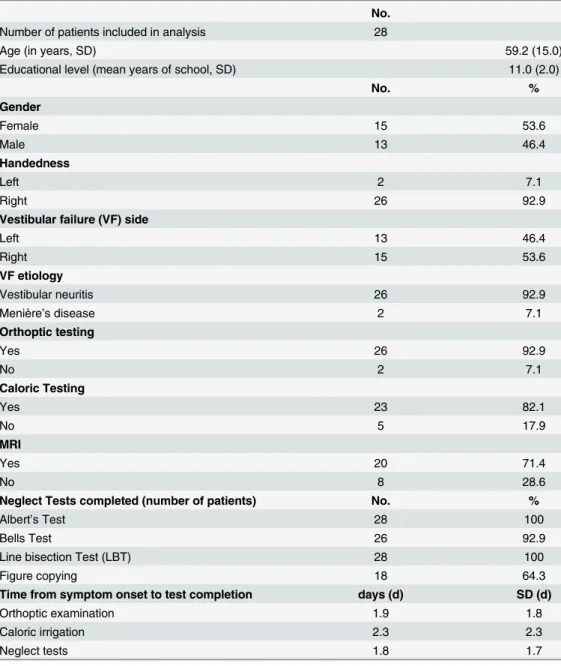
Twenty-nine patients with acute unilateral VF who were admitted to the Department of Neu-rology, Munich University hospital, were studied prospectively. The diagnosis was based on the clinical findings of horizontal rotatory spontaneous nystagmus with the fast phase beating to the contralateral ear and a deficient vestibulo-ocular reflex (VOR) in the head-impulse test. Patients with clinical signs of central involvement (skew deviation, bilateral gaze-evoked nys-tagmus, disturbed fixation-suppression of the VOR) were not included. One of these patients was excluded due to prior sensory deficits (polyneuropathy, bilateral vestibulopathy). The remaining 28 patients were included in the study. Clinical testing was carried out by an experi-enced neuro-otological expert. Patients underwent orthoptic testing, which included measure-ments of the subjective visual vertical (SVV) in 26 patients. The degree of canal paresis was analyzed in 23 patients using Jongkees’s formula for the nystagmus induced by caloric
irrigation.[20] Orthoptic testing could not be performed in two patients, and caloric testing, not in five patients due to severe vertigo and vomiting.
All patients showed clinical signs of VF at the time of testing. The mean time from symptom onset to the completion of the neglect tests was 1.8 days (SD 1.7, max. 6 days). None of the patients had aphasia, hemianopia, or clinically significant cognitive deficits. Patient character-istics are given inTable 1.
For the measurement of SVV a mean of more than 2.5° of the seven measurements of the static SVV determined binocularly was considered pathological.[21]
Spatial neglect was diagnosed on the basis of spontaneous behavior, for example, if patients showed a constant deviation of head and eyes to one side or failed to properly respond and ori-ent themselves to stimuli presori-ented on the contralateral side when approached or addressed verbally. Visual extinction was tested using a confrontation technique. Patients had to
Table 1. Patient demographic and clinical data.
No.
Number of patients included in analysis 28
Age (in years, SD) 59.2 (15.0)
Educational level (mean years of school, SD) 11.0 (2.0)
No. %
Gender
Female 15 53.6
Male 13 46.4
Handedness
Left 2 7.1
Right 26 92.9
Vestibular failure (VF) side
Left 13 46.4
Right 15 53.6
VF etiology
Vestibular neuritis 26 92.9
Menière’s disease 2 7.1
Orthoptic testing
Yes 26 92.9
No 2 7.1
Caloric Testing
Yes 23 82.1
No 5 17.9
MRI
Yes 20 71.4
No 8 28.6
Neglect Tests completed (number of patients) No. %
Albert’s Test 28 100
Bells Test 26 92.9
Line bisection Test (LBT) 28 100
Figure copying 18 64.3
Time from symptom onset to test completion days (d) SD (d)
Orthoptic examination 1.9 1.8
Caloric irrigation 2.3 2.3
Neglect tests 1.8 1.7
determine whether the examiner’s index finger moved in the left, right, or both hemifields, in either the upper or lower quadrants. To determine somatosensory extinction, patients were lightly touched on their forearm, either on the left, right, or both sides simultaneously. Audi-tory stimuli (snipping sounds) were presented on either side or simultaneously directly in front of the ears. All stimulus conditions were repeated in random order three times for each presen-tation (left, right, both). Patients were asked to identify the side of the stimulus. Extinction was diagnosed if patients failed to detect bilateral presentation of the stimuli in any of the runs.
Visuo-spatial functions were further tested by a standardized paper-pencil line bisection task,[22] and two cancellation tasks, the modified Albert’s test,[23,24] and the Bells test.[25] In addition, a figure-copying task was performed by 18 patients.[26]
In the line bisection task three solid lines have to be bisected on a standard sheet of paper. A score from 1–3 is given for the distance from the true center of each line (a mark farther from the midline leads to a lower score; the highest score is 9 and reflects normal peripersonal spatial function). A score<7 was considered pathological.[22] The cancellation tasks were evaluated
as follows: one or more omissions on one side in Albert’s test,[24] and six or more omissions on one side in the Bells test were considered pathological.[25] The figure-copying task was evaluated visually. The results of the cancellation tasks were further analyzed with the Center of Cancellation (CoC) method established by Binder and co-workers, which was recently vali-dated by Rorden and Karnath.[27,28] The continuous scoring of this method is sensitive to both the number as well as the location of these omissions. Scores approaching positive or neg-ative one indicate left- or right-sided neglect, respectively. The cut-off level for pathological test scores was set at 0.081; this was also proposed for right hemisphere stroke.[28]
The Bells test could not be carried out in two patients who were unable to distinguish the bells from the distractors on the test sheet due to impaired vision.
Statistics
To relate omissions on the Bells test to vestibular dysfunction a Kendall’s tau correlation was carried out. Normal distribution was tested using the Shapiro Wilk test, equality of variances was determined using Levene’s test. Differences between the groups with any pathological neglect test score and those without were tested using t-test statistics for independent variables. The values of the deviation of SVV were log-transformed to achieve normal distribution. Sig-nificance level was set to p<0.05.
Standard protocol approvals, registrations and patient consent
Results
On the basis of the clinical criteria none of the patients showed spatial neglect or tactile, visual, or auditory extinction. In the paper-pencil testing one patient fulfilled the criteria for contrale-sional spatial hemineglect (one omission) in Albert’s test. Another patient fulfilled the criteria for contralesional spatial hemineglect in the Bells test. When measuring neglect severity in the Bells test, the mean CoC value was +/- 0.023 (SD 0.027) in 26 patients; seven patients had a CoC score of 0. One patient showed a pathological orientation bias in the Bells test (score 0.088) to the ipsilesional side (see alsoS1 Table).
The mean score was 8.65 in the line-bisection task (mean distance from the center +/- 0.31 cm, max: 2.3 cm). One patient had a pathological score of 4 but showed a leftward and right-ward deviation within the same test. There was no inter-test predictability of a pathological result.
A pathological tilt of SVV was found in 23 of 26 patients; the mean SVV tilt (of 7 measure-ments binocularly) was 8.3° (SD 6.7°). The percentage of canal paresis was 51.3% (SD 26.8%); the mean slow phase velocity (SPV) of spontaneous nystagmus was 6.3°/s. Minor central ocular motor pathology was observed in five patients (disturbed vertical smooth pursuit only).
There was no correlation between the severity of the vestibular tone imbalance and the number of omissions in the Bells test (for deviation of SVV: Kendall’s Tau-b = 0.12, p = 0.43, normal distribution of omissions in the Bells test p = 0.005; for canal paresis: Kendall’s Tau-b = -0.2, p = 0.17, for slow phase velocity of spontaneous nystagmus: Kendall’s Tau-b = -0.19, p = 0.23). The mean deviation of the SVV was 11.27 (SD 4.9°) in patients with any pathological test result compared to 7.9° (SD 6.8°) in patients without such findings. Mean reduction in caloric excitability was 55.2% (SD 5.4%) and 50.2% (SD 28.5%) in the respective groups (see alsoS2 Table). There was no significant difference in the means of the tree measurements (t-test for independent variables: p = 0.27 for deviation of SVV, for canal paresis p = 0.84, for slow phase velocity of SPN p = 0.56). It has to be noted that the group with pathologic scores consisted of only three (two for canal paresis statistics) patients. Therefore the results should be interpreted with caution.
Discussion
Our results pose two major questions: (1) What are the different functional consequences of a peripheral vestibular tone imbalance compared to a cortical imbalance for multisensory spa-tial orientation? (2) Are the methods used sufficient to exclude subtle neglect symptoms, e.g. possibly restricted to the periphery of the visual field, far space or mental representation of space? In the presence of temporo-parietal lesions including the multisensory vestibular net-work, the processing of sensory stimuli in the contralateral visual hemifield is impaired, i.e., right hemispheric lesions impair awareness of visual stimuli in the left hemifield. In contrast, if an acute unilateral peripheral vestibular failure occurs, both hemispheres perceive the vestibu-lar tone imbalance simivestibu-larly because the ascending pathways transmit signals via uncrossed ipsilateral as well as crossed contralateral pathways to both temporo-parietal cortices. There-fore, the vestibular signals from both sides are processed in each hemisphere and do not lead to a functionally relevant cortical asymmetry of“vestibular”spatial orientation. [31,32] This may impair attention in both hemifields, but obviously it does not cause signs and symptoms of a typical spatial hemineglect. Indeed, the pathology of the peripheral vestibular endorgan can cause a range of cognitive deficits, not only spatial but also non-spatial such as a deficiency in object recognition memory.[33,34] The attentional deficits can be attributed to the highly dis-tressing vertigo and nausea in the acute stage. Further, significant deficits in spatial memory and navigation have been demonstrated in rodents as well as in patients with chronicbilateral vestibular deafferentiation.[35,36]
It might be worthwhile to test the deviation of the subjective straight-ahead in these patients rather than the tilts of SVV and to look with more specific methods for attentional deficits especially in the periphery of the visual field contralateral to the side of the lesion. The latter appears promising, since vestibular stimulation like caloric irrigation was able to shift the meridian of the hemineglect in the direction of the slow phase of caloric nystagmus.[16] We also focused on the occurrence of personal and peripersonal neglect in patients with acute ves-tibular failure. We cannot exclude the possibility, however, that subtle spatial deficits involving other subtypes of neglect and more complex tasks might also rely on vestibular cues such as spatial navigation, the mental representation of space (representational neglect), or spatial ori-entation in far space.
Supporting Information
S1 Table. Results neglect tests.
(DOCX)
S2 Table. Results: Subjective visual vertical (SVV), caloric irrigation and mean slow phase velocity of spontaneous nystagmus (SPN).
(DOCX)
Acknowledgments
We thank Judy Benson for copyediting the manuscript.
Author Contributions
References
1. Adair JC, Barrett AM. Spatial neglect: A clinical and neuroscience review: A wealth of information on the poverty of spatial attention. Ann N Y Acad Sci 2008; 1142: 21–43. doi:10.1196/annals.1444.008
PMID:18990119
2. Mort DJ, Malhotra P, Mannan SK, Rorden C, Pambakian A, Kennard C, et al. The anatomy of visual neglect. Brain. 2003; 126: 1986–1997. PMID:12821519
3. De Schotten MT, Tomaiuolo F, Aiello M, Merola S, Silvetti M, Lecce F, et al. Damage to white matter pathways in subacute and chronic spatial neglect: A group study and 2 single-case studies with com-plete virtual“in vivo”tractography dissection. Cereb Cortex. 2012; 24: 691–706. doi:10.1093/cercor/ bhs351PMID:23162045
4. Karnath HO, Fruhmann Berger M, Küker W, Rorden C. The anatomy of spatial neglect based on voxel-wise statistical analysis: a study of 140 patients. Cereb Cortex. 2004; 14(10): 1164–1172. PMID:
15142954
5. Karnath HO, Dieterich M. Spatial neglect–a vestibular disorder? Brain. 2006; 129: 293–305. PMID:
16371409
6. Bartolomeo P, de Schotten MT, Doricchi F. Left unilateral neglect as a disconnection syndrome. Cereb Cortex. 2007; 17: 2479–2490. PMID:17272263
7. Brandt T, Dieterich M, Danek A. Vestibular cortex lesions affect the perception of verticality. Ann Neu-rol. 1994; 35(4): 403–412. PMID:8154866
8. zu Eulenburg P, Caspers S, Roski C, Eickhoff SB. Meta-analytical definition and functional connectivity of the human vestibular cortex. Neuroimage. 2012; 60(1): 162–169. doi:10.1016/j.neuroimage.2011. 12.032PMID:22209784
9. Lopez C, Blanke O, Mast FW. The vestibular cortex in the human brain revealed by coordinate-based activation likelihood estimation meta-analysis. Neuroscience. 2012; 212: 159–179. doi:10.1016/j. neuroscience.2012.03.028PMID:22516007
10. Ventre J, Faugier-Grimaud S. Effects of posterior parietal lesions (area 7) on VOR in monkeys. Exp Brain Res. 1986; 62: 654–658. PMID:3487465
11. Ventre-Dominey J, Vighetto A, Denise P. Vestibulo-ocular dysfunction induced by cortical damage in man: A case report. Neuropsychologia. 1999; 37: 715–721. PMID:10390033
12. Doricchi F, Siegler I, Iaria G, Berthoz A. Vestibulo-ocular and optokinetic impairments in left unilateral neglect. Neuropsychologia. 2002; 40: 2084–2099. PMID:12208005
13. Cappa S, Sterzi R, Vallar G, Bisiach E. Remission of hemineglect and anosognosia during vestibular stimulation. Neuropsychologia. 1987; 25(5): 775–782. PMID:3501552
14. Vallar G, Bottini G, Rusconi ML, Sterzi R. Exploring somatosensory hemineglect by vestibular stimula-tion. Brain. 1993; 116(1): 71–86.
15. Sturt R, David Punt T. Caloric vestibular stimulation and postural control in patients with spatial neglect following stroke. Neuropsychol Rehabil. 2013; 23(2): 299–316. doi:10.1080/09602011.2012.755831
PMID:23305103
16. Karnath HO. Subjective body orientation in neglect and the interactive contribution of neck muscle pro-prioception and vestibular stimulation. Brain. 1994; 117: 1001–1012. PMID:7953584
17. Brandt T, Strupp M, Dieterich M. Towards a concept of disorders of“higher vestibular function”. Front Integr Neurosci. 2014; 8: 47. doi:10.3389/fnint.2014.00047PMID:24917796
18. Brandt T, Dieterich M, Strupp M, Glasauer S. Model approach to neurological variants of visuo-spatial neglect. Biol Cybern. 2012; 106: 681–690. doi:10.1007/s00422-012-0517-3PMID:22941239 19. Choi KD, Jung DS, Jo MK, Kim MJ, Kim JS, Na DL, et al. Vestibular spatial neglect: patterns and
possi-ble mechanism. Neurol Sci. 2014; 35: 341–347. doi:10.1007/s10072-013-1472-zPMID:23812765 20. Jongkees JB, Maas JP, Philipszoon AJ. Clinical nystagmography. A detailed study of
electro-nystag-mography in 341 patients with vertigo. Pract Otorhinolaryngol. 1962; 24: 65–93.
21. Dieterich M, Brandt T. Ocular torsion and tilt of subjective visual vertical are sensitive brainstem signs. Ann Neurol. 1993; 33: 292–299. PMID:8498813
22. Schenkenberg T, Bradford DC, Ajax ET. Line bisection and unilateral visual neglect in patients with neurologic impairment. Neurology. 1980; 30(5): 509–517. PMID:7189256
23. Albert ML. A simple test of visual neglect. Neurology. 1973; 23: 658–664. PMID:4736313
24. Fullerton KJ, McSherry D, Stout RW. Albert's test: a neglected test of perceptual neglect. Lancet. 1986; 1: 430–432. PMID:2868349
26. Wilson B, Cockburn J, Halligan P. Development of a behavioural test of visuo-spatial neglect. Arch Phys Med. 1987; 68: 98–102. PMID:3813864
27. Binder J, Marshall R, Lazar R, Benjamin J, Mohr JP. Distinct syndromes of hemineglect. Arch Neurol. 1992; 49: 1187–1194. PMID:1444886
28. Rorden C, Karnath HO. A simple measure of neglect severity. Neuropsychologia. 2010; 48: 2758– 2763. doi:10.1016/j.neuropsychologia.2010.04.018PMID:20433859
29. Jewell G, McCourt ME. Pseudoneglect: A review and meta-analysis of performance factors in line bisection tasks. Neuropsychologia 2000; 38(1): 93–110. PMID:10617294
30. Bowers D, Heilman KM. Pseudoneglect: Effects of hemispace on a tactile line bisection task. Neuropsy-chologia 1980; 18:491–498. PMID:6777712
31. Conrad J, Baier B, Dieterich M. The thalamus in the human subcortical vestibular system. J Vestib Res. 2014; 24(5): 375–385.
32. Kirsch V, Keeser D, Hergenroeder T, Erat O, Ertl-Wagner B, Brandt T, et al. Structural and functional connectivity mapping of the vestibular circuitry from human brainstem to cortex. Brain Struct Funct. 2015; online first: 1 January 2015. doi:10.1007/s00429-014-0971-x
33. Zheng Y, Balabhadrapatruni S, Masumura C, Munro O, Darlington CL, Smith PF. Bilateral vestibular deafferentation causes deficits in a 5-choice serial reaction time task in rats. Behav Brain Res. 2009; 203(1): 113–117. doi:10.1016/j.bbr.2009.04.027PMID:19397937
34. Andersson G, Hagman J, Talianzadeh R, Svedberg A, Larsen HC. Dual-task study of cognitive and postural interference in patients with vestibular disorders. Otol Neurotol. 2003; 24(2): 289–293. PMID:
12621346
35. Zheng Y, Goddard M, Darlington CL, Smith PF. Long term deficits on a foraging task after bilateral ves-tibular deafferentation in rats. Hippocampus. 2009; 19(5): 480–486. doi:10.1002/hipo.20533PMID:
19072773
36. Brandt T, Schautzer F, Hamilton DA, Brüning R, Markowitsch HJ, Kalla R, et al. Vestibular loss causes hippocampal atrophy and impaired spatial memory in humans. Brain. 2005; 128: 2732–2741. PMID: