Criteria for diagnosing and treating
anterior open bite with stability
Alderico Artese*, Stephanie Drummond**,
Juliana Mendes do Nascimento***, Flavia Artese****
Introduction: Anterior open bite is considered a malocclusion that still defies correc-tion, especially in terms of stability. The literature reports numerous studies on the sub-ject but with controversial and conflicting information. Disagreement revolves around the definition of open bite, its etiological factors and available treatments. It is probably due to a lack of consensus over the etiology of anterior open bite that a wide range of treatments has emerged, which may explain the high rate of instability following the treatment of this malocclusion. Objective: Review the concepts of etiology, treatment and stability of anterior open bite and present criteria for diagnosing and treating this malocclusion based on its etiology, and provide examples of treated cases that have re-mained stable in the long term.
Abstract
Keywords: Open bite. Etiology. Treatment. Stability.
* MSc in Orthodontics, University of Washington. Associate Professor of Orthodontics, UFRJ (Retired). ** Specialist and Masters Student in Orthodontics, UERJ.
*** Specialist in Orthodontics, UERJ.
**** MSc and PhD in Orthodontics, UFRJ. Associate Professor of Orthodontics, UERJ. Brazilian Board of Orthodontics and Facial Orthopedics Diplomate.
IntrOductIOn
The term “open bite” was coined by Caravelli in 1842 as a distinct classification of malocclu-sion1 and can be defined in different manners.2
Some authors have determined that open bite, or a tendency toward open bite, occurs when over-bite is smaller than what is considered normal. Others argue that open bite is characterized by end-on incisal relationships. Finally, others re-quire that no incisal contact be present before diagnosing open bite. For semantic reasons, and because it is in agreement with most definitions
in the literature,2,3,4,5 anterior open bite (AOB) is
herein defined as the lack of incisal contact be-tween anterior teeth in centric relation.
Given these different definitions for AOB, its prevalence varies considerably among studies de-pending on how authors define it. Prevalence in the population ranges from 1.5% to 11%.6 The
age factor, however, affects prevalence, since sucking habits decrease and oral function ma-tures with age. At six years old 4.2% present with AOB whereas at age 14 the prevalence decreases to 2%.5 In the US population, differences in
prev-How to cite this article: Artese A, Drummond S, Nascimento JM, Artese F. Criteria for diagnosing and treating anterior open bite with stability. Dental Press J
alence were detected between the different eth-nicities, with 3.5% occurring in Caucasian
chil-dren and 16.5% in Afro-descendant chilchil-dren.5
Despite its low prevalence, the demand for treat-ment of this malocclusion is very common as ap-proximately 17% of orthodontic patients have
AOB,6 which means that professionals should
treat it in an effective and stable manner.
AOB EtIOlOgIcAl FActOrS: FunctIOnAl Or SKElEtAl?
Teeth and alveolar bones are exposed to an-tagonistic forces and pressures stemming most-ly from muscle function, which may partmost-ly de-termine the position of the teeth. On the other hand, the intrinsic forces of the lips and tongue at rest generate the balance required to posi-tion the teeth (Fig 1). By definiposi-tion, balance
occurs when a body at rest is subjected to forc-es in various directions but doforc-es not undergo acceleration or — in the case of teeth — is not displaced.7 Every time this balance is altered,
changes occur, such as for example contrac-tion of the dental arches in animals subjected to glossectomy when compared to control ani-mals.8 Thus, when a tooth is extracted its
an-tagonist continues the process of passive tion, indicating that the mechanism of erup-tion remains basically unchanged throughout life and that the tooth seeks occlusal or incisal contact until balance is reached.7
Based on this idea of balance several etio-logical factors related to oral function have been associated with AOB. For example, sucking hab-its, presence of hypertrophic lymphoid tissues, mouth breathing, atypical phonation and swal-lowing, and anterior posture of the tongue at rest.2,3,9,10,11 It should be noted, however, that
not all of these etiological factors exhibit a per-fectly clear cause and effect relationship.
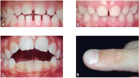
The causal relationship between AOB and nonnutritive sucking habits, such as the suck-ing of fsuck-ingers and pacifiers, has been very well established.12 In such cases, AOB self-corrects
consistently after removal of the sucking habit, provided that no other secondary dysfunctions have set in2 (Fig 2). These secondary
dysfunc-tions may develop from maxillary incisor pro-trusion generated by the sucking habit, thereby hindering the lip seal required for swallowing, and causing the tongue to be abnormally posi-tioned, especially atrest.11
During childhood the tongue is proportion-ally larger than the oral cavity and it therefore protrudes beyond the alveolar ridges. The jaw bones grow faster than the tongue during child-hood and eventually the size of the oral cavity adapts to tongue size.10 In fact, longitudinal
studies in children showed that the prevalence of tongue protrusion in speech and swallowing is significantly reduced starting at 8 years of
A
C
B
D
age. It is approximately 51.7% at 4 years of age and 38.9% at age 12.14
Some authors believe that the forces gen-erated during swallowing and phonation can cause changes in the shape of the dental arch-es.4 Although these disorders are associated in
the literature with AOB etiology, other stud-ies show that these functions are short lived and not sufficient to cause dental changes.7,11
Frequency of atypical speech and swallowing is much higher than AOB prevalence, which may explain the tenuous causal link between the presence of atypical speech and swallowing, and the presence of this malocclusion.11
Hypertrophic adenoids and tonsils are the most common cause of nasal obstruction and,
consequently, mouth breathing in children.4
The effect of airway obstruction on the occlu-sion was demonstrated by Harvold et al16 who,
after placing acrylic blocks in the posterior re-gion of the palate of rhesus monkeys, found that AOB had developed. Induced nasal obstruction was also performed using nasal splints in rhesus
monkeys, which, in an attempt to secure an oral air passage, developed open mouth posture and protruded tongue.17
Therefore, hypertrophic lymphoid tissues and nasal obstruction may force the tongue to remain in a position designed to allow breath-ing to occur through the oropharyngeal rath-er than nasopharyngeal space.12,18 In general,
lymphoid tissues undergo involution during puberty, allowing the tongue to adopt a posi-tion more posterior than what is deemed nor-mal.2 However, Linder-Aronson et al19 found
that dentoalveolar response to adenoidectomy is highly variable and therefore should not be considered as a prophylactic procedure for the development of AOB. Indeed, not all patients with mouth breathing due to partial nasal
blockage develop AOB.4
Most investigations of AOB etiology agree on the existence of secondary dysfunctions, which remain after the correction of an abnormal func-tion, such as, especially, poor tongue posture at rest.4,7,12 It is believed that a gentle but continuous FIGURE 2 - A) AOB in primary teeth caused by pacifier sucking and B) spontaneous correction after
A B
pressure exerted by the tongue against the teeth can move such teeth, yielding significant effects. If a patient has a previous posture in which they have positioned their tongue, the duration of this pressure — even if very light — can affect the eruption process, or move anterior teeth, result-ing in an open bite.10,11
Tongue posture at rest is long lasting (several hours a day), which makes it clinically impor-tant as it can prevent the eruption of incisors, thereby causing and maintaining AOB (Fig 3). In addition, a low tongue posture may encour-age the eruption of posterior teeth and constrict the upper arch since the tongue does not touch the palate.7 This etiological factor has not been
studied enough and is generally overlooked dur-ing AOB treatment. Failure to eliminate this fac-tor may be the primary reason of AOB relapse.10
In 1964, Subtelny and Sakuda2 published an
article on the diagnosis and treatment of AOB. Based on the premise that abnormal functional habits either decrease or are absent in adoles-cents, these authors sought out an explanation for the existence of what they called “persistent open bites,” i.e., those that persist after child-hood. They conducted a cephalometric study in 25 patients with “persistent open bite” and compared them with 30 patients with normal
occlusion. All subjects were over 12 years of age. Basically, in cases of open bite the follow-ing significant differences were found: Greater eruption of maxillary molars, extrusion of max-illary incisors and overly increased mandibular planes and gonial angles. This facial pattern was named “skeletal open bite.” Its primary etiologi-cal factor is an unfavorable growth pattern with divergent basal bones and therefore no contact between the incisors. These etiological factors are associated with growth and not function, and can thus be defined as skeletal factors.
Over the years, vertical facial pattern was ultimately considered as the main risk factor for AOB and its treatment instability. How-ever, other studies10,20 have reported that most
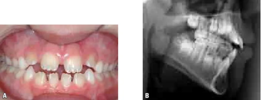
hyperdivergent patients exhibit a normal or excessive overbite (Fig 4) while patients with normal facial patterns display a “persistent open bite”4 (Fig 5).
One can therefore infer that skeletal pattern per se cannot be the cause of AOB.7 In
revisit-ing the aforementioned idea of balance of forces between teeth, the presence of a physical barrier prevents the incisors from coming into occlusal contact. Since an abnormal posture of the tongue at rest may occur in different situations,4,10 this
may be the key etiological factor in AOB.
A
D
B
E F
C
FIGURE 4 - Profile photograph (A), cephalometric radiograph (B), casts (C) and intraoral photographs (D, E and F) of a patient with hyperdivergent facial pattern (SNGoGn=49º), showing excessive overbite, which can be seen more clearly in a posterior view of the plaster casts in occlusion. The lower inci-sors touch the palate seeking occlusal contact since there is no structure preventing its eruption.
AOB trEAtMEnt And StABIlItY
Due to numerous etiological factors de-scribed in the literature various types of treat-ment have been postulated for correcting AOB. No consensus has been reached, however, as to what would be the best treatment for this mal-occlusion:6 (a) Changes in behavior to eliminate
habits or abnormal functions, (b) Orthodontic movement by extruding the anterior teeth or intruding the molars, or (c) Surgical treatment of the basal bones.21 The only consensus that
seems to exist is that AOB treatment is chal-lenging3,6 and has poor stability.6,9,22
Functional treatments
Myofunctional therapy is used to alter func-tion and consists of a set of exercises to reedu-cate orofacial muscles in swallowing, speech and
resting posture.11,12,15 It is believed that
volun-tary activities such as swallowing and speech are easier to correct using myofunctional exercises while involuntary activities such as tongue pos-ture habits are hard to automate.11,14
Another way to correct functional habits is through mechanisms that prevent the tongue from resting on the teeth.23 The best known are
palatal or lingual cribs24 and spurs10,25 There is
a consensus that these devices should be fixed with the purpose of re-educating the function until automatic movements are attained.25,26
Palatal or lingual cribs are aimed at correct-ing AOB by preventcorrect-ing the tongue from restcorrect-ing on the teeth. They must be long to prevent the
tongue from positioning itself below them.24
A
C
B
D E
that in some cases this may prevent the func-tional re-education of the tongue. In these cas-es, the tongue returns to its original position as shown by the cinefluoroscopic method,28 thus
leading to AOB relapse.
The use of spurs was described by Rogers28 in
1927 in the treatment of three AOB cases. The spurs were welded to a palatal arch and placed from canine to canine. All cases were corrected by normalizing the tongue posture. Several types of similar devices were later described in which spurs can be soldered to the lingual surfaces of maxillary incisor bands or attached to palatal10
or lingual29 arches or, alternately, bonded to the
lingual or palatal surfaces of the incisors.26
Despite their efficacy, treatments using spurs are sometimes regarded as punitive,1,2 although
there are no reports of pain or injury to the
tongue.10 Furthermore, Haryett et al23
conclud-ed that any type of device usconclud-ed to break the finger sucking habit, including spurs, can cause psychological disorders.
Spurs induce a change in the resting posi-tion of the tongue, thus allowing tooth erup-tion and open bite closure. This change in tongue position alters sensory perception by the brain, thereby producing a new motor re-sponse. This response can be imprinted manently in the brain, which explains the per-manent change in tongue posture produced by spurs. This is one of the factors responsible for AOB treatment stability.10,25
Huang et al3 evaluated AOB treatment
sta-bility using cribs or spurs in 33 patients di-vided into two groups, one with and one with-out growth. These authors found that AOB
correction occurred in both groups but 17.4% of cases showed relapse. Since no comparison be-tween different treatment types was performed, one could argue that patients whose overbite is corrected with the use of cribs or spurs stand a good chance of maintaining long-term treat-ment outcome. However, comparative studies between these two types of treatment would be invaluable for the prognosis of AOB treatment.
Orthodontic treatments
There are several types of treatment involv-ing orthodontic movement for correction of open bite, with different therapeutic goals. Ex-traoral appliances, vertical chincups, bite-blocks and functional appliances are designed to reduce the extrusion of molars, allowing a counterclock-wise rotation of the mandible.6,9,22 More
recent-ly, the same mechanism was implemented with the aid of anchorage to intrude molars.6,21
Me-chanics with intraoral elastics are used both for incisor extrusion2 and molar intrusion, as well
as for rotation of the occlusal plane combined with multiloop archwires.30 Although there are
many successful reports of these therapies few studies have been conducted to investigate their long-term stability, which precludes any reliable prognoses for these treatments.4,6,22
Stability in the correction of AOB in pa-tients treated orthodontically with fixed appli-ances associated with high-pull and combined headgear was evaluated 10 years after
treat-ment.9 AOB relapse was greater than 3 mm in
35% of the cases. The sample was then stratified into stable and relapse groups for comparison of cephalometric variables. All variables were similar between the groups at the beginning of treatment, except for anterior dental height in the mandibular arch, which was lower in the relapse group at all treatment times.
Zuroff et al6 assessed AOB stability 10
years after treatment. Sixty-four patients were divided into three groups: One with incisal
contact, one with open bite and overlap, and one with open bite. All patients were only treated orthodontically. After treatment, 4% of the group with incisal contact had overjet relapse; 20% of the group with open bite and overlap had overjet relapse but preserved in-cisal contact; and 40% of the open bite group had overjet, with 60% displaying no incisal contact. These results indicate that a lack of vertical overlap prior to treatment exerts a greater adverse effect on AOB stability com-pared to open bite with overlap.
Surgical treatments
Surgical treatments for AOB began in the 70s and were indicated for extremely severe cases with mandibular plane above 50 degrees. Thereafter, these treatments have become more common and usually include LeFort I osteot-omy for superior repositioning of the maxilla. This allows a counterclockwise rotation of the mandible, thus correcting AOB.22
Denison et al22 assessed the stability of AOB
surgical treatment in 66 adult patients fol-lowed up for at least 1 year after surgery. These patients were stratified according to preopera-tive vertical overlap, namely: Open bite, open bite with overlap, and normal overlap. Open bite recurred in 42.9% of cases in the open bite group while the groups with open bite and overlap, and normal overlap showed no chang-es in postoperative overbite. It was found that the instability found in patients in the open bite group was due to dentoalveolar changes and not to skeletal changes.
Greenlee et al21 published a meta-analysis
which evaluated AOB treatment stability in surgical and nonsurgical studies. A 75% stabil-ity rate was found in both types of treatment. However, these results should be viewed with caution since these various treatments were examined in different studies and applied to different populations. Moreover, these studies lacked control groups.
Nowadays there are not enough evidence-based findings to support the effectiveness of
AOB21 treatment or stability of AOB
correc-tion. Randomized trials evaluating different
therapies are thus necessary.5 However, the
outcomes of the stability studies described above indicate that AOB relapse is linked to two factors: Dentoalveolar changes and open bites with no vertical overlap prior to treat-ment.3,6,9,22 These data suggest that AOB
re-lapse is generally caused by the anterior posi-tion of the tongue at rest, an etiological fac-tor that has not merited due attention in both orthodontic and surgical treatment.3,10
dIFFErEnt POSturES OF tHE tOnguE At rESt
AOB morphology is directly associated with etiological factors,7 which differ for each type of
habit (Fig 2). In AOB cases that do not result from sucking habits one can use this logic to dif-ferentiate between the resting positions of the tongue, as there may be more than one type of resting position.
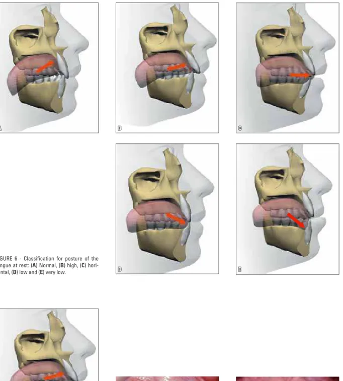
The position considered normal for the tongue at rest is one in which the tip of the tongue rests on the incisal papilla and its back lies along the palate (Figs 1 and 6A), keeping the anterior teeth in balance while preserving
the transverse dimension of the upper arch.7
However, some AOBs show changes in the positions assumed by maxillary incisors and others display changes in the positions of man-dibular incisors. Based on these morphological
characteristics some different resting positions of the tongue are suggested: High, horizontal, low and very low (Fig 6).
A high posture of the tongue at rest is associ-ated with slightly protruded maxillary incisors and AOB may exhibit vertical overlap and positive horizontal overlap. Since the tongue rests on the palatal surface of the incisors, beneath the incisal papilla, upper incisors are positioned above the occlusal plane. Leveling of the mandibular arch is unaffected and displays a single occlusal plane (Fig 7). Posterior crossbites are not present as the back of the tongue rests on the palate while maintaining the transverse dimension of the upper arch.
In the horizontal posture of the tongue at rest, the tongue appears lower than in the high position, although with greater protrusion, rest-ing on the palatal surface of the upper incisors and on the incisal edges of the lower incisors. The major effect in this case can only be seen in the upper arch, where protrusion of maxillary incisors was more prominent, which prevented their extrusion, thereby causing AOB. Also due to the greater protrusion of the incisors, a posi-tive and increased horizontal overlap was noted. As the tongue positions itself lower, its back turns away from the palate allowing transverse changes to occur in the maxillary arch, which may cause posterior crossbites (Fig 8).
D
A B C
E
A B C
FIGURE 6 - Classification for posture of the tongue at rest: (A) Normal, (B) high, (C) hori-zontal, (D) low and (E) very low.
A B D C A very low tongue posture occurs when the
tongue rests below the crowns of the mandibu-lar incisors in the lingual region of the lower alveolar ridge. The direction of tongue pressure produces retroclination of mandibular incisors and prevents their eruption, positioning them below the occlusal level. The open bite is more severe and associated with posterior crossbite due to the fact that the tongue moves away from the palate. The tongue sprawls across the mouth floor, expanding the lower arch in the transverse direction (Fig 10).
trEAtMEnt cHOIcE BASEd On tOnguE POSItIOn At rESt: rEStrAInIng And OrIEntIng trEAtMEntS
Understanding AOB etiology in each patient may help in their treatment and long-term sta-bility.4 These various postures of the tongue at
rest will guide orthodontists in choosing the treatment capable of bringing the tongue back to a correct resting posture, thus removing the causative agent of the malocclusion.
Once the AOB causative agent has been
iden-tified and ascribed to an abnormal posture of the tongue, orthodontists should classify tongue posture through an analysis of the morphologi-cal features of the malocclusion.
High and horizontal tongue postures are positioned very close to normal posture and re-quire control in the horizontal direction only. It is suggested that blocking mechanisms such as cribs are sufficient to produce this tongue retrac-tion and adapt it to its correct posture at rest. This type of treatment will be referred to as re-straining treatment.
However, in the low and very low tongue postures, the tongue is not only protruded but it is positioned below its correct position and needs to be retracted and elevated. This process is difficult to learn and automate,25 requiring
ed-ucating devices which force the direction of the tongue, such as spurs. This type of treatment will be referred to as orienting treatment.
To illustrate these types of treatment, and in particular their stability, AOB cases caused by each type of tongue posture at rest, which were monitored in the long-term, will be presented.
A B D C
A B D
C
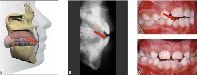
FIGURE 9 - Schematic (A), radiograph (B) and photographs (C and D) of low posture of the tongue at rest, associated with a moderate AOB. The mandibular incisors display a pronounced protrusion. Lower arch leveling is changed, with mandibular incisors positioned below the occlusal level. Due to the distance between the back of the tongue and the palate, posterior crossbites may emerge. The arrows represent the direction of the force exerted by the tongue.
FIGURE 10 - Schematic (A), radiograph (B) and photographs (C and D) of very low posture of the tongue at rest, associated with a severe AOB. The mandibular incisors appear uprighted or retroclined. Lower arch leveling is changed, with mandibular incisors well below the occlusal level. Due to the distance between the back of the tongue and the palate, posterior crossbites are bound to emerge. The arrows represent the direction of the force exerted by the tongue.
APPlYIng crItErIA FOr AOB dIAgnOSIS And trEAtMEnt: cASE rEPOrtS
case 1: High Posture of tongue at rest This is an 8-year-old female patient in the mixed dentition stage. She presented with an Angle Class I malocclusion with AOB, slightly increased overjet, protruded maxillary incisors and interincisal diastemas in the upper arch. The
C
A
D
B
E
A B
the maxillary incisors were protruded and po-sitioned above the occlusal plane (Figs 11C, D and E). Since the treatment goal was to restrain the tongue in the horizontal direction, placing it further back, restraining treatment was pre-ferred and a Hawley retainer was therefore used, combined with a crib (Fig 12A).
The retainer was used for a period of two years until the patient was in the final stage of mixed dentition (Fig 12B). She was monitored until the permanent dentition phase. The open bite was
closed, overjet and interincisal diastemas reduced (Figs 13C, D and E). No other treatment was per-formed on this patient, who achieved a stable re-sult as can be seen from the records obtained 32 years after treatment (Fig 14).
It was only thanks to the removal of a poor tongue posture that establishing a normal hori-zontal overlap became possible and, more im-portantly, the AOB etiological factor was elim-inated, thus ensuring a stable result for many years (Fig 15).
FIGURE 11 - Initial facial (A and B) and intraoral photographs (C, D and E).
C D E
A B
C D E
A B
FIGURE 13 - Extraoral (A and B) and intraoral photographs (C, D and E) at the end of treatment. The patient was not subjected to any other type of orth-odontic treatment.
A B
C D
FIGURE 15 - A) Initial AOB condition, B) during treatment with Hawley retainer with crib, C) end of treatment and D) 32 years after treatment, demonstrating stability of AOB correction.
case 2:
Horizontal Posture of tongue at rest
A female patient aged 9 years, in the mixed dentition period presenting with an Angle Class II, Division 1 malocclusion, 8 mm overjet, cross bite of teeth 16 and 46, AOB and less than 2 mm midline shift to the right (Figs 16E, F and G). She had a Class II skeletal pattern with 10º ANB (SNA=88° and SNB=78°) and normal mandibular plane (SNGoGn=34º) (Fig 16D). Facial evaluation showed a symmetrical face and convex profile (Figs 16A, B and C).
Patient history revealed that she had no sucking habits, suggesting that AOB etiol-ogy was related to abnormal tongue posture. To determine what sort of tongue posture the patient had it was observed that lower arch leveling was normal while the upper incisors were protruded and positioned above the oc-clusal level. These features suggest a horizontal posture of the tongue associated with marked overjet. Therefore, restraining treatment would be indicated in this case.
It was decided the use of a modified Thu-row appliance with expansion screw and
pala-tal crib (Fig 17), which was worn for six con-secutive months. After this period, an Angle Class I molar relationship was attained with 3 mm overjet, the crossbite was corrected as well as the AOB (Figs 18E, F and G) and there was improvement in the skeletal relationship (SNA=83°, SNB=78º and ANB=5º) (Fig 18D). The face remained symmetrical and the profile slightly convex (Figs 18A, B and C). The appli-ance was then worn only at night for another six months for retention purposes.
E F G
A B C D
E F G
A B C D
FIGURE 16 - Initial extraoral photographs (A, B, C), cephalometric radiograph (D) and intraoral photographs (E, F and G).
A B
D E F
A B C
FIGURE 20 - Extraoral (A, B and C) and intraoral photographs (D, E and F) at the end of the second treatment phase.
FIGURE 19 - Total (A) and partial (B) cephalo-metric superimpositions comparing the begin-ning and end of the first treatment phase. It is noteworthy that AOB correction occurred by extrusion of the maxillary incisors.
In this case, AOB correction occurred thanks to a spontaneous extrusion of the incisors (Fig 19) after using a palatal crib and correcting the tongue posture. The results were stable as can be seen in the follow-up photographs 10 years after treatment (Fig 21). Stability of AOB correction was accomplished because the etiological factor
D E F
A B C
A B
C D
FIGURE 21 - Extraoral (A, B and C) and intraoral photographs (D, E and F) 10 years after treatment.
E F G
A B C D
case 3: High Posture of tongue at rest A 7-year-old female patient with mixed den-tition presented with a Class I molar relation-ship, without horizontal overlap, with AOB and tendency toward posterior crossbite (Figs 23E, F and G). No sucking habit was reported. She had a typical skeletal Class I (SNA=78°, SNB=77° and ANB=1º) with increased mandibular plane (SNGoGn=37) (Fig 23D). The face was balanced with no apparent asymmetries, with lip incompe-tence and a convex profile (Figs 23A, B and C).
The morphological features of this AOB included slightly protruded maxillary incisors with deficiently erupted and protruded man-dibular incisors (IMPA=100º) (Figs 23D and F). These effects in the lower arch suggest a low posture of the tongue at rest. Since this tongue had to be retracted and elevated, it was decided to conduct orienting treatment with spurs on the lingual arch (Fig 24).
The spurs were worn for a period of two years and the patient monitored for another two years until the permanent dentition stage. By then the patient had developed a Class I molar relationship, severe lack of space in both arches, posterior crossbite on the right side, and normal overbite (Fig 25). The mandibular inci-sors were uprighted and extruded through the use of spurs (IMPA=92º) (Fig 26). The skeletal Class I relationship was maintained (ANB=1º). Corrective treatment was then initiated with extraction of first premolars.
Corrective treatment was performed with canine distalization followed by retraction of the incisors. No anchorage mechanism was used, nor any vertical elastics, which attests to the sta-bility of the AOB correction. Dental alignment was attained as well as vertical and horizontal overlaps, and adequate intercuspation. The pro-file remained balanced (Fig 27).
E F G
A B C D
A B C
A B
FIGURE 24 - Panoramic radiograph of patient with spurs in place, reori-enting the tongue backwards and upwards.
FIGURE 25 - Extraoral photographs (A, B and C), cephalometric radiograph (D) and intraoral photographs (E, F and G) after use of spurs in permanent dentition.
D E F
A B C
FIGURE 27 - Extraoral (A, B and C) and intraoral photographs (D, E and F) at the end of corrective treatment after 7 years of spur use, showing stability of AOB correction.
case 4: Very low Posture of tongue at rest A female patient aged 9 years, showing se-vere anterior open bite and sese-vere lack of space in the lower arch (Figs 28E, F and G). The pa-tient was a mouth breather and undergoing speech therapy. She had a Class III skeletal pattern (ANB=-1°), a tendency toward verti-cal growth, and an increased mandibular plane (SNGoGn=49º) (Fig 28D). The face showed no clear asymmetry and had an adequate pro-file (Figs 28A, B and C).
According to the morphological character-istics of the open bite, the patient had a very low position of the tongue at rest, clearly char-acterized by retroclination of mandibular in-cisors (IMPA=70°) and posterior crossbite. To perform the correction it would be necessary to move the tongue upward and backward with orienting treatment. The appliance of choice was a lower lingual arch with spurs. Firstly, a
single spur was placed in the midline region, then other spurs were gradually inserted in the canine-to-canine region (Fig 29).
Use of lingual arch with spurs was suspended four years later. At this time a significant im-provement in vertical overlap was observed as well as the presence of diastemas in the mandib-ular incisor region (Figs 30D, E and F) due to the protrusion of these teeth. The profile remained balanced and the face symmetrical (Figs 30A, B and C). At this stage, it was decided to place a fixed orthodontic appliance in the mandibular arch in order to close spaces.
E F G
A B C D
A B
FIGURE 28 - Initial extraoral photographs (A, B, C), lateral cephalometric radiograph (D) and intraoral photographs (E, F and G).
FIGURE 29 - Spurs used on lingual arch, start-ing with one spur at arch center (A) and in-creasing number and size of spurs (B) in order to reorient tongue posture backwards and upwards.
No expansion was performed in the upper arch and crossbite was corrected by posi-tioning the tongue higher, thus changing the transverse dimension of the arch. The face remained symmetrical with a balanced fa-cial profile (Figs 31A, B and C). At this stage, fixed appliances were installed in the upper jaw to finish the case.
At the end of treatment an excellent occlu-sal outcome was accomplished, with the estab-lishment of a Class I relationship and correct horizontal and vertical overlap (Figs 32E, F and G). A skeletal Class I relationship was attained (ANB=1º) (Fig 31D). Despite the high mandib-ular plane (SNGoGn=50) the face was balanced
with a good profile and adequate lip seal (Figs 32A, B and C).
Correction of this AOB was achieved mostly by a significant extrusion of the mandibular in-cisors (Figs 33A and B). The backward and up-ward change in tongue posture allowed eruption of the incisors, thereby lengthening the alveolar process (Figs 33C, D, E and F), as reported by Meyer-Marcotty et al.25 The skeletal features of
this face would have one believe that the cause of the AOB might be an unfavorable growth pat-tern.2 However, this case suggests that AOB
D E F
A B C
D E F
A B C
FIGURE 30 - Extraoral (A, B and C) and intraoral (D, E and F) photographs after 4 years of spur use.
E F G
A B C D
A B
C D E F
FIGURE 32 - Extraoral photographs (A, B and C), lateral cephalometric radiograph (D) and intraoral photographs (E, F and G) at the end of treatment.
D E F
A B C
A B
C D
skeletal pattern would not play an etiological role in AOB.
Removal of the causative agent of this AOB ensured outcome stability 10 years after treat-ment, as shown in Figure 34. Treatment of these cases requires patience and the long-term use
FIGURE 34 - Extraoral (A, B and C) and intraoral (D, E and F) photographs 10 years after treatment.
FIGURE 35 - A) Initial open bite position, B) Intermediate treatment stage after adjusting overbite with spurs and placement of appli-ance in the lower arch, C) Overbite achieved after corrective treatment and D) Overbite stability 10 years after treatment.
FInAl cOnSIdErAtIOnS
The difficulties encountered in obtaining stable results for AOB correction can be justified by the fact that their true etiology still defies understand-ing. The posture of the tongue at rest is not highly regarded in AOB treatments. Some evidence sug-gests that the posture of the tongue may be one of the most important etiological factors in AOB. Therefore, it must be analyzed and addressed when it is abnormal.
There is more than one possible resting posi-tion for the tongue. It can posiposi-tion itself on a high-er or lowhigh-er level, producing open bite with dif-ferent morphological characteristics and severity.
Appropriate treatment should be selected based on these characteristics, and can be conducted by either restraining or orienting the tongue. Once the posture of the tongue has been corrected, the etiological factor is extinguished and treatment stability is ensured.
contact address Flavia Artese
Rua Santa Clara, 75/1110
CEP: 22.041-011 - Copacabana / RJ, Brazil E-mail: [email protected]
1. Parker JH. The interception of the open bite in the early growth period. Angle Orthod. 1971 Jan;41(1):24-44. 2. Subtelny HD, Sakuda M. Open bite: diagnosis and
treatment. Am J Orthod. 1964 May;50(5):337-58. 3. Huang GJ, Justus R, Kennedy DB, Kokich VG. Stability of
anterior openbite treated with crib therapy. Angle Orthod. 1990 Jun;10(1):17-24.
4. Shapiro PA. Stability of open bite treatment. Am J Orthod Dentofacial Orthop. 2002 June;121(6):566-8.
5. Cozza P, Mucedero M, Baccetti T, Franchi L. Early orthodontic treatment of skeletal open bite malocclusion: a systematic review. Angle Orthod. 2005 Sept;75(5):707-13. 6. Zuroff JP, Chen SH, Shapiro PA, Little RM, Joondeph DR,
Huang GJ. Orthodontic treatment of anterior open-bite malocclusion: stability 10 years postretention. Am J Orthod Dentofacial Orthop. 2010 Mar;137(3):302.e1-302.e8.
7. Profit WR. Equilibrium theory revisited: factors inluencing
position of the teeth. Angle Orthod. 1978 July;48(3)175-86.
8. Negri PL, Croce G. Inluence of the tongue on development
of the dental arches. Dental Abstr. 1965;10:453.
9. Lopez-Gavito G, Wallen T, Little RM, Joondeph DR. Anterior open-bite malocclusion: a longitudinal 10-year postretention evaluation of orthodontically treated patients. Am J Orthod. 1985 Mar;87(3):175-86.
10. Justus R. Correction of anterior open bite with spurs: long-term stability. World J Orthod. 2001;2(3):219-31. 11. Franco FC, Araújo TM, Habib F. Pontas ativas: um recurso
para o tratamento da mordida aberta anterior. Ortodon Gaúch. 2001 jan-jun;5(1):5-12.
12. Miller H. The early treatment of anterior open bite. Int J Orthod. 1969 Mar;7(1):5-14.
13. Andrianopoulos MV, Hanson ML. Tongue-thrust and the stability of overjet correction. Angle Orthod. 1987 Apr;57(2):121-35.
14. Yashiro K, Takada K. Tongue muscle activity after orthodontic treatment of anterior open bite: a case report. Am J Orthod Dentofacial Orthop. 1999 June;115(6):660-6.
15. Subtelny JD, Subtelny JD. Malocclusion, speech, and deglutition. Am J Orthod. 1962 Sept;48(9):685-97. 16. Harvold EP, Vagervik K, Chierici G. Primate experiments on
oral sensation and dental malocclusion Am J Orthod. 1973 May;63(5):494-508.
17. Harvold EP, Tomer BS, Vagervik K, Chierici G. Primate experiments on oral respiration. Am J Orthod. 1981 Apr;79(4):359-72.
rEFErEncES
18. Brauer JS, Holt TV. Tongue thrust classiication. Angle
Orthod. 1965 Apr;35(2):106-12.
19. Linder-Aronson S, Woodside D, Hellsing E, Emerson W. Normalization of incisor position after adenoidectomy. Am J Orthod Dentofacial Orthop. 1993 May;103(5):412-27. 20. Dung J, Smith R. Cephalometric and clinical diagnosis of
open bite tendency. Am J Orthod. 1998 Dec;94(6):484-90. 21. Greenlee GM, Huang GJ, Chen SS, Chen J, Koepsell T,
Hujoel P. Stability of treatment for anterior open-bite malocclusion: a meta-analysis. Am J Orthod Dentofacial Orthop. 2011 Feb;139(2):154-69.
22. Denison TF, Kokich VG, Shapiro PA. Stability of maxillary surgery in openbite versus nonopenbite malocclusions. Angle Orthod. 1989 Spring;59(1):5-10.
23. Haryett RD, Hansen FC, Davidson PO, Sandilands ML. Chronic thumb-sucking: the psychologic effects and the relative effectiveness of various methods of treatment. Am J Orthod. 1967 Aug;53(8):569-85.
24. Subtelny JD. Examination of current philosophies associated with swallowing behavior. Am J Orthod. 1965 Mar;51(3):161-82. 25. Meyer-Marcotty P, Hartmann J, Stellzig-Eisenhauer A.
Dentoalveolar open bite treatment with spur appliances. J Orofac Orthop. 2007 Nov;68(6):510-21.
26. Nogueira FF, Mota LM, Nouer PRA, Nouer DF. Esporão lingual colado Nogueira®: tratamento coadjuvante da
deglutição atípica por pressionamento lingual. Rev Dental Press Ortod Ortop Facial. 2005 mar-abr;10(2):129-56. 27. Cleall JF. Deglutition: a study of form and function Am J
Orthod. 1965 Aug;51(8):587-94.
28. Rogers AP. Open bite cases involving tongue habits. Int J Orthod. 1927;13:837-44.
29. Hickham JH. Maxillary protraction therapy: diagnosis and treatment. J Clin Orthod. 1991 Feb;25(2):102-13. 30. Kim YH, Han UK, Lim DD, Serraon ML. Stability of anterior
openbite correction with multiloop edgewise archwire therapy: a cephalometric follow up study. Am J Orthod Dentofacial Orthop. 2000 July;118(1):43-54.